Acupuncture is an ancient Chinese therapy involving the stimulation of specific trigger points along the body’s 18 meridian lines to help regulate the flow of Qi (energy). The meridian lines represent the normal flow of Qi through the body. It is believed that when this energy is disrupted, disease ensues. The use of thin metal needles or other acupuncture techniques is proposed to conduct Qi through its correct paths. The trigger points used are areas of the skin where Qi flows close to the surface and thus can be reached by the various acupuncture therapies.
While the exact mechanisms are not well defined in terms of Western medicine, there are biological responses that occur directly at the stimulus point and indirectly at other parts of the body. In addition to the use of fine needles, other methods of acupuncture include:
electro-acupuncture (current through the needles),Pictures courtesy of Ricardo Miranda,L.Ac
cupping (suction cups on trigger points),Pictures courtesy of Ricardo Miranda,L.Ac
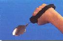
acupressure using trigger points (applying pressure with fingers or instruments),
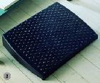
reflexology (using pressure on the soles of the feet and inferior ankle to stimulate various parts of the body),
moxibustion (heat at trigger points, often combined with needles),Pictures courtesy of Ricardo Miranda,L.Ac
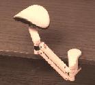
auriculotherapy (stimulating trigger points on the ear to affect other parts of the body),
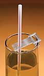
laserpuncture and sonopuncture (using sound waves over trigger points).
Acupuncture has been used to treat many types of health problems and in the past decade has been advocated by some for the treatment of stroke. Recently, a number of studies have explored the use of acupuncture in stroke rehabilitation.
Acupuncture comes from ancient Chinese medicine. It has been used to treat pain in China for about 3000 years. The Chinese explanation involves Qi (pronounced Chee), an energy that flows through the body. The belief is that when this Qi is balanced (Yin and Yang), then the body is healthy. Qi flows through different lines within your body called “meridians”. With the most common form of acupuncture, an expert puts very small needles into specific areas of your body where Qi flows close to the surface of the skin.
There is some evidence that acupuncture works after operations to stop pain, after chemotherapy to stop feeling sick and vomiting, during pregnancy to stop feeling sick and after dental surgery for dental pain. It has also been used to treat headaches, tennis elbow, fibromyalgia (general muscle pain), low back pain, carpal tunnel syndrome and asthma.
While we are not sure exactly how it works, 3 possible explanations have been given:
Acupuncture blocks pain from traveling in your nerves
Acupuncture causes your body to make chemicals that prevent pain
Acupuncture opens or closes your veins and arteries in important areas of the body
Are there different kinds of acupuncture?
The most popular acupuncture is performed by putting thin metal needles into the skin. Other forms of acupuncture include:
electro-acupuncture, which again uses needles through which very small electrical currents are passed;Pictures courtesy of Ricardo Miranda,L.Ac
auriculotherapy, which uses either needles or pressure on different spots of the ear which are trigger points for the entire body;
moxibustion, which uses heat at different spots on the body;Pictures courtesy of Ricardo Miranda,L.Ac
sonopuncture, which uses sound waves at different spots on the body
cupping, which uses suction cups over areas such as the back or the legs to pull blood and other fluids in the area under the skin;Pictures courtesy of Ricardo Miranda,L.Ac
acupressure, which uses pressure on different spots on the body;
reflexology, which uses pressure under the feet or the back part of the ankles.
Why use acupuncture after a stroke?
Acupuncture has been used after a stroke to treat spasticity (stiffness of muscles caused by the stroke), loss of function, loss of mobility, depression, aphasia (loss of speaking and writing skills), hemiplegia (loss of feeling and/or power to move one side of the body) and for pain reduction.
Does it work for stroke?
Experts have done some experiments to compare acupuncture with other treatments to see whether acupuncture helps people who have had a stroke.
In individuals with ACUTE stroke (< 4 weeks after stroke) Thirteen high quality studies and 7 fair quality studies found that acupuncture:
Was not more helpful than other treatments for improving cognitive skills (e.g. memory, language); mood (e.g. depression); self-care skills (e.g. dressing, shopping); quality of life; physical skills (e.g. strength, range of motion, sensation, motor function of arms and legs); or mobility (e.g. balance, walking speed); but
Was more helpful than the usual treatment for improving swallowing skills and swallowing safety.
In individuals with SUBACUTE stroke (1 to 6 months after stroke) One high quality study found that acupuncture:
Was not more helpful than pretend acupuncture for improving range of motion.
In individuals with CHRONIC stroke (> 6 months after stroke) Three high quality studies and 1 low quality study found that acupuncture:
Was not more helpful than pretend acupuncture for improving mood (e.g. depression); self-care skills (e.g. dressing); mobility (e.g. walking endurance); physical skills (e.g. spasticity, range of motion, strength) or pain.
What can I expect?
Most people find that having acupuncture treatment causes very little pain, if any. In most cases you feel the needle going in, but it doesn’t hurt. Some people say they feel cramping, heaviness or tingling at the needle site or up the “meridian”.
The acupuncturist may use other treatments once the needles are in place. This depends on his/her training.
Side effects/risks?
As with any other use of needles, sanitation is very important to not spread germs. All acupuncturists should use new, individually packaged, disposable needles. If these are not used, don’t agree to treatment.
There is little risk related to acupuncture if done by a qualified professional. Side effects could include dizziness, feeling sick and feeling tired after treatment. There could also be a little bleeding at the needle site and some slight bruising. There is always a slight risk of infection when putting needles in the skin.
Who provides the treatment?
Acupuncture should be practiced by a trained health professional. For example, in Quebec (Canada) the practice of acupuncture is regulated by a professional Order and only members of the Order can practice it. Different health care professionals such as physicians and physiotherapists may use the trigger point needle technique as part of their treatment.
How many treatments?
This depends on the reason you are getting acupuncture. You should discuss the treatment plan with the acupuncturist before starting treatment. You might receive anywhere from one to 15 treatment sessions.
How much does it cost? Does insurance pay for It?
Acupuncture is not paid for by provincial insurance plans. However, it is covered by some private insurance plans. The cost for each session may vary from $40.00 to $90.00.
Is acupuncture for me?
Although the benefits of acupuncture have been talked about for hundreds of years, there is no strong scientific evidence that it works to reduce spasticity, loss of function, loss of mobility, depression, aphasia or pain. Yet, there are some people who say they have found it helpful.
Clinician Information
Note: When reviewing the findings, it is important to note that they are always made according to randomized clinical trial (RCT) criteria – specifically as compared to a control group. To clarify, if a treatment is “effective” it implies that it is more effective than the control treatment to which it was compared. Non-randomized studies are no longer included when there is sufficient research to indicate strong evidence (level 1a) for an outcome.
The current module includes 35 RCTs including 25 high quality RCTs, nine fair quality RCTs and one poor quality RCT. Numerous outcome measures were used throughout studies and outcomes include balance, cognitive function, dexterity, depression, functional independence, motor function, quality of life, swallowing function, etc. Studies conducted with patients in one phase of stroke recovery, be it the acute, subacute, or chronic phases of stroke recovery, predominantly reported that acupuncture was not more effective than comparison interventions in improving most outcomes (with the exception of dysphagia and swallowing function). By comparison, studies that included patients across stages of stroke recovery (e.g. patients in the acute or subacute phases of stroke recovery) generally reported that acupuncture was more effective than comparison interventions in improving outcomes (especially those related to cognitive function, health related quality of life, insomnia, mobility and swallowing function).
One high quality RCT (Hsieh et al., 2007) and one fair quality RCT (Johansson et al., 1993) investigated the effect of acupuncture on balance in patients with acute stroke.
The high quality RCT (Hsieh et al., 2007) randomized patients to receive electroacupuncture or no acupuncture; both groups received conventional rehabilitation. Balance was measured by the Fugl-Meyer Assessment (FMA – Balance) during treatment (2 weeks), at post-treatment (4 weeks), and follow-up (3 and 6 months post-stroke). No significant between-group differences were found at any time point.
The fair quality RCT (Johansson et al., 1993) randomized patients to receive electroacupuncture or no acupuncture; both groups received conventional rehabilitation. Balance was measured by the modified Chart for Motor Capacity Assessment – Balance at mid-treatment (1 month post-stroke), and follow-up (3 months post-stroke); measures were not taken at post-treatment (10 weeks). Significant between-group differences were found at both time points, favoring electroacupuncture vs. no acupuncture.
Conclusion: There is moderate evidence (Level 1b) from one high quality RCT that acupuncture is not more effective than a comparison intervention (conventional rehabilitation with no acupuncture) in improving balance in patients with acute stroke.
Note: However, one fair quality RCT found that acupuncture was more effective than no acupuncture in improving balance in patients with acute stroke; the studies differed in duration of the intervention (4 weeks vs. 10 weeks) and outcome measures used to assess balance.
The first high quality RCT (Rorsman & Johansson, 2006) randomized patients to receive acupuncture (including electroacupuncture), high intensity/low frequency transcutaneous electrical nerve stimulation TENS) or low intensity (subliminal)/high frequency TENS. Cognitive function was measured by the Mini-Mental State Examination (MMSE) at follow-up (3 and 12 months post-stroke); measures were not taken at post-treatment (10 weeks). No significant between-group differences were found at either time point.
The second high quality RCT (Chen et al., 2016) randomized patients to receive electroacupuncture or no acupuncture; both groups received conventional rehabilitation. Cognitive function was measured by the MMSE and the Montreal Cognitive Assessment (MOCA) at baseline, at post-treatment (3 weeks) and at follow-up (7 weeks). There were no significant between-group differences on either measure at post-treatment. There were significant differences in change scores on both measures from baseline to follow-up, favoring acupuncture vs. no acupuncture.
Conclusion: There is strong evidence (Level 1a) from 2 high quality RCTs that acupuncture is not more effective than comparison interventions (TENS, conventional rehabilitation with no acupuncture) for improving cognitive function in patients with acute stroke.
Note: However, one of the high quality RCTs reported gains in favour of acupuncture at follow-up.
Depression
Not effective
1b
One high quality RCT (Rorsman & Johansson, 2006) investigated the effect of acupuncture on depression in patients with acute stroke. The high quality RCT randomized patients to receive acupuncture (including electroacupuncture), high intensity/low frequency TENS or low intensity (subliminal)/high frequency TENS. Depression was measured at follow-up (3- and 12-months post-stroke) by the Hospital Anxiety and Depression Scale and the Comprehensive Psychiatric Rating Scale; measures were not taken at post-treatment (10 weeks). No significant between-group differences were found on either measure at either follow-up time point.
Conclusion: There is moderate evidence (Level 1b) from one high quality RCT that acupuncture is not more effective than comparison interventions (high intensity/low frequency TENS, low intensity/high frequency TENS) in improving depression in patients with acute stroke.
The first high quality RCT (Johansson et al., 2001) randomized patients to receive electroacupuncture, high intensity/low frequency TENS or low intensity (subliminal)/high frequency TENS; all groups received conventional rehabilitation. Dexterity was measured by the Nine Hole Peg Test (NHPT) at follow-up (3 and 12 months post-stroke); measures were not taken at post-treatment (10 weeks). No significant between group differences were found at either follow-up time point.
The second high quality RCT (Park et al., 2005) randomized patients to receive manual acupuncture or sham acupuncture. Dexterity was measured by the NHPT at post-treatment (2 weeks). No significant between-group differences were found.
Conclusion: There is strong evidence (Level 1a) from two high quality RCTs that acupuncture is not more effective than comparison interventions (TENS, sham acupuncture) in improving dexterity in patients with acute stroke.
Dysphagia
Effective
1b
One high quality RCT (Xia et al., 2016) investigated the effect of acupuncture on functional severity of dysphagia in patients with acute stroke and subsequent dysphagia. This high quality RCT randomized patients to receive acupuncture or no acupuncture; both groups received standard swallowing training. Functional severity of dysphagia was measured by the Dysphagia Outcome and Severity Scale at post-treatment (4 weeks). Significant between-group differences were found, favoring acupuncture vs. no acupuncture.
Conclusion: There is moderate evidence (Level 1b) from one high quality RCT that swallowing training with acupuncture is more effective than a comparison intervention (swallowing training with no acupuncture) in improving functional severity of dysphagia in patients with acute stroke and subsequent dysphagia.
The first quality RCT(Gosman-Hedstrom et al., 1998) randomized patients to receive deep electroacupuncture, superficial acupuncture or no acupuncture; all groups received conventional rehabilitation. Functional independence was measured by the Barthel Index (BI) and Sunnaas Index at post-treatment (3 months) and at follow-up (12 months). No significant between-group differences were found on any measure at either time point.
The second high quality RCT(Johansson et al., 2001) randomized patients to receive electroacupuncture, high intensity/low frequency TENS or low intensity (subliminal)/high frequency TENS; all groups received conventional rehabilitation. Functional independence was measured by the BI at follow-up (3 and 12 months post-stroke); measures were not taken at post-treatment (10 weeks). No significant between group differences were found at either follow-up time point.
The third high quality RCT(Sze et al., 2002) randomized patients to receive manual acupuncture or no acupuncture; both groups received conventional rehabilitation. Functional independence was measured by the BI and the Functional Independence Measure (FIM) at post-treatment (10 weeks). No significant between-group differences were found on any measure.
The forth high quality RCT (Park et al., 2005) randomized patients to receive manual acupuncture or sham acupuncture. Functional independence was measured by the BI at post-treatment (2 weeks). No significant between-group differences were found.
The fifth high quality RCT (Hsieh et al., 2007) randomized patients to receive electroacupuncture or no acupuncture; both groups received conventional rehabilitation. Functional independence was measured by the FIM (total, self-care, social, mobility, locomotion, sphincter control, communication) during treatment (2 weeks), at post-treatment (4 weeks), and follow-up (3- and 6-months post-stroke). A significant between-group difference was found on only one score (FIM – social) during treatment (2 weeks), favoring electroacupuncture vs. no acupuncture. There were no other significant between-group differences on any measure, at any time point.
The sixth high quality RCT (Hopwood et al., 2008) randomized patients to receive electroacupuncture or placebo electroacupuncture. Functional independence was measured by the BI during treatment (3 weeks) and at several follow-up time points (6, 12, 25, and 52 weeks); measures were not taken at post-treatment (4 weeks). No significant between-group differences were found at any time point.
The seventh high quality RCT(Zhu et al., 2013) randomized patients to receive acupuncture or no acupuncture; both groups received conventional rehabilitation. Functional independence was measured by the BI at mid-treatment (1 month), post-treatment (3 months) and follow-up (6 months). No significant between-group differences were found at any time point.
The eighth high quality RCT (Li et al., 2014) randomized patients to receive verum acupuncture or sham acupuncture. Functional independence was measured by the modified BI and the modified Rankin Scale (mRS) at baseline, at mid-treatment (2 weeks), post-treatment (4 weeks), and follow-up (12 weeks). Significant between-group differences were found at post-treatment (both measures) and at follow-up (BI only), favoring verum acupuncture vs. sham acupuncture.
Note: Differences at post-treatment reflect change scores from baseline to post-treatment; differences at follow-up reflect scores at that time point as well as change scores from baseline to follow-up.
The ninth high quality RCT (Liu et al., 2016) randomized patients to receive manual acupuncture or no acupuncture. Functional independence was measured by the BI,the mRS and the FIM at post-treatment (2 weeks: FIM) and at follow-up (3 weeks: FIM; 1 month: FIM; 3 months: MRS, BI). No significant between-group differences were found on any measure at any time point.
The tenth high quality RCT (Xia et al., 2016) randomized patients to receive acupuncture or no acupuncture; both groups received standard swallowing training. Functional independence was measured by the modified BI at post-treatment (4 weeks). Significant between group differences were found, favoring acupuncture vs. no acupuncture.
The first fair quality RCT (Hu et al., 1993) randomized patients to receive acupuncture or no acupuncture; both groups received conventional rehabilitation. Functional independence was measured by the BI at post-treatment (4 weeks) and at follow-up (3 months). No significant between-group differences were found at either time point.
The second fair quality RCT (Johansson et al., 1993) randomized patients to receive electroacupuncture or no acupuncture; both groups received conventional rehabilitation. Functional independence was measured by the BI at mid-treatment (1 month post-stroke) and at two follow-up timepoints (3 and 12 months post-stroke); measures were not taken at post-treatment (10 weeks). Significant between-group differences were found at all time points, favoring electroacupuncture vs. no acupuncture.
The third fair quality RCT (Wong et al., 1999) randomized patients to receive electroacupuncture or no acupuncture. Functional independence was measured by the FIM (total, self-care, locomotion, sphincter control, transfers, communication, social interaction) at post-treatment (2 weeks). Significant between-group differences were found (FIM total, self-care, locomotion), favoring electroacupuncture vs. no acupuncture.
The forth fair quality RCT (Pei et al., 2001) randomized patients to receive electroacupuncture or no acupuncture; both groups received conventional rehabilitation. Functional independence was measured by the BI mid-treatment (1 and 2 weeks), at post-treatment (4 weeks) and at follow-up (3 months). Significant between-group differences were found at all time points, favoring electroacupuncture vs. no acupuncture.
The fifth fair quality RCT (Min et al., 2008) randomized patients to receive acupuncture or no acupuncture; both groups received conventional rehabilitation. Functional independence was measured by the modified BI at post-treatment (3 months). Significant between-group differences were found, favoring acupuncture vs. no acupuncture.
The sixth fair quality RCT (Wang et al., 2014) randomized patients to receive electroacupuncture or no electroacupuncture; both groups received conventional rehabilitation. Functional independence was measured by the BI at follow-up (3 and 6 months); measures were not taken at post-treatment (4 weeks). Significant between-group differences were found at 6-month follow-up only, favoring electroacupuncture vs. no electroacupuncture.
Conclusion: There is strong evidence (Level 1a) from eight high quality RCTs and one fair quality RCT that acupuncture is not more effective than comparison interventions (superficial acupuncture, no acupuncture, TENS, conventional rehabilitation, sham or placebo acupuncture) in improving functional independence in patients with acute stroke.
Note: However, two high quality RCTs and five fair quality RCTs found that acupuncture was more effective than comparison interventions (sham acupuncture, standard swallowing training, no acupuncture, conventional rehabilitation) in improving functional independence in patients with acute stroke.
The first high quality RCT (Gosman-Hedstrom et al., 1998) randomized patients to receive deep electroacupuncture, superficial acupuncture or no acupuncture; all groups received conventional rehabilitation. HRQoL was measured by the Nottingham Health Profile (NHP – energy level, pain, emotional reaction, sleep, social isolation, physical abilities) at post-treatment (3 months) and at follow-up (12 months). There were no significant between-group differences at post-treatment; there was a significant between-group difference in one component of HRQoL (physical abilities) at follow-up, favoring deep electroacupuncture vs. no acupuncture.
The second high quality RCT (Johansson et al., 2001) randomized patients to receive electroacupuncture, high intensity/low TENS or low intensity (subliminal)/high frequency TENS; all groups received conventional rehabilitation. HRQoL was measured by the NHP at follow-up (3 and 12 months post-stroke); measures were not taken at post-treatment (10 weeks). No significant between group differences were found at both follow-up time points.
The third high quality RCT (Park et al., 2005) randomized patients to receive manual acupuncture or sham acupuncture. HRQoL was measured by the EuroQoL (EuroQoL5 – Visual Analogue Scale) at post-treatment (2 weeks). No significant between-group differences were found.
The forth high quality RCT (Hopwood et al., 2008) randomized patients to receive electroacupuncture or placebo electroacupuncture. HRQoL was measured by the NHP during treatment (3 weeks) and at follow-up (6, 12, 25, and 52 weeks). There was a significant between-group difference in one score (NHP – Energy) during treatment and at all follow-up time points, favoring electroacupuncture vs. placebo acupuncture.
The fifth high quality RCT (Li et al., 2014) randomized patients to receive verum acupuncture or sham acupuncture. HRQoL was measured by the Stroke Specialization Quality of Life Scale (SS-QoL) at baseline, at mid-treatment (2 weeks), post-treatment (4 weeks), and at follow-up (12 weeks). Significant between-group differences were found at post-treatment and at follow-up, favoring verum acupuncture vs. sham acupuncture.
Note: Differences at post-treatment reflect change scores from baseline to post-treatment; differences at follow-up reflect scores at that time point as well as change scores from baseline to follow-up.
The fair quality RCT (Johansson et al., 1993) randomized patients to receive electroacupuncture or no acupuncture; both groups received conventional rehabilitation. HRQoL was measured by the modified NHP at follow-up (3, 6 and 12 months post-stroke); measures were not taken at post-treatment (10 weeks). There were significant between-group differences in some components of HRQoL at 3 months post-stroke (energy, mobility, emotion, social isolation), at 6 months post-stroke (energy, mobility, emotion, social isolation, sleep), and at 12 months post-stroke (mobility, emotion), favoring electroacupuncture vs. no acupuncture.
Conclusion: There is strong evidence (Level 1a) from four high quality RCTs that acupuncture is not more effective than comparison interventions (superficial acupuncture, no acupuncture, TENS, sham or placebo acupuncture) in improving health-related quality of life in patients with acute stroke.
Note: However, one high quality RCT found that acupuncture was more effective than a comparison intervention (sham acupuncture); this study used the SS-QoL to measure quality of life, rather than the NHP used by most other studies. In addition, one fair quality RCT found that acupuncture was more effective than no acupuncture in improving some components of the health-related quality of life.
Instrumental activities of daily living (IADLs)
Not effective
1b
One high quality RCT (Park et al., 2005) investigated the effect of acupuncture on IADLs in patients with acute stroke. This high quality RCT randomized patients to receive manual acupuncture or sham acupuncture. IADLs were measured by the Nottingham Extended ADL scale at post-treatment (2 weeks). No significant between-group differences were found.
Conclusion: There is moderate evidence (Level 1b) from one high quality RCT that acupuncture is not more effective than a comparison intervention (sham acupuncture) in improving IADLs in patients with acute stroke.
Language function
Not effective
1b
One high quality RCT (Rorsman & Johansson, 2006) investigated the effect of acupuncture on language function with acute stroke. This high quality RCT randomized patients to receive acupuncture (including electroacupuncture), high intensity/low frequency TENS or low intensity (subliminal)/high frequency TENS. Language function was measured by the Token Test and FAS Word Fluency Test at follow-up (3 and 12 months post-stroke); measures were not taken at post-treatment (10 weeks). No significant between-group differences were found on any measure at either follow-up time point.
Conclusion: There is moderate evidence (Level 1b) from one high quality RCT that acupuncture is not more effective than comparison interventions (TENS) in improving language function in patients with acute stroke.
Memory
Not effective
1b
One high quality RCT (Rorsman & Johansson, 2006) investigated the effect of acupuncture on memory in patients with acute stroke. This high quality RCT randomized patients to receive acupuncture (including electroacupuncture), high intensity/low frequency TENS or low intensity (subliminal)/high frequency TENS. Memory was measured by the Rey Auditory Verbal Learning Test and Facial Recognition Memory Test at follow-up (3 and 12 months post-stroke); measures were not taken at post-treatment (10 weeks). No significant between-group differences were found on either measure of memory at either time point.
Conclusion: There is moderate evidence (Level 1b) from one high quality RCT that acupuncture is not more effective than comparison interventions (TENS) in improving memory in patients with acute stroke.
Mobility
Not effective
1b
One high quality RCT (Johansson et al., 2001) and one fair quality RCT (Johansson et al., 1993) investigated the effect of acupuncture on mobility in patients with acute stroke.
The high quality RCT (Johansson et al., 2001) randomized patients to receive electroacupuncture, high intensity/low TENS or low intensity (subliminal)/high frequency TENS; all groups received conventional rehabilitation. Mobility was measured by the Rivermead Mobility Index at follow-up (3 and 12 months post-stroke); measures were not taken at post-treatment (10 weeks). No significant between-group differences were found at either follow-up time point.
The fair quality RCT (Johansson et al., 1993) randomized patients to receive electroacupuncture or no acupuncture; both groups received conventional rehabilitation. Mobility was measured by the modified Chart for Motor Capacity Assessment (Walking) at mid-treatment (1 month post-stroke) and at follow-up (3 months post-stroke); measures were not taken at post-treatment (10 weeks). Significant between-group differences were found at both time points, favoring electroacupuncture vs. no acupuncture.
Conclusion: There is moderate evidence (Level 1b) from one high quality RCT that electroacupuncture is not more effective than comparison interventions (TENS) in improving mobility in patients with acute stroke.
Note: However, one RCT found that acupuncture was more effective than no acupuncture in improving mobility in patients with acute stroke.
The first high quality RCT (Sze et al., 2002) randomized patients to receive manual acupuncture or no acupuncture; both groups received conventional rehabilitation. Motor function measured by the Fugl-Meyer Assessment (FMA) at post-treatment (10 weeks). No significant between-group differences were found.
The second high quality RCT (Hsieh et al., 2007) randomized patients to receive electroacupuncture or no acupuncture; both groups received conventional rehabilitation. Motor function was measured by the FMA (total score) at mid-treatment (2 weeks), post-treatment (4 weeks), and follow-up (3 and 6 months post-stroke). Significant between-group differences were found at mid-treatment, post-treatment and at 3 months post-stroke, favoring electroacupuncture vs. no acupuncture.
The third high quality RCT (Tan et al., 2013) randomized patients to receive electroacupuncture or no electroacupuncture. Motor function was measured by the FMA at post-treatment (14 days). Significant between-group differences were found at post-treatment, favoring electroacupuncture vs. no electroacupuncture.
The fourth high quality RCT (Li et al., 2014) randomized patients to receive verum acupuncture or sham acupuncture. Motor function was measured by the FMA – Upper and Lower Extremity scores combined at baseline, at mid-treatment (2 weeks), at post-treatment (4 weeks), and at follow-up (12 weeks). Significant between-group differences were found at post-treatment and at follow-up, favoring verum acupuncture vs. sham acupuncture.
Note: Differences at post-treatment reflect change scores from baseline to post-treatment; differences at follow-up reflect scores at that time point as well as change scores from baseline to follow-up.
The fifth high quality RCT (Liu et al., 2016) randomized patients to receive manual acupuncture or no acupuncture. Motor function was measured by the FMA at follow-up (1 month); measures were not taken at post-treatment (2 weeks). No significant between-group differences were found.
The first fair quality RCT (Johansson et al., 1993) randomized patients to receive electroacupuncture or no acupuncture; both groups received conventional rehabilitation. Motor function was measured by the modified Chart for Motor Capacity Assessment (motor function) at 1 and 3 months post-stroke (follow-up); measures were not taken at post-treatment (10 weeks). No significant between group differences were found at either time point.
The second fair quality RCT (Pei et al., 2001) randomized patients to receive electroacupuncture or no acupuncture; both groups received conventional rehabilitation. Motor function was measured by the FMA at mid-treatment (1 and 2 weeks), post-treatment (4 weeks) and at follow-up (3 months). Significant between-group differences were found at all time points, favoring electroacupuncture vs. no acupuncture.
The third fair quality RCT (Min et al., 2008) randomized patients to receive acupuncture or no acupuncture; both groups received conventional rehabilitation. Motor function was measured by the FMA at post-treatment (3 months). A significant between-group difference was found at post-treatment, favoring acupuncture vs. no acupuncture.
Conclusion: There is conflicting evidence (Level 4) regarding the effect of acupuncture on motor function. Two high quality RCTs and one fair quality RCT reported that acupuncture is not more effective than no acupuncture, whereas two other high quality RCTs and two fairquality RCTs found that acupuncture was more effective than comparison interventions (no/sham acupuncture) in improving motor function in patients with acute stroke. A fifth high quality RCT also reported of significant differences in change scores at post-treatment and follow-up.
Note: There was significant variation between studies in type, frequency and duration of acupuncture.
The first quality RCT (Hsieh et al., 2007) randomized patients to receive electroacupuncture or no acupuncture; both groups received conventional rehabilitation. Lower extremity motor function was measured by the Fugl Meyer Assessment (FMA – hip/knee/ankle motor function, lower extremity coordination and speed) at mid-treatment (2 weeks), post-treatment (4 weeks), and follow-up (3 and 6 months post-stroke). No significant between-group differences were found at any time point.
The second high quality RCT (Zhu et al., 2013) randomized patients to receive acupuncture or no acupuncture; both groups received conventional rehabilitation. Lower extremity motor function was measured by the Fugl-Meyer Assessment – Lower Extremity (FMA-LE) at mid-treatment (1 month), post-treatment (3 months), and at follow-up (6 months). No significant between-group differences were found at any time point.
The third high quality RCT (Chen et al., 2016) randomized patients to receive electroacupuncture or no acupuncture; both groups received conventional rehabilitation. Lower extremity motor function was measured by the FMA-LE at baseline, at post-treatment (3 weeks) and at follow-up (7 weeks). There were no significant differences at post-treatment; there were significant differences in change scores from baseline to follow-up, favoring acupuncture vs. no acupuncture.
The first fair quality RCT (Wong et al., 1999) randomized patients to receive electroacupuncture or no acupuncture. Lower extremity motor function was measured using Brunnstrom’s lower limb motor recovery at post-treatment (2 weeks). Significant between-group differences were found, favoring electroacupuncture vs. no acupuncture.
The second fair quality RCT (Min et al., 2008) randomized patients to receive acupuncture or no acupuncture; both groups received conventional rehabilitation. Lower extremity motor function was measured by the FMA–LE at post-treatment (3 months). Significant between-group difference were found, favoring acupuncture vs. no acupuncture.
Conclusion: There is strong evidence (level 1a) from 3 high quality RCTs that acupuncture is not more effective than a comparison intervention (no acupuncture) for improving lower extremity motor function in patients with acute stroke.
Note: One of the high quality RCTs reported a significant difference in change scores at follow-up, in favour of acupuncture vs. no acupuncture. Further, two fair quality RCTs reported that acupuncture was more effective than no acupuncture. There was significant variation in the frequency and duration of interventions.
The first high quality RCT (Hsieh et al., 2007) randomized patients to receive electroacupuncture or no acupuncture; both groups received conventional rehabilitation. Upper extremity motor function was measured by the Fugl Meyer Assessment (FMA – shoulder / elbow / wrist / hand motor function, upper extremity coordination and speed) during treatment (2 weeks), at post-treatment (4 weeks), and follow-up (3 and 6 months post-stroke). Significant between-group differences were found during treatment (FMA – hand motor function, upper extremity coordination and speed), post-treatment (FMA – wrist motor function, hand motor function, upper extremity coordination and speed), and at both follow-up time points (FMA – wrist motor function, hand motor function, upper extremity coordination and speed), favoring electroacupuncture vs. no acupuncture.
The second high quality RCT (Zhu et al., 2013) randomized patients to receive acupuncture or no acupuncture; both groups received conventional rehabilitation. Upper extremity motor function was measured by the Fugl-Meyer Assessment – Upper Extremity scale (FMA-UE) at mid-treatment (1 month), post-treatment (3 months) and follow-up (6 months). No significant between-group differences were found at any time point.
The third high quality RCT (Chen et al., 2016) randomized patients to receive electroacupuncture or no acupuncture; both groups received conventional rehabilitation. Upper extremity motor function was measured by the FMA-UE at post-treatment (3 weeks) and follow-up (7 weeks). No significant between-group differences were found at either time point.
The first fair quality RCT (Wong et al., 1999) randomized patients to receive electroacupuncture or no acupuncture. Upper extremity motor function was measured by Brunnstrom’s upper limb motor recovery at post-treatment (2 weeks). Significant between-group differences were found, favoring electroacupuncture vs. no acupuncture.
The second fair quality RCT (Min et al., 2008) randomized patients to receive acupuncture or no acupuncture; both groups received conventional rehabilitation. Upper extremity motor function was measured by the FMA-UE at post-treatment (3 months). A significant between-group difference was found, favoring acupuncture vs. no acupuncture.
Conclusion: There is strong evidence (Level 1a) from two high quality RCTs that acupuncture is not more effective than a comparison intervention (no acupuncture) in improving upper extremity motor function in patients with acute stroke.
Note: However; one high quality RCT and two fair quality RCTs found that acupuncture was more effective than a comparison intervention (no acupuncture) in improving upper extremity motor function in patients with acute stroke. Studies varied in terms of the intervention, frequency (2-6 times/week) and duration (2 weeks – 3 months) of the intervention, and outcome measures used.
Range of motion
No effective
1b
One high quality RCT (Hsieh et al., 2007) investigated the effect of acupuncture on range of motion in patients with acute stroke. This high quality RCT randomized patients to receive electroacupuncture or no acupuncture; both groups received conventional rehabilitation. Range of motion was measured by the Fugl Meyer Assessment (FMA – range of motion) at mid-treatment (2 weeks), post-treatment (4 weeks), and follow-up (3 and 6 months post-stroke). There was a significant between-group difference in range of motion at 3 months post-stroke only, favoring electroacupuncture vs. no acupuncture.
Conclusion: There is moderate evidence (Level 1b) from one high quality RCT that electroacupuncture is not more effective than a comparison intervention (no acupuncture) in improving range of motion in patients with acute stroke.
Sensation
Not effective
1b
One high quality RCT (Hsieh et al., 2007) investigated the effects of acupuncture on sensation in patients with acute stroke. The high quality RCT randomized patients to receive electroacupuncture or no acupuncture; both groups received conventional rehabilitation. Sensation was measured by the Fugl Meyer Assessment (FMA – sensation) at mid-treatment (2 weeks), post-treatment (4 weeks), and follow-up (3 and 6 months post-stroke). No significant between-group differences were found at any time point.
Conclusion: There is moderate evidence (Level 1b) from one high quality RCT that acupuncture is not more effective than a comparison intervention (no acupuncture) in improving sensation in patients with acute stroke.
Spasticity
Conflicting
4
Two high quality RCTs (Park et al., 2005; Li et al., 2014) investigated the effect of acupuncture on spasticity in patients with acute stroke.
The first high quality RCT (Park et al., 2005) randomized patients to receive manual acupuncture or sham acupuncture. Spasticity was measured by the Modified Ashworth Scale (MAS) at post-treatment (2 weeks). No significant between-group differences were found.
The second high quality RCT (Li et al., 2014) randomized patients to receive verum acupuncture or sham acupuncture. Spasticity was measured by the MAS at baseline, at mid-treatment (2 weeks), post-treatment (4 weeks), and follow-up (12 weeks). Significant between-group differences in spasticity were found at post-treatment and follow-up, favoring verum acupuncture vs. sham acupuncture. Note: Differences at post-treatment reflect change scores from baseline to post-treatment; differences at follow-up reflect scores at that time point as well as change scores from baseline to follow-up.
Conclusion: There is conflicting evidence (Level 4) regarding the effect of acupuncture on spasticity in patients with acute stroke. While one high quality RCT found manual acupuncture (2 weeks duration) was not more effective than sham acupuncture, a second high quality RCT reported a significant difference in change scores following verum acupuncture (4 weeks duration), in improving spasticity in patients with acute stroke.
The first high quality RCT (Park et al., 2005) randomized patients to receive manual acupuncture or sham acupuncture. Strength was measured by the Motricity Index (MI) at post-treatment (2 weeks). No significant between-group differences were found.
The second quality RCT (Hopwood et al., 2008) randomized patients to receive electroacupuncture or placebo electroacupuncture. Strength was measured by the MI at mid-treatment (3 weeks) and at follow-up (6, 12, 25, and 52 weeks); measures were not taken at post-treatment (4 weeks). No significant between-group differences were found at any time point.
Conclusion: There is strong evidence (Level 1a) from two high quality RCTs that acupuncture is not more effective than comparison interventions (sham acupuncture, placebo electroacupuncture) in improving strength in patients with acute stroke.
The first high quality RCT (Gosman-Hedstrom et al., 1998) randomized patients to receive deep electroacupuncture, superficial acupuncture or no acupuncture; all groups received conventional rehabilitation. Stroke outcomes were measured by the Scandinavian Stroke Study Group – Neurological score at post-treatment (3 months) and follow-up (12 months). No significant between-group differences were found at either time point.
The second high quality RCT (Park et al., 2005) randomized patients to receive manual acupuncture or sham acupuncture. Stroke outcomes were measured by the National Institutes of Health Stroke Scale (NIHSS) at post-treatment (2 weeks). No significant between-group differences were found.
The third high quality RCT (Tan et al., 2013) randomized patients to receive electroacupuncture or no electroacupuncture. Stroke outcomes were measured by the Modified Edinburg Scandinavian Stroke Scale and the NIHSS at post-treatment (14 days). Significant between-group differences were found on both measures at post-treatment, favoring electroacupuncture vs. no electroacupuncture.
The forth high quality RCT (Li et al., 2014) randomized patients to receive verum acupuncture or sham acupuncture. Stroke outcomes were measured by the NIHSS at mid-treatment (2 weeks), post-treatment (4 weeks), and follow-up (12 weeks). No significant between-group differences were found at any time point.
The fifth high quality RCT (Zhang et al., 2015) randomized patients to receive acupuncture or no acupuncture. Stroke outcomes were measured by the Scandinavian Stroke Scale at post-treatment (3 weeks). Significant between-group differences were found, favoring acupuncture vs. no acupuncture. Note: Results were significant only for participants who had received 10 or more acupuncture sessions.
The sixth high quality RCT (Chen et al., 2016) randomized patients to receive electroacupuncture or no acupuncture; both groups received conventional rehabilitation. Stroke outcomes were measured by the NIHSS at baseline, during treatment (1 week), at post-treatment (3 weeks), and follow-up (7 weeks). There were no significant differences between groups during treatment or at post-treatment. There was a significant between-group difference in change scores from baseline to follow-up, favoring acupuncture vs. no acupuncture.
The seventh high quality RCT (Liu et al., 2016) randomized patients to receive manual acupuncture or no acupuncture. Stroke outcomes were measured by the NIHSS at post-treatment (2 weeks) and follow-up (3, 4, 12 weeks). No significant between-group differences were found at any time point.
The first fair quality RCT (Si et al., 1998) randomized patients to receive electroacupuncture or no acupuncture. Stroke outcomes were measured by the Chinese Stroke Scale (CSS – total score, motor shoulder/hand/leg, level of consciousness, extraocular movements, facial palsy, speech, walking capacity) at discharge from hospital (average of 37±12 days). Significant between group differences in some stroke outcomes (CSS – total, motor shoulder/hand/leg) were found at discharge, favoring electroacupuncture vs. no acupuncture.
The second fair quality RCT (Pei et al., 2001) randomized patients to receive electroacupuncture or no acupuncture; both groups received conventional rehabilitation. Stroke outcomes were measured by the CSS during treatment (1 and 2 weeks), at post-treatment (4 weeks) and at follow-up (3 months). Significant between-group differences in stroke outcomes were found at 2 weeks, 4 weeks and 3 months, favoring electroacupuncture vs. no acupuncture.
The third fair quality RCT (Wang et al., 2014) randomized patients to receive electroacupuncture or no acupuncture; both groups received conventional rehabilitation. Stroke outcomes were measured by the NIHSS at post-treatment (4 weeks) and at follow-up (3 months). Significant between-group differences were found at post-treatment, favoring electroacupuncture vs. no electroacupuncture. These differences were not maintained at follow-up.
Conclusion: There is strong evidence (Level 1a) from five high quality RCTs that acupuncture is not more effective than comparison interventions (superficial/no/sham acupuncture) in improving stroke outcomes in patients with acute stroke. Note: However, two high quality RCTs and three fair quality RCTs found that acupuncture is more effective than a comparison intervention (no acupuncture) in improving stroke outcomes in patients with acute stroke. Differences between studies, including variation in the type of acupuncture, treatment frequency/duration and outcome measures used may account for this discrepancy in findings.
The first high quality RCT (Park et al., 2005) randomized patients to receive manual acupuncture or sham acupuncture. Swallowing function was measured by the Bedside Swallowing Assessment (BSA) at post-treatment (2 weeks). Significant between group differences were found, favoring sham acupuncture vs. manual acupuncture (i.e. participants who received manual acupuncture presented with a higher incidence of unsafe swallow than participants who received sham acupuncture).
The second high quality RCT (Chen et al., 2016) randomized patients to receive electroacupuncture or no acupuncture; both groups received conventional rehabilitation. Swallowing function was measured by the BSA at post-treatment (3 weeks) and follow-up (7 weeks), and by Videofluoroscopic Swallowing Study (VFSS) at follow-up (7 weeks). Significant between-group differences were found at post-treatment (BSA) and at follow-up (BSA, VFDSS), favoring acupuncture vs. no acupuncture.
The third high quality RCT (Xia et al., 2016) randomized patients to receive acupuncture or no acupuncture; both groups received standard swallowing training. Swallowing function was measured by the Standardized Swallowing Assessment at post-treatment (4 weeks). Significant between-group differences were found, favoring acupuncture vs. no acupuncture.
Conclusion: There is strong evidence (Level 1a) from two high quality RCTs that acupuncture is more effective than a comparison intervention (no acupuncture) in improving swallowing function in patients with acute stroke. Note: However, one high quality RCT found that acupuncture was LESS effective than a comparison intervention (sham acupuncture) in improving swallowing function in patients with acute stroke.
Swallowing-related quality of life
Effective
1b
One high quality RCT (Xia et al., 2016) investigated the effects of acupuncture on swallowing-related quality of life in patients with acute stroke and subsequent dysphagia. This high quality RCT randomized patients to receive acupuncture or no acupuncture; both groups received standard swallowing training. Swallowing-related quality of life was measured with the Swallowing Related Quality of Life scale at post-treatment (4 weeks). Significant between-group differences were found, favoring acupuncture vs. no acupuncture.
Conclusion: There is moderate evidence (Level 1b) from one high quality RCT that acupuncture is more effective than a comparison intervention (no acupuncture with standard swallowing training) in improving swallowing related quality of life in patients with acute stroke and subsequent dysphagia.
Unilateral spatial neglect
Not effective
1b
One high quality RCT (Rorsman & Johansson, 2006) investigated the effect of acupuncture on unilateral spatial neglect in patients with acute stroke. This high quality RCT randomized patients to receive acupuncture (including electroacupuncture), high intensity/low frequency TENS or low intensity (subliminal)/high frequency TENS. Unilateral spatial neglect was measured by the Star Cancellation Test and Time Perception Test at follow-up (3 and 12 months post-stroke); measures were not taken at post-treatment (10 weeks). No significant between-group differences were found on any measure at either time point.
Conclusion: There is moderate evidence (Level 1b) from one high quality RCT that acupuncture is not more effective than comparison interventions (TENS) in improving unilateral spatial neglect in patients with acute stroke.
Walking speed
Not effective
1b
One high quality RCT (Park et al., 2005) investigated the effect of acupuncture on walking speed in patients with acute stroke. This high quality RCT randomized patients to receive manual acupuncture or sham acupuncture. Walking speed was measured by the 10 Meter Walk Test at post-treatment (2 weeks). No significant between-group differences were found.
Conclusion: There is moderate evidence (Level 1b) from one high quality RCT that acupuncture is not more effective than a comparison intervention (sham acupuncture) in improving walking speed in patients with acute stroke.
Subacute phase
Range of motion
Not effective
1b
One high quality RCT (Naeser et al., 1992) investigated the effect of acupuncture on range of motion in patients with subacute stroke. This high quality RCT randomized patients to receive electroacupuncture or sham acupuncture. Isolated active range of motion was measured at post-treatment (4 weeks). No significant between-group differences were found. Note: A subgroup analysis of patients with the lesion in half or less than half of the motor pathway areas revealed significant between-group differences, favoring electroacupuncture vs. sham acupuncture.
Conclusion: There is moderate evidence (Level 1b) from one high quality RCT that electroacupuncture is not more effective than a comparison intervention (sham acupuncture) in improving isolated active range of motion in patients with subacute stroke.
Chronic phase
Depression
Not effective
1a
Two high quality RCTs (Fink et al., 2004; Wayne et al., 2005) investigated the effect of acupuncture on depression in patients with chronic stroke. This first high quality RCT (Fink et al., 2004) randomized patients to receive acupuncture or placebo acupuncture. Depression was measured by the von Zerssen Depression Scale at post-treatment (4 weeks) and follow-up (3 months). No significant between-group differences were found at either time point.
The second high quality RCT (Wayne et al., 2005) randomized patients to receive acupuncture or sham acupuncture. Depression was measured by the Center for Epidemiological Surveys Depression at post-treatment (12 weeks). No significant between-group differences were found.
Conclusion: There is strong evidence (Level 1a) from two high quality RCTs that acupuncture is not more effective than a comparison intervention (placebo/sham acupuncture) in improving depression in patients with chronic stroke.
Functional independence
Not effective
1b
One high quality RCT (Wayne et al, 2005) investigated the effect of acupuncture on functional independence in patients with chronic stroke. This high quality RCT randomized patients to receive acupuncture or sham acupuncture. Functional independence was measured by the Barthel Index at post-treatment (12 weeks). No significant between-group differences were found.
Conclusion: There is moderate evidence (Level 1b) from one high quality RCT that acupuncture is not more effective than a comparison intervention (sham acupuncture) in improving functional independence in patients with chronic stroke.
Gait parameters
Not effective
1b
One high quality RCT (Fink et al., 2004) investigated the effect of acupuncture on gait parameters in patients with chronic stroke. This high quality RCT randomized patients to receive acupuncture or placebo acupuncture. Gait parameters (step length, cadence, mode of initial foot contact) were measured at first treatment, post-treatment (4 weeks), and follow-up (3 months). No significant between-group differences were found at any time point.
Conclusion: There is moderate evidence (Level 1b) from one high quality RCT that acupuncture is not more effective than a comparison intervention (placebo acupuncture) in improving gait parameters in patients with chronic stroke.
Grip strength
Not effective
1b
One high quality RCT (Wayne et al, 2005) investigated the effect of acupuncture on grip strength in patients with chronic stroke. This high quality RCT randomized patients to receive acupuncture or sham acupuncture. Grip strength was measured by Jamar dynamometer at post-treatment (12 weeks). No significant between-group differences were found.
Conclusion: There is moderate evidence (Level 1b) from one high quality RCT that acupuncture is not more effective than a comparison intervention (sham acupuncture) in improving grip strength in patients with chronic stroke.
Health-related quality of life (HRQoL)
Not effective
1a
Two high quality RCTs (Fink et al., 2004; Wayne et al., 2005) investigated the effect of acupuncture on HRQoL in patients with chronic stroke.
This first high quality RCT (Fink et al., 2004) randomized patients to receive acupuncture or placebo acupuncture. HRQoL was measured by the Nottingham Health Profile and the Everyday Life Questionnaire at post-treatment (4 weeks) and follow-up (3 months). No significant between-group differences were found on either measure at either time point.
The second high quality RCT (Wayne et al, 2005) randomized patients to receive acupuncture or sham acupuncture. HRQoL was measured by the Nottingham Health Profile at post-treatment (12 weeks). No significant between-group differences were found.
Conclusion: There is strong evidence (Level 1a) from two high quality RCTs that acupuncture is not more effective than a comparison intervention (placebo/sham acupuncture) in improving health-related quality of life in patients with chronic stroke.
Impression of improvement
Not effective
1b
One high quality RCT (Fink et al., 2004) investigated the effect of acupuncture on impression of improvement in patients with chronic stroke. This high quality RCT randomized patients to receive acupuncture or placebo acupuncture. Impression of improvement was measured by the Clinical Global Impressions Scale at first treatment, post-treatment (4 weeks), and follow-up (3 months). Significant between-group differences in patients’ impression of improvement were found at post-treatment, favoring placebo acupuncture vs. acupuncture.
Conclusion: There is moderate evidence (Level 1b) from one high quality RCT that acupuncture is not more effective than a comparison intervention (placebo acupuncture) in increasing the impression of improvement in patients with chronic stroke. In fact, patients who received acupuncture showed lower impression of improvement as compared to those who received placebo acupuncture.
Mobility
Not effective
1b
One high quality RCT (Fink et al., 2004) investigated the effect of acupuncture on mobility in patients with chronic stroke. This high quality RCT randomized patients to receive acupuncture or placebo acupuncture. Mobility was measured by the Rivermead Mobility Index at first treatment, post-treatment (4 weeks), and follow-up (3 months). No significant between-group differences were found at any time point.
Conclusion: There is moderate evidence (Level 1b) from one high quality RCT that acupuncture is not more effective than a comparison intervention (placebo acupuncture) in improving mobility in patients with chronic stroke.
Motor function
Not effective
1a
Two high quality RCTs (Fink et al., 2004, Wayne et al., 2005) investigated the effect of acupuncture on motor function in patients with chronic stroke.
This first high quality RCT (Fink et al., 2004) randomized patients to receive acupuncture or placebo acupuncture. Motor function was measured by the Rivermead Motor Assessment at first treatment, post-treatment (4 weeks), and follow-up (3 months). No significant between-group differences were found at any time point.
The second high quality RCT (Wayne et al., 2005) randomized patients to receive acupuncture or sham acupuncture. Motor function was measured by the Fugl-Meyer Assessment at post-treatment (12 weeks). No significant between-group differences were found.
Conclusion: There is strong evidence (Level 1a) from two high quality RCTs that acupuncture is not more effective than a comparison intervention (placebo/sham acupuncture) in improving motor function in patients with chronic stroke.
Pain
Not effective
1b
One high quality RCT (Fink et al., 2004) investigated the effect of acupuncture on pain in patients with chronic stroke. This high quality RCT randomized patients to receive acupuncture or placebo acupuncture. Pain was measured by Visual Analogue Scale at first treatment, post-treatment (4 weeks), and follow-up (3 months). No significant between-group differences were found at any time point.
Conclusion: There is moderate evidence (Level 1b) from one high quality RCT that acupuncture is not more effective than a comparison intervention (placebo acupuncture) in improving pain in patients with chronic stroke.
Range of motion - upper extremity
Not effective
1a
Two high quality RCTs (Wayne et al., 2005, Schaechter et al., 2007) investigated the effect of acupuncture on upper extremity range of motion in patients with chronic stroke.
The first high quality RCT (Wayne et al., 2005) randomized patients to receive acupuncture or sham acupuncture. Upper extremity range of motion (shoulder, elbow, forearm, wrist, thumb, digits) was measured at post-treatment (12 weeks). No significant between-group differences were found.
The second high quality RCT (Schaechter et al., 2007) randomized patients to receive acupuncture with electroacupuncture or sham acupuncture with sham electroacupuncture. Upper extremity active assisted range of motion was measured at 2 weeks post-treatment (12 weeks). No significant between-group differences were found.
Conclusion: There is strong evidence (Level 1a) from two high quality RCTs that acupuncture is not more effective than comparison interventions (sham acupuncture, sham electroacupuncture) in improving upper extremity range of motion in patients with chronic stroke.
Spasticity - lower extermity
Not effective
1b
One high quality RCT (Fink et al., 2004) investigated the effect of acupuncture on lower extremity spasticity in patients with chronic stroke. This high quality RCT randomized patients to receive acupuncture or placebo acupuncture. Ankle spasticity was measured by the Modified Ashworth Scale and the Hoffman’s reflex (Hmax/Mmax ratio of the spastic leg) using the Nicolet Viking II device at first treatment, post-treatment (4 weeks), and follow-up (3 months). Significant between-group differences in spasticity (Hoffman’s reflex) were found at post-treatment, favoring placebo acupuncture vs. acupuncture. These differences were not maintained at follow-up.
Conclusion: There is moderate evidence (Level 1b) from one high quality RCT that acupuncture is not more effective than a comparison intervention (placebo acupuncture) in reducing ankle spasticity in patients with chronic stroke. In fact, patients who received acupuncture showed greater spasticity in their affected ankle as compared to those who received placebo acupuncture.
The first high quality RCT (Wayne et al., 2005) randomized patients to receive acupuncture or sham acupuncture. Spasticity in the elbow and wrist was measured by the Modified Ashworth Scale at post-treatment (12 weeks). No significant between-group differences were found.
The second high quality RCT (Schaechter et al., 2007) randomized patients to receive acupuncture with electroacupuncture or sham acupuncture with sham electroacupuncture. Upper extremity spasticity was measured by the Modified Ashworth Scale at 2 weeks post-treatment (12 weeks). No significant between-group differences were found.
The poor quality crossover RCT (Mukherjee et al., 2007) randomized patients to receive electroacupuncture or no electroacupuncture; both groups received strengthening exercises. Spasticity of the wrist was measured at post-treatment (6 weeks). Significant between-group differences on one measure of wrist spasticity were found, favoring electroacupuncture vs. no electroacupuncture. Note: Other measures of spasticity were taken, however between-group analyses were not performed.
Conclusion: There is strong evidence (Level 1a) from two high quality RCTs that acupuncture is not more effective than comparison interventions (sham acupuncture, sham electroacupuncture) in reducing upper extremity spasticity in patients with chronic stroke. Note: However, a poor quality crossover RCT found a significant difference on one measure of wrist spasticity, in favour of electroacupuncture + strengthening exercises alone vs. strengthening exercises alone.
Walking endurance
Not effective
1b
One high quality RCT (Fink et al., 2004) investigated the effect of acupuncture on walking endurance in patients with chronic stroke. This high quality RCT randomized patients to receive acupuncture or placebo acupuncture. Walking endurance was measured by the 2-Minute Walk Test at first treatment, post-treatment (4 weeks), and follow-up (3 months). No significant between-group differences were found at any time point.
Conclusion: There is moderate evidence (Level 1b) from one high quality RCT that acupuncture is not more effective than a comparison intervention (placebo acupuncture) in improving walking endurance in patients with chronic stroke.
Phase not specific to one period
Balance
Not effective
1b
One high quality RCT (Alexander et al., 2004) investigated the effect of acupuncture on balance in patients with stroke. This high quality RCT randomized patients with acute/subacute stroke to receive acupuncture or no acupuncture for 2 weeks; both groups received conventional rehabilitation. Balance was measured by the Fugl-Meyer Assessment (FMA – Balance) at discharge from hospital. No significant between-group differences were found.
Conclusion: There is moderate evidence (Level 1b) from one high quality RCT that acupuncture is not more effective than a comparison intervention (no acupuncture) in improving balance in patients with stroke.
Cognitive function
Effective
1b
One high quality RCT (Jiang et al., 2016) investigated the effect of acupuncture on cognitive function in patients with stroke. This high quality RCT randomized patients with acute/subacute stroke to receive acupuncture (AC) + conventional rehabilitation (CR), computerized cognitive rehabilitation (COG) + CR, combined AC+COG+CR, or CR alone. Cognitive function was measured by the Mini Mental State Examination and the Montreal Cognitive Assessment (MOCA) at baseline and at post-treatment (12 weeks). Significant between-group differences in change scores from baseline to post-treatment were found on both measures, favoring AC+CR vs. CR alone. There were no significant between-group differences between AC+CR vs. COG+CR. Note: Significant between-group differences in change scores of both measures were also found in favour of COG+CR vs. CR alone; AC+COG+CR vs. CR alone; AC+COG+CR vs. AC+CR; and AC+COG+CR vs. COG+CR.
Conclusion: There is moderate evidence (Level 1b) from one high quality RCT that acupuncture is more effective than a comparison intervention (conventional rehabilitation) in improving cognitive function in patients with stroke. Note: Combined acupuncture + computerized cognitive training was also found to be more effective than comparison interventions (acupuncture alone, computerized cognitive training alone, conventional rehabilitation) in improving cognitive function in patients with stroke.
The first high quality RCT (Sallstrom et al., 1996) randomized patients with acute/subacute stroke to receive electroacupuncture or no acupuncture; both groups received conventional rehabilitation. Functional independence was measured by the Sunnaas Index at post-treatment (6 weeks) and at 1 year post-discharge from hospital (Kjendahl et al., 1997, follow-up study). Significant between-group differences were found at post-treatment and at follow-up, favoring electroacupuncture vs. no acupuncture.
The second high quality RCT (Alexander et al., 2004) randomized patients with acute/subacute stroke to receive acupuncture or no acupuncture for 2 weeks; both groups received conventional rehabilitation. Functional independence was measured by the Functional Independence Measure (FIM) at discharge from hospital. A significant between-group difference was found in only one measure of functional independence (tub/shower transfer), favoring acupuncture vs. no acupuncture.
The third high quality RCT (Schuler et al., 2005) randomized patients with acute/subacute stroke to receive electroacupuncture or placebo acupuncture. Functional independence was measured by the Barthel Index at post-treatment (4 weeks) and at follow-up (6 months). No significant between-group differences were found at either time point.
The forth high quality RCT (Zhuang et al., 2012) randomized patients with acute/subacute stroke to receive acupuncture, conventional rehabilitation or combined acupuncture with conventional rehabilitation. Functional independence was measured by the modified Barthel Index at mid-treatment (2 weeks) and at post-treatment (4 weeks). No significant between-group differences were found at either time point.
The fifth high quality RCT (Jiang et al., 2016) randomized patients with acute/subacute stroke to receive acupuncture (AC) + conventional rehabilitation (CR), computerized cognitive rehabilitation (COG) + CR, combined AC+COG+CR, or CR alone. Functional independence was measured at baseline and at post-treatment (12 weeks) by the FIM. Significant between-group differences were found in FIM change scores from baseline to post-treatment, favoring AC+CR vs. CR alone. There were no significant differences between AC+CR vs. COG+CR. Note: Significant differences in FIM change scores were also found in favour of COG+CR vs. CR alone; AC+COG+CR vs. CR alone; AC+COG+CR vs. AC+CR; and AC+COG+CR vs. COG+CR.
The fair quality RCT (Hegyi & Szigeti, 2012) randomized patients with acute/subacute stroke to receive acupuncture or no acupuncture for the time of hospitalization (duration not specified); both groups received conventional physical therapy. Functional independence was measured by the Barthel Index at 2 years post-stroke. Significant between-group differences were found, favoring acupuncture vs. no acupuncture.
Conclusion: There is strong evidence (Level 1a) from three high quality RCTs that acupuncture is not more effective than comparison interventions (no/placebo acupuncture, conventional rehabilitation) in improving functional independence in patients with stroke. Note: However, two high quality RCTs and one fair quality RCT found that acupuncture was more effective than a comparison intervention (no acupuncture, conventional rehabilitation alone) in improving functional independence in patients with stroke.
The high quality RCT (Sallstrom et al., 1996) randomized patients with acute/subacute stroke to receive electroacupuncture or no acupuncture; both groups received conventional rehabilitation. HRQoL was measured by the Nottingham Health Profile (NHP – Part I, Part II) at post-treatment (6 weeks) and at 1 year post-discharge from hospital (Kjendahl et al., 1997 follow-up study). Significant between-group differences were found at post-treatment (NHP Part I: sleep, energy) and at follow-up (NHP Part I: emotion, sleep, physical movement, energy; Part II), favoring electroacupuncture vs. no acupuncture.
The fair quality RCT (Hegyi & Szigeti, 2012) randomized patients with acute/subacute stroke to receive acupuncture or no acupuncture for the time of hospitalization (duration not specified); both groups received conventional physical therapy. HRQoL (general and physical statuses) was measured by Visual Analogue Scale at 2 years post-stroke. A significant between-group difference was found, favoring acupuncture vs. no acupuncture.
Conclusion: There is moderate evidence (Level 1b) from one high quality RCT and one fair quality RCT that electroacupuncture is more effective than a comparison intervention (no acupuncture) in improving health-related quality of life in patients with stroke.
Insomnia
Effective
1b
One high quality RCT (Kim et al., 2004) investigated the effect of acupuncture on insomnia in patients with stroke. This high quality RCT randomized patients with stroke (stage of recovery not specified) and insomnia to receive intradermal acupuncture or sham acupuncture. Symptoms of insomnia were measured by the Morning Questionnaire (MQ – sleep latency, sleep quality, condition upon awakening, ability to concentrate, ease of falling asleep, morning sleepiness), the Insomnia Severity Index (ISI) and the Athens Insomnia Scale (AIS) at mid-treatment (1 day) and post-treatment (2 days). Significant between-group differences were found at both time points (MQ – sleep quality, condition upon awakening, ability to concentrate, morning sleepiness; ISI; AIS), favoring intradermal acupuncture vs. sham acupuncture.
Conclusion: There is moderate evidence (Level 1b) from one high quality RCT that acupuncture is more effective than a comparison intervention (sham acupuncture) in improving symptoms of insomnia in patients with stroke and insomnia.
Joint pain
Not effective
1b
One high quality RCT (Alexander et al., 2004) investigated the effect of acupuncture on joint pain in patients with stroke. This high quality RCT randomized patients with acute/subacute stroke to receive acupuncture for 2 weeks or no acupuncture; both groups received conventional rehabilitation. Joint pain was measured by the Fugl-Meyer Assessment (FMA – upper and lower extremity joint pain) at discharge from hospital. No significant between-group differences were found.
Conclusion: There is moderate evidence (Level 1b) from one high quality RCT that acupuncture is not more effective than a comparison intervention (no acupuncture) in improving joint pain in patients with stroke.
Mobility
Effective
2a
One fair quality RCT (Hegyi & Szigeti, 2012) investigated the effect of acupuncture on mobility in patients with stroke. This fair quality RCT randomized patients with acute/subacute stroke to receive acupuncture or no acupuncture for the time of hospitalization (duration not specified); both groups received conventional physical therapy. Mobility was measured by the Rivermead Mobility Index at 2 years post-stroke. Significant between-group differences were found, favoring acupuncture vs. no acupuncture.
Conclusion: There is limited evidence (Level 2a) from one fair quality RCT that acupuncture is more effective than a comparison intervention (no acupuncture) in improving mobility in patients with stroke.
The first high quality RCT (Sallstrom et al., 1996) randomized patients with acute/subacute stroke to receive electroacupuncture or no acupuncture; both groups received conventional rehabilitation. Motor function was measured by the Motor Assessment Scale at post-treatment (6 weeks) and at 1 year post-discharge from hospital (Kjendahl et al., 1997 follow-up study). Significant between-group differences were found, at both time points, favoring electroacupuncture vs. no acupuncture.
The second high quality RCT (Alexander et al., 2004) randomized patients with acute/subacute stroketo receive acupuncture for 2 weeks or no acupuncture; both groups received conventional rehabilitation. Motor function was measured by the Fugl-Meyer Assessment (FMA-total) at discharge from hospital. No significant between-group differences were found.
The third high quality RCT (Zhuang et al., 2012) randomized patients with acute/subacute stroke to receive acupuncture, conventional rehabilitation or combined acupuncture with conventional rehabilitation. Motor function was measured by the FMA at mid-treatment (2 weeks) and at post-treatment (4 weeks). No significant between-group differences were found at either time point.
Conclusion: There is strong evidence (Level 1a) from two high quality RCTs that acupuncture is not more effective than a comparison intervention (no acupuncture, conventional rehabilitation) in improving motor function in patients with stroke. Note: However, one high quality RCT found that acupuncture was more effective than a comparison intervention (no acupuncture) in improving motor function in patients with stroke.
Motor function - lower extremity
Effective
1b
One high quality RCT (Alexander et al., 2004) investigated the effect of acupuncture on lower extremity motor function in patients with stroke. This high quality RCT randomized patients with acute/subacute stroke to receive acupuncture for 2 weeks or no acupuncture; both groups received conventional rehabilitation. Lower extremity motor function was measured by the Fugl-Meyer Assessment (FMA – lower extremity motor function) at discharge from hospital. Significant between-group differences were found, favoring acupuncture vs. no acupuncture.
Conclusion: There is moderate evidence (Level 1b) from one high quality RCT that acupuncture is more effective than a comparison intervention (no acupuncture) in improving lower extremity motor function in patients with stroke.
Motor function - upper extremity
Not effective
1b
One high quality RCT (Alexander et al., 2004) investigated the effects of acupuncture on upper extremity motor function in patients with stroke. This high quality RCT randomized patients with acute/subacute stroke to receive acupuncture for 2 weeks or no acupuncture; both groups received conventional rehabilitation. Upper extremity motor function was measured by the Fugl-Meyer Assessment (FMA – Upper extremity motor function) at discharge from hospital. No significant between-group differences were found.
Conclusion: There is moderate evidence (Level 1b) from one high quality RCT that acupuncture is not more effective than a comparison intervention (no acupuncture) in improving upper extremity motor function in patients with stroke.
Range of motion
Not effective
1b
One high quality RCT (Alexander et al., 2004) investigated the effect of acupuncture on range of motion in patients with stroke. This high quality RCT randomized patients with acute/subacute stroke to receive acupuncture for 2 weeks or no acupuncture; both groups received conventional rehabilitation. Joint motion was measured by the Fugl-Meyer Assessment (FMA – upper/lower extremity joint motion) at discharge from hospital. No significant between-group differences were found.
Conclusion: There is moderate evidence (Level 1b) from one high quality RCT that acupuncture is not more effective than no acupuncture in improving upper and lower extremity range of motion in patients with stroke.
Sensation
Not effective
1b
One high quality RCT (Alexander et al., 2004) investigated the effect of acupuncture on sensation in patients with stroke. This high quality RCT randomized patients with acute/subacute stroke to receive acupuncture for 2 weeks or no acupuncture; both groups received conventional rehabilitation. Sensation was measured by the Fugl-Meyer Assessment (FMA – upper/lower extremity sensation) at discharge from hospital. No significant between-group differences were found.
Conclusion: There is moderate evidence (Level 1b) from one high quality RCT that acupuncture is not more effective than a comparison intervention (no acupuncture) in improving sensation in patients with stroke.
The first high quality RCT (Schuler et al., 2005) randomized patients with acute/subacute stroke to receive electroacupuncture or placebo acupuncture. Stroke outcomes were measured by the European Stroke Scale at post-treatment (4 weeks) and at follow-up (6 months). No significant between-group differences were found at either time point.
The second high quality RCT (Zhuang et al., 2012) randomized patients with acute/subacute stroke to receive acupuncture, conventional rehabilitation or combined acupuncture with conventional rehabilitation. Stroke outcomes were measured by the Neurologic Defect Scale at mid-treatment (2 weeks) and at post-treatment (4 weeks). No significant between-group differences were found at either time point.
Conclusion: There is strong evidence (Level 1a) from two high quality RCTs that acupuncture is not more effective than comparison interventions (placebo acupuncture, conventional rehabilitation) in improving stroke outcomes in patients with stroke.
Swallowing function
Effective
2b
One fair quality RCT (Mao et al., 2016) investigated the effect of acupuncture on swallowing function in patients with stroke. This fair quality RCT randomized patients with acute/subacute stroke and dysphagia to receive acupuncture + standard swallowing training or standard swallowing training alone. Swallowing function was measured by the Video Fluoroscopic Swallowing Study (VFSS), Standardized Swallowing Assessment (SSA) and the Royal Brisbane Hospital Outcome Measure for Swallowing (RBHOMS) at post-treatment (4 weeks). Significant between-group differences were found in two measures of swallowing function (VSFF, SSA), favoring acupuncture + standard swallowing training vs. standard swallowing training alone.
Conclusion: There is limited evidence (Level 2b) from one fair quality RCT that acupuncture with swallowing training is more effective than a comparison intervention (standard swallowing training alone) in improving swallowing function in patients with stroke.
References
Alexander D.N., Cen S., Sullivan K.J., Bhavnani G., Ma X., & Azen S.P. (2004). Effects of acupuncture treatment on poststroke motor recovery and physical function: a pilot study. Neurorehabilitation and Neural Repair, 18(4), 259-267. https://www.ncbi.nlm.nih.gov/pubmed/15537996
Chen, L., Fang, J., Ma, R., Gu, X., Chen, L., Li, J., & Xu, S. (2016). Additional effects of acupuncture on early comprehensive rehabilitation in patients with mild to moderate acute ischemic stroke: a multicenter randomized controlled trial. BMC complementary and alternative medicine, 16(1), 226. https://bmccomplementalternmed.biomedcentral.com/articles/10.1186/s12906-016-1193-y
Fink M., Rollnik J. D., Bijak M., Borstadt C., Dauper J., Guergueltcheva V., Dengler, Reinhard D., & Karst M. (2004). Needle acupuncture in chronic poststroke leg spasticity. Archives of Physical Medicine and Rehabilitation, 85(4), 667-672. http://www.archives-pmr.org/article/S0003-9993(03)00941-9/abstract
Gosman – Hedstrom G., Claesson L., Klingenstierna U., Carlsson J., Olausson B., Frizell M., Fagerberg B., & Blomstrand C. (1998). Effects of acupuncture treatment on daily life activities and quality of life: a controlled, prospective, and randomized study of acute stroke patients. Stroke, 29, 2100-2108. https://www.ncbi.nlm.nih.gov/pubmed/9756589
Hegyi, G., & Szigeti, G. P. (2012). Rehabilitation of stroke patients using yamamoto new scalp acupuncture: a pilot study. The Journal of Alternative and Complementary Medicine, 18(10), 971-977. http://online.liebertpub.com/doi/abs/10.1089/acm.2011.0047
Hopwood V, Lewith G., Prescott P., & Campbell M.J. (2008). Evaluating the efficacy of acupuncture in defined aspects of stroke recovery: A randomized, placebo controlled single blind study, Journal of Neurology, 255, 858-866. https://www.ncbi.nlm.nih.gov/pubmed/18465110
Hsieh R.L., Wang L.Y., & Lee W.C. (2007). Additional therapeutic effects of electro-acupuncture in conjunction with conventional rehabilitation for patients with first-ever ischaemic stroke. Journal of Rehabilitation Medicine, 39, 205-211. https://www.medicaljournals.se/jrm/content/html/10.2340/16501977-0032
Hu H.H., Chung C., Li T.J., Chen R.C., Chen C.H., Chou P., Huang W.S., Lin J.C.T., & Tsuei J.J. (1993). A randomized controlled trial on the treatment for acute partial ischemic stroke with acupuncture. Neuroepidemiology, 12, 106-113. https://www.ncbi.nlm.nih.gov/pubmed/8232703
Jiang, C., Yang, S., Tao, J., Huang, J., Li, Y., Ye, H., … & Chen, L. (2016). Clinical efficacy of acupuncture treatment in combination with rehacom cognitive training for improving cognitive function in stroke: a 2× 2 factorial design randomized controlled trial. Journal of the American Medical Directors Association, 17(12), 1114-1122. http://www.sciencedirect.com/science/article/pii/S1525861016302997
Johansson K., Lindgren I., Widner H., Wiklund I., & Johansson B.B. (1993). Can sensory stimulation improve the functional outcome in stroke patients? Neurology, 43, 2189-2192. https://www.ncbi.nlm.nih.gov/pubmed/8232927
Johansson B.B., Haker E., von Arbin M., Britton M., Langstrom G., Teréent A, Ursing D., & Asplund K. (2001). Acupuncture and transcutaneous nerve stimulation in stroke rehabilitation: a randomized, controlled trial. Stroke, 32, 707-713. http://stroke.ahajournals.org/content/strokeaha/32/3/707.full.pdf
Kim Y.S., Lee S.H., Jung W.S., Park S.U., Moon S.K., Ko C.N., Cho K.H., Bae H.S. (2004). Intradermal acupuncture on shen-men and nei-kuan acupoints in patients with insomnia after stroke. American Journal of Chinese Medicine, 32, 771-778. https://www.ncbi.nlm.nih.gov/pubmed/15633811
Kjendahl A., Sallstrom S., Egil Osten P., & Kvalvik Stanghelle J. (1997). A one year follow-up study on the effects of acupuncture in the treatment of stroke patients in the subacute stage: a randomized, controlled study. Clinical Rehabilitation, 11, 192-200. https://www.ncbi.nlm.nih.gov/pubmed/9360031
Li, H., Liu, H., Liu, C., Shi, G., Zhou, W., Zhao, C., … & Sun, J. (2014). Effect of “Deqi” during the study of needling “Wang’s Jiaji” acupoints treating spasticity after stroke. Evidence-Based Complementary and Alternative Medicine, 2014. https://www.hindawi.com/journals/ecam/2014/715351/abs/
Liu, C. H., Hsieh, Y. T., Tseng, H. P., Lin, H. C., Lin, C. L., Wu, T. Y., … & Zhang, H. (2016). Acupuncture for a first episode of acute ischaemic stroke: an observer-blinded randomised controlled pilot study. Acupuncture in Medicine, 34(5), 349-355. http://aim.bmj.com/content/34/5/349
Mao, L. Y., Li, L. L., Mao, Z. N., Han, Y. P., Zhang, X. L., Yao, J. X., & Li, M. (2016). Therapeutic effect of acupuncture combining standard swallowing training for post-stroke dysphagia: A prospective cohort study. Chinese Journal of Integrative Medicine, 22(7), 525-531. https://link.springer.com/article/10.1007/s11655-016-2457-6
Min, M., Xin, C., Yuefeng, C., Ping, R., & Jian, L. (2008). Stage-oriented comprehensive acupuncture treatment plus rehabilitation training for apoplectic hemiplegia. Journal of Traditional Chinese Medicine, 28(2), 90-93. http://www.sciencedirect.com/science/article/pii/S0254627208600229
Mukherjee M., McPeak L.K., Redford J. B., Sun C., & Liu W. (2007). The effect of electro-acupuncture on spasticity of the wrist joint in chronic stroke survivors. Archives of Physical Medicine and Rehabilitation, 88, 159-166. https://www.ncbi.nlm.nih.gov/pubmed/17270512
Naeser M.A., Alexander M.P., Stiassny-Eder D., Galler V., Hobbs J., & Bachman D. (1992). Real versus sham acupuncture in the treatment of paralysis in acute stroke patients: a CT scan lesion site study. Journal of Neuroengineering and Rehabilitation 6(4), 163-173. http://www.bu.edu/naeser/acupuncture/publications/Naeser_RealvsSham_1992.pdf
Park J., White A.R., James M.A., Hemsley A.G., Johnson P., Chambers J., & Ernst E. (2005). Acupuncture for subacute stroke rehabilitation. A sham-controlled, subject and assessor-blind, randomized trial. Archives of Internal Medicine, 165, 2026-2031. https://www.ncbi.nlm.nih.gov/pubmed/16186474
Pei, J., Sun, L., Chen, R., Zhu, T., Qian, Y., & Yuan, D. (2001). The effect of electro-acupuncture on motor function recovery in patients with acute cerebral infarction: a randomly controlled trial. Journal of Traditional Chinese Medicine, 21(4), 270-272. http://europepmc.org/abstract/med/12014128
Rorsman I. & Johansson B. (2006). Can electro-acupuncture or transcutaneous nerve stimulation influence cognitive and emotional outcome after stroke? Journal of Rehabilitation Medicine, 38, 13-19. https://www.ncbi.nlm.nih.gov/pubmed/16548081
Sallstrom S., Kjendahl A., Osten P.E., Stanghelle J.H., & Borchgrevink C.F. (1996). Acupuncture in the treatment of stroke patients in the subacute stage: a randomized, controlled study. Complementary Therapies in Medicine, 4, 193-197. http://www.sciencedirect.com/science/article/pii/S0965229996800094
Schaechter J.D., Connell B.D., Stason W.B., Kaptchuk T.J., Krebs D.E., Macklin E.A., Schnyer R.N., Stein J., Scarborough D.M., Parker S.W., McGibbon C.A., & Wayne P.M. (2007). Correlated change in upper limb function and motor cortex activation after verum and sham acupuncture in patients with chronic stroke. Journal of Alternative and Complementary Medicine. 13, 527-532. https://www.ncbi.nlm.nih.gov/pubmed/17604556
Schuler M.S., Durdak C., Hosl N.M., Klink A., Hauer K.A., & Oster P. (2005). Acupuncture treatment of geriatric patients with ischemic stroke: a randomized, double-controlled, single-blind study. Journal of the American Geriatrics Society, 53, 549-550. https://www.ncbi.nlm.nih.gov/pubmed/15743311
Si Q.C., Wu G.C., & Cao X.D. (1998). Effects of electroacupuncture on acute cerebral infarction. Acupuncture and Electro-therapeutics Research, 23, 117-124. https://www.ncbi.nlm.nih.gov/pubmed/9789586
Sze F.K., Wong E., Yi X., & Woo J. (2002). Does acupuncture have additional value to standard poststroke motor rehabilitation? Stroke, 33, 186-194. https://www.ncbi.nlm.nih.gov/pubmed/11779909
Tan, F., Wang, X., Li, H. Q., Lu, L., Li, M., Li, J. H., … & Zheng, G. Q. (2013). A randomized controlled pilot study of the triple stimulation technique in the assessment of electroacupuncture for motor function recovery in patients with acute ischemic stroke. Evidence-Based Complementary and Alternative Medicine, 2013. https://www.hindawi.com/journals/ecam/2013/431986/abs/
Wang, C., Wu, Z., Li, N., Zhao, Y., Tian, F., Zhou, X., & Wang, Z. (2014). Clinical curative effect of electric acupuncture on acute cerebral infarction: a randomized controlled multicenter trial. Journal of Traditional Chinese Medicine, 34(6), 635-640. http://www.sciencedirect.com/science/article/pii/S0254627215300753
Wayne P. M., Krebs D. E., Macklin E. A., Schnyer R., Kaptchuk T. J., Parker S. W., Scarborough D.M., McGibbon C.A., Schaechter J.D., Stein J., & Stason W.B. (2005). Acupuncture for upper-extremity rehabilitation in chronic stroke: a randomized sham-controlled study. Archive of Physical Medicine Rehabilitation, 86, 2248-2255. https://www.ncbi.nlm.nih.gov/pubmed/16344019
Wong A.M., Su T.Y., Tang F.T., Cheng P.T., & Liaw M.Y. (1999). Clinical trial of electrical acupuncture on hemiplegic stroke patients. American Journal of Physical Medicine and Rehabilitation, 78, 117-122. https://www.ncbi.nlm.nih.gov/pubmed/10088585
Xia, W., Zheng, C., Zhu, S., & Tang, Z. (2016). Does the addition of specific acupuncture to standard swallowing training improve outcomes in patients with dysphagia after stroke? a randomized controlled trial. Clinical Rehabilitation, 30(3), 237-246. http://journals.sagepub.com/doi/abs/10.1177/0269215515578698
Zhuang, L. X., Xu, S. F., D’Adamo, C. R., Jia, C., He, J., Han, D. X., & Lao, L. X. (2012). An Effectiveness Study Comparing Acupuncture, Physiotherapy, and Their Combination in Poststroke Rehabifitation: A Multicentered, Randomized, Controlled Clinical Trial. Alternative Therapies in Health & Medicine, 18(3). https://www.ncbi.nlm.nih.gov/pubmed/22875557
Agnosia is defined as the inability to recognize, identify and name familiar objects using one or more senses, or the inability to recognize one’s own deficits (anosognosia). This inability is not associated with a sensory impairment, but may be expressed specifically in one or more senses, such as sight (visual agnosia), hearing (auditory agnosia) or touch (tactile agnosia or astereognosia). Agnosia can also be characterized according to the nature of the object rather than the modality; for example, prosopagnosia is a form of visual agnosia where the person is unable to recognize faces. Agnosia affects less than 1% of the neurologically impaired population.
Patient/Family Information
What is agnosia?
Agnosia is defined as the inability to recognize, identify and name familiar objects using one or more of the senses, or the inability to recognize physical, cognitive and/or affective impairments (anosognosia). Agnosias are rare deficits, with less than 1% of people with neurological disorders suffering from agnosia.
Are there different types of agnosia?
Agnosia can be described as specific to the stimulus modality, such as sight (visual agnosia), hearing (auditory agnosia), smell (olfactory agnosia) or touch (tactile agnosia or astereognosia).
Visual agnosias are the most common and best-understood forms, divided into two main classes: aperceptive and associative visual agnosias.
Visual aperceptive agnosias are characterized by an inability to perceive the primary characteristics of objects. Sufferers are unable to copy shapes or objects. As for people with associative visual agnosia, they are unable to recognize the object despite perceiving all its features. So, even if they are able to copy or describe an object, they remain incapable of recognizing it, naming it, describing its function or using it.
Agnosia can also be characterized according to the nature of the object or stimulus rather than the modality, for example, prosopagnosia is a form of visual agnosia specific to the inability to recognize faces.
Another example of stimulus-specific agnosia is anosognosia, which is the inability to recognize the presence or severity of cognitive, sensory, motor or affective deficits.
The following table provides a non-exhaustive list of several forms of visual agnosia documented in the literature.
Table 1: Different forms of visual agnosia found in the literature
Stimulus-specific visual agnosia
Description
Achromatopsia
Inability to recognize colors.
Shape agnosia
Inability to perceive the shape, orientation, length and contours of an object.
Integrative agnosia
Inability to integrate features such as contours, shape, color and orientation as a whole to form an object.
Visuospatial agnosia
Difficulty perceiving the spatial relationship between objects or between the object and oneself.
Akinetopsia
Inability to perceive movement.
Agnostic alexia
Language disorder consisting of the inability to recognize a word visually.
Topographical disorientation/ landmark agnosia
Inability to orientate oneself in familiar surroundings due to inability to recognize landmarks once known.
Prosopagnosia
Inability to recognize a familiar face or even one’s own face. In some cases, the person can deduce information such as age, gender and emotion, while others cannot recognize a face as one.
Simultagnosia
Inability to perceive more than one object or object component at a time.
Balint syndrome
Triad of symptoms including simultagnosia, ocular apraxia and ataxia.
Why do people become agnostic after a stroke?
The information captured by our senses is interpreted in different places in the brain according to its auditory, tactile, visual, olfactory or gustatory modality. In the event of a stroke, a brain lesion may occur in the regions linking the different primary sensory areas that interpret information from a specific modality, resulting in an inability to interpret the information that is perceived and, therefore, to recognize it. Depending on the location of the lesion, different forms of agnosia may occur.
For example, visual information captured by the eyes, such as colors, shapes, contours or movement, is interpreted in the brain by the primary visual cortex located in the occipital lobe. A lesion in the temporal, occipital or parietal lobes can result in visual agnosia. A lesion in the right temporal lobe could result in auditory agnosia.
What impact does agnosia have on my daily life?
All people carry out their activities of daily living by interacting with different elements in their environment. People with post-stroke visual agnosia may perceive the characteristics of familiar objects and environments differently from before the stroke. This can lead to feelings of confusion and insecurity when interacting with their environment on a daily basis, as they may perceive objects as obstacles rather than tools. Agnosia can also present a safety issue. For example, if a person with visual agnosia fails to recognize the sharp edge of a knife or road signs (making it impossible to drive safely), or if a person with olfactory agnosia fails to recognize the smell of a gas leak, smoke or burnt food.
Here are some other possible examples:
Difficulty recognizing familiar objects: can make simple daily activities difficult. For example, feeding oneself, choosing food at the grocery store, using tools at work, or getting dressed if the person doesn’t recognize his or her clothes.
Difficulties in social interaction: difficulty recognizing familiar faces (prosopagnosia) or understanding language due to difficulty recognizing words (auditory agnosia). This can lead to social isolation if not addressed by a professional.
Emotional impact: Living with agnosia can be stressful for the person, due to the constant challenges encountered. This can eventually lead to anxiety, depression and loss of self-confidence.
In short, agnosia can reduce a person’s autonomy, as they are unable to analyze their environment and interact with it adequately. Impacts vary according to the specific type of agnosia, and can have a significant impact on an individual’s quality of life.
Who diagnoses and treats agnosia?
The diagnosis of agnosia is made following medical imaging and neurological examination by a physician, most often a specialist in neurology. Agnosias can also be diagnosed following a neuropsychological examination, in which case the diagnosis is made by a neuropsychologist.
The resulting difficulties can be addressed in rehabilitation by the occupational therapist and speech language therapist.
Will my agnosia improve?
Few people recover their perceptual abilities. However, significant improvement may occur in the first 3 months post-stroke, and may continue to progress up to a year later. Recovery depends on a number of factors, including age, extent of disability, type, severity and location of stroke, and the effectiveness of therapies.
What therapies are available for agnosia?
Agnosia is a perceptual deficit for which the literature on interventions is scarce compared with other deficits such as hemineglect.
There are two types of approach, namely remedial and compensatory. The remedial approach consists in training the person’s cognitive abilities through exercises. The effectiveness of this approach has not been demonstrated in the literature.
There is currently no cure for agnosia. However, there are compensatory strategies that can help limit the impact on daily life. These strategies mainly involve using the other senses to compensate for modality-specific agnosia. Occupational therapists and speech therapists can help sufferers to adapt their environment and use compensatory strategies to assist recognition of environmental elements. In general, the use of compensatory strategies is accompanied by teaching about them, and training in the task to use them effectively.
Visual agnosias
These strategies include modifying the environment to facilitate recognition of objects relevant to the task, and to reduce risk. The strategies used must be adapted to the individual’s needs.
The organization of the environment is also a strategy that can help the person interact with his or her surroundings by purifying the space and organizing it in a way that assists recognition. Here are a few examples:
Lock rooms considered to be at risk, such as the garage;
Adding tactile cues to help the person recognize certain elements, such as a rough texture, can help identify dangerous objects by touch;
Organize the refrigerator so that fruits and vegetables can be found in an easily accessible place;
etc.
Auditory agnosia
In the case of auditory agnosias, compensatory strategies to improve the ability to communicate aim to compensate via the visual modality. This may involve using non-verbal cues such as intonation, facial expression or gestures to deduce the meaning of the conversation. They can also learn to lip-read. Reducing ambient noise can help the person to better understand their interlocutor. Adapting the environment can also help the person with auditory agnosia to identify risks in their environment, for example, by replacing an audible alarm with a flashing one.
Tactile agnosia
For tactile agnosias, it is generally recommended to compensate for object recognition using vision.
Anosognosia
For anosognosia, self-awareness training is the most common type of intervention. Training can take the form of a formal intensive program, the modalities of which are indicated by the therapist, or recurrent follow-up with the therapist, in which the teaching and application of compensatory strategies are prioritized (e.g., presenting a stimulus in the attained direction and then in an unattained direction, splitting the task into small steps, etc.). The use of video self-observation can serve as a relevant tool for precipitating the patient’s awareness. In both cases, the aim is to improve the person’s ability to become aware of his or her difficulties in order to better compensate for them (e.g., using visual scanning methods to encourage the person to become aware of objects to his or her left in the case of hemineglect). In addition to training, some interventions combine personal training with education for the patient and those around him/her.
What can I expect from agnosia therapies?
Studies on the effectiveness of agnosia-specific interventions are few and far between. Thus, the level of scientific evidence is insufficient to date. What’s more, interventions are mainly aimed at compensating for agnosia on a day-to-day basis, rather than recovering perceptual skills.
How does agnosia affect my stroke recovery?
Anosognosia can limit a person’s ability to benefit from rehabilitation. Indeed, since they do not recognize the presence of their disabilities, they cannot devote their efforts to compensatory strategies, or see the relevance of therapy. However, there are strategies that can be used to help the person become aware of their difficulties.
A member of my family is agnostic. How can I help them?
Educate yourself: it’s beneficial for family members to learn about agnosia, its symptoms and its impact on daily life. Understanding the condition can help them better support their loved one.
Emotional support: Offering emotional support and encouragement is important. Listen to their frustrations and reassure your loved one. Being patient, understanding and empathetic is essential to overcoming the challenges associated with agnosia. In the case of anosognosia, it’s important to remain patient and avoid rushing your loved one and confronting them with their difficulties. This is even more likely to upset him/her.
Don’t forget to seek support for yourself, as a loved one.
Help, but not too much!
Depending on the severity of the agnosia, help may be needed with everyday tasks such as meal preparation, grocery shopping, personal care and leisure activities. Be careful not to do everything for your loved one.
It is possible to help the person with agnosia by adapting the environment to his or her needs to promote independence. This may involve organizing the refrigerator in a logical way, while helping the person to understand how his or her environment is organized. As a loved one, you have an important role to play in the person’s rehabilitation, and can help integrate the strategies learned in home therapy into everyday life. Therapists can guide you in the optimal strategies to use.
When working with a person with auditory agnosia, you can facilitate communication by adapting your approach. You could use gestures, writing, or make sure the environment is free of noises that interfere with communication.
Participate in rehabilitation: Interact with therapists and apply the strategies they teach to adapt the environment and/or activities in the community (e.g., reduce clutter, label objects, maintain good lighting to facilitate daily living). Keep them informed of your needs and the strategies that work with your loved one in their environment.
References
Besharati, S., Kopelman, M., Avesani, R., Moro, V. and Fotopoulou, A. (2015, 2015/05/04). Another perspective on anosognosia: Self-observation in video replay improves motor awareness. Neuropsychological Rehabilitation, 25(3), 319-352. https://doi.org/10.1080/09602011.2014.923319
Bouwmeester, L., van de Wege, A., Haaxma, R. and Snoek, J. W. (2015, 2015/01/02). Rehabilitation in a complex case of topographical disorientation. Neuropsychological Rehabilitation, 25(1), 1-14. https://doi.org/10.1080/09602011.2014.923318
Buchmann, I., Finkel, L., Dangel, M., Erz, D., Maren Harscher, K., Kaupp-Merkle, M., Liepert, J., Rockstroh, B. and Randerath, J. (2020). A combined therapy for limb apraxia and related anosognosia. Neuropsychological Rehabilitation, 30(10), 2016-2034. https://doi.org/https://dx.doi.org/10.1080/09602011.2019.1628075
Coslett, H. B. (2018, Jun). Apraxia, Neglect, and Agnosia. Continuum (Minneap Minn), 24(3, behavioral neurology and psychiatry), 768-782. https://doi.org/10.1212/con.0000000000000606
Cuomo, J., Flaster, M. and Biller, J. (2012). Right Brain: A descriptive account of two patients’ experience with and adaptations to Bálint syndrome. Neurology, 79(11), e95-e96. https://doi.org/10.1212/WNL.0b013e3182698d28
Denis, M. (2016). Fragilités. In Petit traité de l’espace (pp. 123-136). Mardaga. https://www.cairn.info/petit-traite-de-l-espace–9782804703226-page-123.htm
Dirette, D. (2010, 2010/07/01). Self-Awareness Enhancement through Learning and Function (SELF): A Theoretically Based Guideline for Practice. British Journal of Occupational Therapy, 73(7), 309-318. https://doi.org/10.4276/030802210X12759925544344
Fotopoulou, A., Rudd, A., Holmes, P. and Kopelman, M. (2009, Apr). Self-observation reinstates motor awareness in anosognosia for hemiplegia. Neuropsychologia, 47(5), 1256-1260. https://doi.org/10.1016/j.neuropsychologia.2009.01.018
Gazzaniga, M. S., Ivry, R. B., Mangun, G. R., Coquery, J. M. and Macar, F. (2000). Cognitive neuroscience: The biology of the mind. De Boeck Supérieur. https://books.google.ca/books?id=P__aswEACAAJ
Gillen, G. (2009). Managing agnosia to optimize function. In Cognitive and Perceptual Rehabilitation: Optimizing Function. Mosby Elsevier.
Hazelton, C., Thomson, K., Todhunter-Brown, A., Campbell, P., Chung, C. S. Y., Dorris, L., Gillespie, D. C., Hunter, S. M., McGill, K., Nicolson, D. J. et al. (2022). Interventions for perceptual disorders following stroke. Cochrane Database of Systematic Reviews, (11). https://doi.org/10.1002/14651858.CD007039.pub3
Heutink, J., Indorf, D. L., & Cordes, C. (2019, 2019/11/26). The neuropsychological rehabilitation of visual agnosia and Balint’s syndrome. Neuropsychological Rehabilitation, 29(10), 1489-1508. https://doi.org/10.1080/09602011.2017.1422272
Lampinen, J. and Tham, K. (2003, 2003/12/01). Interaction with the Physical Environment in Everyday Occupation after Stroke: A Phenomenological Study of Persons with Visuospatial Agnosia. Scandinavian Journal of Occupational Therapy, 10(4), 147-156. https://doi.org/10.1080/11038120310016580
Roberts, S. P. (1992). Visual disorders of higher cortical function. Journal of the American Optometric Association, 63 10.
Rosselli, M., Ardila, A. and Beltran, C. (2001). Rehabilitation of Balint’s syndrome: a single case report. Appl Neuropsychol, 8(4), 242-247. https://doi.org/10.1207/s15324826an0804_7
Sohlberg, M. M. (2000). Assessing and managing unawareness of self. Semin Speech Lang, 21(2), 135-150; quiz 150-131. https://doi.org/10.1055/s-2000-7561
Sohlberg, M. M., Glang, A. and Todis, B. (1998, Apr). Improvement during baseline: three case studies encouraging collaborative research when evaluating caregiver training. Brain Inj, 12(4), 333-346. https://doi.org/10.1080/026990598122638
Ward, J. (2020). The Seeing Brain. In Routledge (ed.), The Student’s Guide to Cognitive Neuroscience (4e ed.).
Wilkinson, D., Ko, P., Kilduff, P., McGlinchey, R. and Milberg, W. (2005). Improvement of a face perception deficit via subsensory galvanic vestibular stimulation. Journal of the International Neuropsychological Society, 11(7), 925-929.https://doi.org/10.1017/S1355617705051076
Assistive Devices
Evidence Reviewed as of before: 10-12-2009
Author(s)*: Wing Chung Chan, BSc OT; Catherine Cusson, BSc OT; Andréane Lalumière Saindon, BSc OT; Nicol Korner-Bitensky, Ph. D OT
Assistive Technology Devices (ATDs) include aids to locomotion such as canes, walkers and wheelchairs, as well as a wide variety of other technologies for accommodating the functional limitations that result from a disabling condition like stroke. They have the potential to reduce residual disability, slow functional declines, lower health care costs and decrease burden of care. Seniors with stroke living at home own a large number of assistive devices (almost 16 devices per person on average; Mann et al., 1995).
The most commonly adopted ATDs following stroke are locomotion aids since locomotor disability, as defined by difficulties in activities of daily living related to lower limb function, is among the first basic components of daily activities to be trained in the early stages of physical rehabilitation (Isaacs, 1985) and is a common consequence of stroke.
Devices such as canes and walkers are frequently used by stroke survivors. The principal reason is that, for those who reside at home following completion of a hospital rehabilitation program, tolerance for walking tends to be significantly reduced (Chiou & Burnett, 1985; McMurdo & Johnstone, 1995; Myers et al., 1996; Overstall et al., 1977).
Patients who suffer a stroke, particularly when associated with hemiplegia, often require the use of an assistive device such as a wheelchair, walking aids or robotics. The major functions of walking aids post-stroke are to increase stability, to improve muscle action. Aids such as canes serve to increase the base of support and improve ambulation for those with impaired balance. Canes and walkers appear to be effective in compensating for their decreased postural sway and for enhancing their participation in activities of daily living (Fernie et al., 1982), especially if they have hemiplegia (Maeda et al., 2001).
The main advantages for early use of wheelchairs post-stroke are related to support for the hemiplegic sides and greater limited functional improvement and independence. The use of robotics to aid stroke rehabilitation of the upper extremities has gained recent attention. Robotic devices for gait training are modeled by evidence that body-weight support locomotion training improves gait.
An assistive device is any piece of equipment that you can use to help make your daily activities easier to perform. Examples of assistive devices include wheelchairs or bath benches, as well as talking, hearing and vision aids. They can assist you with:
walking,
bathing,
dressing,
eating,
communicating with family/friends.
Many devices are commercially available (e.g. bath bench) or home-made by family or health care professionals.
Which assistive devices can help with walking?
One study looked at the effects of walking with a cane after a stroke. In this study, some patients walked with a cane while others walked without one. The authors reported that those who used the cane improved on most aspects of walking. While the results of this study were promising, the study itself was not of good quality. We therefore cannot make any firm recommendations about the use of assistive devices.
Which assistive devices can help with bathing?
Studies show that assistive devices often help people to be more independent. Everyone has their own bathing/showering habits and needs. Usually a combination of the following equipments is helpful.
Grab bars: Grab bars can be installed in the shower and/or next to the bathtub. Holding onto a grab bar will provide support when you:
Stand up and sit down
Go in and out of the shower or bathtub
The number and type of grab bars depends on your needs. Some people only need one, others need more. This diagram shows some of the grab bars that are available. It is very important to install the grab bars firmly and at the right height or place to ensure your safety. You should talk to your occupational therapist or another health care professional for suggestions on the best way to install the bars. Grab bars can be installed onto the wall or clamped directly on the bathtub. Some bars may not be safe for you if you have a weak arm or leg caused by the stroke. Again, make sure to ask a health professional for help or advice before you purchase or install bars. Bath chair/bench: A bath chair or bench allows you to sit during your bath or shower, which will save your personal energy levels. Sitting in the bath or shower is also a good idea if you are experiencing problems with balance. Transfer bath bench: A transfer bath bench can make it easier for you to get in and out of the bathtub. Once you are securely seated on the bench, you can lift your legs over the edge into the bathtub. Most people find this easier than lifting their leg over the edge of the bathtub while standing up. Again, it is important that a therapist or nurse show you the safest way to use this device before you try it on your own or with your family. Anti-slip rubber mats: Anti-slip rubber mats placed inside and outside of the shower/bathtub help prevent falls by providing a surface that is not slippery when it is wet. Tap turners: By adding leverage, tap turners make it easier for you to open and close taps. Long handle brushes and sponges: These devices provide assistance in washing parts of your body that require bending and stretching. Bath mitt: If you cannot use both hands it is often difficult to hold the soap, lather a washcloth, and wring it out. A possible solution: You can put a bar of soap inside a bath mitt so you do not have to hold it. This site does not show all the available assistive devices for bathing. There may be additional devices that could be helpful to you. Further information can be found by asking your health care professional or searching on the Internet by typing key words such as “assistive devices” or “assistive technology”.
Which assistive devices can help with toileting?
Toileting is a task involving many steps:
entering the bathroom
getting on/off the toilet
managing clothing
cleaning yourself
Each of these steps can represent a challenge for an individual who has had a stroke. The use of assistive devices can help you to perform these tasks more easily and more safely.
Bathroom accessibility: If you need to adapt your bathroom because it is not accessible for you, here are some guidelines to follow:
The doorframe should be large enough for you to enter the bathroom easily, especially if you have a wheelchair or walker.
The space available within the bathroom should be large enough to allow you to circulate easily with a walker or a wheelchair if you have one.
To reduce the risk of falling, make sure the floor is free from unstable carpets and other objects that may cause you to slip.
If you can not access the bathroom, there are other solutions available for you. You can use a urinal and/or a commode in another room.
Getting on/off the toilet:Many assistive devices can help if you have difficulty getting on/off the toilet.
Fixed grab bars:
These are recommended for someone who can walk into the bathroom and who can sit on the toilet while using only one hand for support. The grab bar has to be installed on a wall close to the toilet. The types of grab bars needed may differ from one person to another. The height of the grab bars is also variable depending on your needs. Consult an occupational therapist to ensure that the type of grab bar and the place where you will install the grab bar fit your needs.
Fixed toilet frame:
If there is no wall close to the toilet, or if the person needs two hands to hold on to something to get on/off the toilet, a toilet frame can be fixed to the floor around the toilet.
Raised toilet seat: A raised toilet seat increases the height of the toilet pan. It makes it easier for you to sit down on the toilet and stand up. Raised toilet seats of different heights are available commercially. The adequate height should allow the user to place his/her foot flat on the floor while seated on the toilet. If you live with other people who use the same bathroom, they will need to know how to remove the raised toilet seat. NOTE: It is important to make sure that it is securely attached to the toilet each time you use it. Incorrect fitting of the raised toilet seat increases risks of falls since it can move. It can also damage the seat if it is placed incorrectly.
Toilet frame with seat /Toilet seat with handles:
This device has both a toilet frame that allows you to hold onto something with two hands as well as a raised toilet seat. All the assistive devices listed above should have non-slippery surfaces to maximize safety when using them.Managing clothing: Refer to “Which assistive devices can help with dressing?” section to learn safe techniques to undress/dress the lower body. Do not rush the task and keep the bathroom floor dry at all times to reduce risks of falling.
Cleaning yourself:
Sheets of toilet paper or wet wipe sheets: If you have difficulty separating the sheets off a roll of toilet paper, you can try to use already separated sheets or wet wipe sheets, as they are easier to manipulate with one hand.
Portable bidet/bidet-toilets: If using toilet paper is hard for you because you have difficulty moving one arm, you can also try to use a bidet, which is similar in appearance to a toilet bowl, however it projects water for personal cleaning. In addition to being expensive, bidets are not recommended if you have difficulty standing safely.
Other assistive devices:
Urinals
If it is difficult for you to get into the bathroom or to transfer to the toilet, you can try using a urinal, which can be used in the lying or sitting positions. They may be convenient for day and/or night time use. Two types of urinals are available: male urinals (bottles) and female urinals. Urinals should not be used for bowel movements.
Bed pans:
A bed pan can be used if you have difficulty getting up from your bed safely to go to the bathroom. Bed pans are designed to be used for bowel movements or urinating.Bed pans and urinals should be washed regularly for cleanliness and to prevent odours.
Bedside commode:
A bedside commode can be used in the bedroom when it is difficult for you to access the bathroom or if you have difficulty reaching the bathroom on time. It is possible to adjust the height of a commode. It is important that your feet touch the ground (preferably flat) when you sit on the commode. Make sure that the brakes are on when transferring onto your commode. This will ensure safety and reduce the risk of injury. The bathroom is the most frequent site of falls for people who have had a stroke. Hence, it is important to identify the potential dangers and to correct them. Also, try to make sure that you do not hurry to the bathroom and that the floor is dry to reduce the risks of falls. This site does not show all the available assistive devices for toileting. There may be additional devices that could be helpful to you. Further information can be found by asking your health care professional or searching on the Internet by typing key words such as “assistive devices” or “assistive technology”.
Which assistive devices can help with grooming?
Individuals who have had a stroke may find it difficult to groom. This section will give you ideas about what assistive devices can be used to make it easier to:
Cut your nails:
If you have difficulty cutting your nails, you can try to soak them in warm water before doing so. This will make them softer and easier to cut. Clippers can be easier to use than scissors as their design makes them easier to manipulate. If you have difficulty using a regular clipper because you lack strength in your hand, the clipper on the left may help you. It is designed to help you cut your nails using a minimal amount of force. Also, its shape makes it easier for you to grasp.
Shaving:If you feel that the sensation on your face is not the same as it was before you had your stroke or that your hand is less coordinated, it may be safer for you to use an electric shaver instead of a manual stick razor as they decrease the risks of cutting yourself. Also, it is recommended that you shave in front of a mirror so that you can see what you are doing.
Cleaning your teeth: If you have difficulty squeezing the toothpaste out of the tube, you can use a tube dispenser that will allow you to put toothpaste on your toothbrush while using only one hand.While brushing your teeth, you may find that using your strong hand to hold the toothbrush is easier. However, it is sometimes recommended that you still try to use your weak hand in daily tasks if regaining function is one of your rehabilitation goals. If you have dentures, you can use a one-handed technique to soak them. For example, you can open the container and put them in water or in a denture cleansing liquid using only your strong hand.
Applying makeup: Applying makeup can boost your morale. Try to continue wearing makeup as you would normally. If you have difficulty grasping the brush, a padded handle may help.
Brushing and combing your hair:
Brushing your hair can be difficult after having a stroke, especially if you have lost strength in your arms and hands.Using a brush with a padded handle or adding a universal cuff will make it easier to hold. A long-handle comb can help you to reach the hair at the back of your head and does not require as much lifting and stretching of the arm as does the use of a regular comb. If you feel that these tasks are too difficult to do by yourself, ask the help of your friends or family members.This site does not show all the available assistive devices for grooming. There may be additional devices that could be helpful to you. Further information can be found by asking your health care professional or searching on the Internet by typing key words such as “assistive devices” or “assistive technology”.
Which assistive devices can help with dressing?
There are many reasons why people who have had a stroke have problems getting dressed and undressed. Assistive devices can help them to maintain their independence.
General Rule:Dressing techniques:dress the weaker side first.Undressing techniques:techniques are always reversed for undressing.
Click here to see videos showing techniques as to how to dress after a stroke
General advice:
Dressing and undressing should be done in a warm, comfortable environment. This will help you to focus on the task.
It is recommended to sit on a chair while dressing.
Contribute as much as you can to the task.Do not rush, and rest as needed.
Avoid tight-fitting clothes.
Reduce the amount of clothes worn.
Choose clothes made from cotton and natural fibres. This is especially important for someone who spends much of their time sitting. These fabrics are also easier to launder.
Putting on a Sweater/Shirt Dressing techniques:
Lay the garment on your lap or in front of you on a bed, with the back facing upwards.
Put in your weaker arm first. Pull the sleeve of the sweater or shirt over your elbow. It will prevent the sleeve from falling.
When the weaker arm is in, place the other arm into the other sleeve.
Use the strong hand to grip the back of the sweater or shirt and pull it over your head.
Undressing techniques: Remove clothing from your stronger arm first and then from the weaker one. Dressing aids:
Button hook:
If you have difficulty buttoning and unbuttoning, a button hook can be of help.
Velcro:Velcro fastening, which can be found at local fabric stores can replace buttons on clothing.
Long-handled reacher:
It can help you:
– if you have difficulty reaching clothes that are placed too high, too low, or too far from you – if you use a wheelchair, since it may help you to take clothes out of the closet or pick up objects that are on the floor.
Putting on Pants:Dressing techniques:
While seated, place the weaker leg in the garment as far down as possible.
Next, place the strong leg in.
Bring your pants up to mid-thigh and stand to pull the top of the garment up.
Extra-care is needed for people who have difficulty with balance when standing. A therapist can show you how to dress without standing up if this is safer for you. For example, put pants on while lying on the bed. Undressing techniques:
Stand only to pull down lower garments.
Sit to remove them from you legs.
Dressing aids:
Dressing stick:
If you have difficulty getting your leg into the pant, a dressing stick may help you.
Footstool:If you have difficulty bending and reaching, a footstool may be of help, as it will raise your feet and legs off the floor.- you may need the help of another individual in getting dressed. Speak with a health professional who knows how to make dressing easier after a stroke. This person is usually an occupational therapist,- to prevent falls when bringing pants up, the individuals who has had a stroke should never stand in socks or barefoot.
Other garments:
Sock aids:
A sock aid can be of help if you have difficulty bending forward to put on socks or holding your sock with one hand.
Long-handled shoe horn:
A long-handled shoe horn can help you if you have difficulty bending and putting on your shoes.
Elastic shoe laces:
Elastic shoelaces make lacing up shoes easier.
Bras: Fastening a bra at the front around your waist, then turning it around, followed by placing your arms in can make it easier to put on a bra. Alternatively, you can buy a front fastening bra.
Neckties:
If you have difficulty putting on a necktie: – Do not loosen the tie completely after wearing it so that you can re-use the knot by just slipping it over your head with one hand. – You can purchase a clip that is put on the tie that saves you from having to tie a knot.
Which assistive devices can help with preparing meals?
Individuals who have had a stroke may find it difficult to prepare meals. This next section will give you ideas about what assistive devices can be used to make it easier to:
Transport Objects:
A kitchen trolley can be used to transport objects from the kitchen to the dining room table. This is a good product to use to make it safer to carry heavy objects. If you choose to use a trolley on wheels, remember that it is not as stable as your walking aid. Make sure that you do not lean on it. Home-made or specialized bags can be attached to your kitchen trolley or walking aid and are useful for carrying items. If you choose to attach bags to your walking aid, make sure that it is not too heavy as carrying a lot of weight in your bag could make your walking aid tip over.
Use of utensils:
Adapted utensils can be useful if you find that you are having more difficulty in using your hands after the stroke. Try combined utensils such as a spoon combined with a fork or a fork with a serrated edge. A rocker knife can help you to cut your food with one hand. The rocking motion of the knife makes it easier to cut without stabilizing the food with the other hand. You can also try a cutting board with spikes to stabilize the food while cutting.
Making toast:
You can use a buttering board to make it easier to spread butter on your toast. The raised edges will keep the bread in place while you are buttering it.
Making hot beverages:
To maximize safety, try using a kettle that turns itself off automatically. You can also try to use a kettle tipper which makes it easier to pour hot liquid into a cup with one hand. In the beginning, pouring hot liquid with one hand can be difficult. It is recommended that you practice using the kettle tipper a few times with a kettle filled with cold water before using it with hot liquids.
Other assistive devices:Many other assistive devices are available to help you prepare meals. For example, you can use a bowl holder to transfer food from one bowl to another. A can opener that is mounted on the wall makes it easier to open a can with one hand. Finally, you can try a grater with suction feet if you want to prepare cheese with one hand.
If you get tired while preparing meals, a perching stool with a back support can help you, since it allows you to sit when you are tired. Using a stool with back support makes preparing meals safer and requires less energy. This site does not show all the available assistive devices for preparing meals. There may be additional devices that could be helpful to you. Further information can be found by asking your health care professional or searching on the Internet by typing key words such as “assistive devices” or “assistive technology”.
Which assistive devices can help with eating/feeding?
First, it is important for you to maintain a good position when eating. Sit in a chair that provides good support. If your trunk is weak and you need extra support, you can use pillows, an arm trough for your weaker arm, or a lap board. Good positioning will make it easier for you to swallow your food safely.
Use cutlery:If it is difficult for you to use cutlery because one of your arms is weak, you can use adapted utensils which require the use of one hand.
Combined utensils (fork and knife, spoon and fork): Using these makes it so that you don’t need to switch from one utensil to another. These utensils have been created for people who have one strong hand.
Rocking knives:The rocking action prevents you from having to hold the food in place with the other hand allowing you to cut your food with only one hand.
Adhesive placemats:These hold the plate in place. People who have had a stroke often use only one hand when eating. Because they do not have the other hand to hold things down, the plate often moves during cutting or eating. By using a mat like the one in the picture, your plate will stay in one place.
Plate guards: These can help you use utensils without pushing food off of your plate. This is helpful for people who cannot use their second hand to hold the knife. http://www.handiplat.fr/
Grasp objects: If it is difficult for you to grasp objects, you can try:
Utensils with built-in or longer handles:If your hand is weak, these are easier to grasp.
Cups with T-shaped handles:These are easy to hold if you have trouble gripping, since you can simply put your fingers around the handle without closing your hand around it.
Attachable handle to add on a glass or soft drink can:These can help you to hold a glass or a can without having to grasp it. They can be attached to any glass or can.
Universal cuff:These are to hold a utensil in place, making it easier for some individuals who have difficulty grasping utensils. It is possible to make the cuff tighter around your hand so that the utensil will not move.
Drink from a glass or cup:The muscles that you use for drinking and swallowing may be weaker since your stroke. This can cause you to have difficulty drinking. If liquid is leaking out of your mouth when you drink, a straw might help you. You should talk to your health professional if you are having difficulty drinking. There are assessments that he or she will do to see if you are having problems with the muscles that are used for swallowing.A straw-holder may also make drinking easier for you as the straw is fixed into the glass and cannot fall out or move.
This site does not show all the available assistive devices for eating/feeding. There may be additional devices that could be helpful to you. Further information can be found by asking your health care professional or searching on the Internet by typing key words such as “assistive devices” or “assistive technology.
Which assistive devices can help with using a computer?
Individuals who have had a stroke may find it difficult to use a computer. This next section will give you ideas about what assistive devices can be used for this task. First, it is important that you consult an occupational therapist if you think that some devices would help you to use your computer more easily. She or he will evaluate your physical and mental capacities as well as your environment, needs, and goals. Based on this evaluation, devices that best fit your needs will be chosen.
Accessibility features on your computer’s operating system ( i.e. Windows): These features are free and are already installed on your computer if you are using more recent versions of your operating system. Here are the main ones:
Sticky keys:This feature was designed for people who have difficulty holding down more than one key at a time i.e. if you can only use one hand to type. Once this function is activated pressing one key only can activate the SHIFT, CTRL, ALT, or Windows logo keys.
Filter keys:This function can help you if you have difficulty controlling the movements of your hands and fingers. It adjusts the keyboard response so that inadvertently repeated keystrokes are ignored. If you activate it, brief or repeated keystrokes will not be taken into consideration. You can also slow the rate at which a key repeats when you hold it down.
Toggle keys:When this feature is turned on, you will hear tones when pressing on the locking keys (CAPS LOCK, NUM LOCK, and SCROLL LOCK). A high sound is played when the keys are switched on and a low sound is played when they are switched off. This feature is helpful to someone with visual impairments.
An on-screen keyboard is also available:This may be helpful for you if typing on a regular keyboard is too difficult. It is a virtual keyboard displayed on the computer screen. It allows you to type by using a pointing device.
A magnifier:This can be used if you have difficulty reading small characters because of decreased vision. A second assistive device for people with low vision is a narrator which can read out load what is displayed on the computer screen. It is designed to work with many programs including: Notepad, WordPad, Control Panel programs, Internet Explorer, the Windows desktop, and some parts of Windows Setup.Click on www.microsoft.com for more information on free accessibility features available to users.Speak to your rehabilitation therapist for additional programs and technologies for using the computer.
Ergonomic equipment: Ergonomic equipment includes accessories that you can use to help you to maintain a good posture and to be more comfortable while using a computer. Here are a few examples of ergonomic equipment that can be helpful to you:
Foot rest:
A foot rest may be useful if your feet are not in contact with the floor when using your computer. This will help you to maintain a better seating position when working at your computer.
Good chair:
If you are using your computer often, it might be important to buy a quality chair with arm rests, adjustable height, and a back rest. It is recommended that you get a chair without wheels to decrease the risk of falls. Many other types of ergonomic supports are available on the market. You can ask your health care professional or search on the Internet for more information.
Keyboard:
A contracted keyboard can help you if your weaker arm has limited movement. This keyboard is small enough to be used easily with only one hand. There are also many different on-screen keyboards that are available and that can help you to type more efficiently. They consist of virtual representations of keyboards displayed on the computer screen. They can also be useful if you are not able to use a regular mouse, since they can be used with other pointing devices, such as a joystick. Many other types of keyboards are available on the market. You can ask your health care professional or search on the Internet for more information.
Mouse and other pointing devices: If the stroke has led to weakness in your dominant hand, you can use the computer mouse with your non-dominant one. Place the mouse on the other side of the keyboard and buttons by going into the control panel of your computer to change the mouse options.
Other types of pointing devices such as trackballs, joysticks, and head pointers are available commercially. You can ask your health care professional or search on the Internet for more information.
Which assistive devices can help you communicate?
Individuals who have had a stroke may find it difficult to communicate if their symptoms include aphasia (Partial or complete loss of the ability to speak, or to understand spoken language). If this is your case, the use of assistive devices for communication can help you to communicate and interact socially. This next section will give you ideas about what assistive devices can be used to help you communicate. First, it is important that you consult an expert, such as a Speech-Language Pathologist or an Occupational Therapist, in order to choose the right device for you. She or he will evaluate your physical and mental capacities, as well as your environment, needs, and goals. Based on this evaluation, devices that best fit your needs will be chosen. There are two types of assistive devices that can help you communicate:
Alternative communication devices are used for people who cannot speak. They utilize other ways of communication such as gestures and writing.
Augmentative communication devices are used for people who are able to speak, but with some limitations in this type of communication. These devices utilize other ways to communicate to support speech.
Some of the many techniques and assistive devices that help with communication are:
Gestures: You can use gestures and simple signing to make yourself more easily understood by your family members and friends.
Communication boards and books:
These may contain pictures, drawings, letters, words, sentences, and/or symbols that represent objects. If you cannot speak, you can communicate your needs to others more easily with these devices by pointing to a specific symbol.
Electronic devices:
These produce speech when you activate the device, either by using a keyboard, switches, or other methods.
There may be additional devices that could be helpful to you. Further information can be found by asking your health care professional or searching on the Internet by typing key words such as “communication” or “augmentative communication”.
Which assistive devices can help with leisure activities (cards, golf,fishing, etc.)?
Cards:
Card shuffling is easier with an automatic card shuffler.
With a cardholder, you can give your hand a break when it feels tired. The cardholder is a useful way of playing cards with only one hand.
Gardening:
You can sit on a stool instead of kneeling. If you purchase a garden hopper (see picture) you can push with your feet to move around. A specially designed garden tool may also be helpful if your arm is weaker after your stroke. For example, the extra support offered by an arm cuff on garden tools makes gardening less strenuous. Look for a handle that is comfortable and easy for you to grasp.
Embroidery:
If you enjoy embroidery, don’t stop just because you have limited use of one hand. An embroidery hood holds the fabric and twists to a convenient angle.
Fishing:
A fishing rod holder straps the fishing rod to your body so you can troll and cast with one strong arm. The fishing rod can also be attached to a wheelchair.
Golf:
You can tee a golf ball standing with a tee tool. A claw is useful to pick up a ball so you do not have to bend down as much. With a specialized golf glove, your golf club is attached firmly with Velcro fastening and allows a controlled swing even if you now have a weak grip.
Who can teach me how to use assistive devices?
Different health care professionals focus on assistive devices in their field:
Hearing specialists (audiologists): For hearing aids such as sound magnifiers.
Low vision specialists: For visual aids such as glasses, magnifiers, etc.
Speech-language pathologists: For communication aids.
Physical therapists: For mobility aids such as canes and walkers.
Occupational therapists: For wheelchair fittings, devices to help things you do everyday activities such as feeding, dressing, toileting, grooming, cooking, and adaptations to help you continue with your hobbies.
You can always ask any health care professional with whom you are in contact and they will refer you to the right health professional.
Where can I find assistive devices?
Before you decide to buy an assistive device (especially an expensive one) it is usually better to ask the advice of a health professional. They can guide you in choosing an appropriate model, help to make sure the device will be useful to you, and most importantly, to minimize risk of injury. You might be able to buy some simple assistive devices, such as a dressing aid, in the hospital or rehabilitation center where you are receiving treatment. Usually, the occupational therapist will have a list of companies supplying assistive devices in your area. There are many private companies who sell different kinds of assistive devices. In addition to your healthcare provider, the Internet is a valuable source of information.
How much do assistive devices cost?
It is difficult to put a price range on assistive devices because there are many different kinds, and the cost varies from country to country. A long-handle sponge might cost less than 15$, while a powered wheelchair can cost more than 3000$ Canadian.
Are they covered by my insurance company or other governmental programs?If you have private insurance, contact your insurance company to find out your personal coverage information as different plans vary.Governmental programs also provide some coverage on assistive devices. Ask your healthcare professional for details. Note: In Canada, programs vary by province.
Which assistive devices can help with going back to work?
It is possible to go back to work after you have had your stroke. Depending on the type of work you want to do, there are many assistive devices that may help you. Assistive devices for driving, walking and communication can help make the transition back to work more successful. Your healthcare provider can assess your needs and can help you choose the proper assistive devices.
Fatigue is a multidimensional, motor-perceptive, cognitive and emotional experience. It is described as “a feeling of early exhaustion with weariness, lack of energy and aversion to effort that develops during physical or mental activity and is usually not ameliorated by rest” (Staub & Bogousslavsky, 2001). Post-stroke fatigue can be distinguished into three types:
1) Physical fatigue (i.e. inability to perform activities at physical lengths and intensities);
2) Cognitive fatigue (i.e. inability to perform activities at concentration, multitasking and/or cognitive load stressors lengths and intensities); and
3) Emotional fatigue (i.e. getting tired when facing demanding interactions or relationships) (Terrill, Schwartz & Belagaje, 2018).
Post-stroke fatigue is a prevalent stroke consequence, affecting more than 50% of stroke survivors (Cumming et al. 2016). Prevalence cannot be explained by type of stroke, side of stroke or lesion location. Prevalence is also not associated to stroke severity, meaning that prevalence is the same in mild stroke as compared to severe stroke (Acciarresi et al., 2014). Fatigue is associated with depressive symptoms but can be present without depression. Its association to cognitive deficits and gender remains unclear. However, higher levels of fatigue are found to be associated with female sex, depression, longer post-stroke time period and greater disability (Cumming et al., 2018).
A documentary (lasting 40 minutes) presenting how fatigue impacts daily life of five individuals and what strategies they use to effectively cope with fatigue was produced in March 2019. The documentary (in French) can be viewed by clicking here.
Patient/Family Information
Author: Tatiana Ogourtsova, PhD OT; Annabel McDermott, OT
Since my stroke I feel tired. Am I normal?
Fatigue is common in patients with stroke. Approximately 50% of stroke survivors will experience fatigue after having a stroke, no matter what the severity of the stroke is.
What is fatigue after stroke?
Fatigue is a feeling of early tiredness, lack of energy and aversion to effort. Fatigue occurs during or after activity that is physically demanding, mentally demanding (i.e. requiring attention and concentration) or emotionally demanding (e.g. conflict with another person). The main difference with regular fatigue is that post-stroke fatigue usually does not get better as fast with rest.
Are there different types of fatigue?
Fatigue after stroke is usually distinguished into three types: 1) physical, 2) mental or cognitive, and 3) emotional.
Physical fatigue is when a person is unusually tired after physical activity, or is unable to perform a physical activity that requires more effort or strength (e.g. walking, going up the stairs) or for a long period of time.
Mental or cognitive fatigue is when a person is unusually tired after or unable to perform an activity that requires attention, concentration or multitasking (e.g. reading, following a movie).
Emotional fatigue is when a person is unusually tired after difficult interactions or conflicts with close ones (e.g. marital conflict, being uncomfortable with someone, difficulty managing emotions).
When would fatigue appear after a stroke?
Fatigue after stroke can appear at different times. Some people experience fatigue shortly after the stroke. Others experience fatigue much later after stroke, even 1 year after stroke.
Is fatigue caused by my stroke?
It is possible that the fatigue you are experiencing is an effect of your stroke. Here is one possible explanation:
Injury to your brain
There are debates on whether the site of the lesion (stroke location) is related to symptoms of fatigue. Some research shows that people who have a stroke in specific parts of the brain (basal ganglia, internal capsule, brain stem, thalamus) are more likely to experience post-stroke fatigue. Other research argues that it is the number of strokes that matter, where fatigue is more common in people who have had several strokes rather than in those who had a stroke for the first time.
How do I know if I have post-stroke fatigue? What are the common signs of fatigue after a stroke?
People who have fatigue after stroke share some common traits such as:
Low energy
Feeling weary soon after starting a physical activity (e.g. walking, exercise), an activity that is mentally demanding (e.g. reading, social event) or an activity that is emotionally demanding (e.g. conflict with another person).
Feeling a loss of self-control
Feeling emotional instability
Feeling of tiredness that becomes greater during physical exercise, during activities that require concentration and/or with stress.
Is it easy to detect fatigue after a stroke?
It is often easy to detect fatigue in a person that has had a stroke. However, it can be difficult to identify the type of fatigue you are experiencing and what causes you to feel tired. Your rehabilitation therapist may often ask about your level of fatigue. However, sometimes people who had a stroke have problems speaking or understanding words; this makes it more difficult to share information about fatigue symptoms.
How is the diagnosis of fatigue after a stroke made?
Your therapist may ask you a series of questions or have you or your caregiver fill out a questionnaire. This will help to identify presence of fatigue.
Are there different kinds of therapies for fatigue?
There are many different therapies available for fatigue after stroke. This module includes the following interventions:
Mindfulness-based stress reduction (MBSR): a program that helps you to calm you mind and body to help cope with illness, pain, and stress.
Inspiratory muscle training (IMT): breathing exercises using a breathing device.
Game-based team therapy: playing games in groups that are competitive in nature (e.g. playing ball with scores).
Multimodal interventions: rehabilitation that combines physical exercises and cognitive exercises together.
Psychoeducation: education, advice, recommendations, and strategies to help change your thoughts and behavior.
There is no known ‘cure’ for post-stroke fatigue. However, when we asked individuals who have had a stroke for their key strategies to cope with fatigue post-stroke, they told us:
To accept that you may need to reduce the frequency or intensity of an activity;
To plan rest periods into your daily routine;
To organise your environment and routine;
To conserve your energy when doing everyday activities by making a task simpler;
To identify the type of fatigue you are prone to and the activities that trigger your fatigue;
To prioritise activities that are meaningful to you and your well-being;
To communicate with your close-ones about your level of fatigue;
To engage in planned exercise such as aerobics to increase endurance;
To practice good sleep patterns.
What fatigue therapies work for stroke?
Fatigue therapies have been examined using high and fair quality research studies. Some therapies were shown to improve mental fatigue and other important domains such as independence in self-care activities, depression, sleep, endurance and respiratory function in some patients after stroke.
In particular, for patients with chronic stroke (more than 6 months after stroke), mindfulness-based stress reduction therapy has been shown to be useful to improve mental fatigue, depression, anxiety, and cognitive abilities (e.g. attention).
For patients with stroke across the recovery continuum (acute, subacute and/or chronic), inspiratory muscle training, game-based team therapy, and multimodal interventions have been shown to be useful to improve fatigue, independence in everyday activities (e.g. dressing, walking), respiratory function (e.g. inspiration and expiration lung capacities), depression, and sleep.
What can I expect in terms of therapy for fatigue?
Your therapist will discuss with you what fatigue therapy is most suitable for you. How often and for how long the therapy is provided for depends on the nature of therapy.
Who provides the treatment?
Different health-care providers can administer fatigue therapies: occupational therapists, physiotherapists, psychologists, neuropsychologists and nurses.
Are there any side effects or risks?
Fatigue therapies are usually administered by a trained health professional at a rehabilitation clinic or at home. Your therapist will monitor your reactions to the therapy closely. It is important to report to your therapist any changes in your state (e.g. more or less fatigue, sleep quality, independence for daily tasks). Your therapist will adjust the nature, intensity and the duration of therapy according to your ability, endurance and progress.
Is it possible to speak to someone who had a stroke?
Support groups are available in some regions for people who have had a stroke. You can also find stories about people who have had problems similar to yours. Consult your National Stroke Association.
How does my fatigue impact on my recovery?
Fatigue after stroke may make you feel less motivated, more tired, and also may cause you to have trouble concentrating. All these symptoms of fatigue will slow down your recovery. Studies have shown that people who have fatigue after stroke do not get better as quickly as people who do not have fatigue.
I would like to know more about fatigue and stroke?
Understanding how fatigue and stroke happen can reassure you. There are many resources online. Your health care provider can help answer your specific questions.
A documentary (lasting 40 minutes) presenting how fatigue impacts daily life of five individuals and what strategies they use to effectively cope with fatigue was produced in March 2019. The documentary (in French) can be viewed by clicking here.
Please click here to access a video on fatigue posted by Canadian Partnership for stroke recovery.
Clinician Information
Note: When reviewing the findings, it is important to note that they are always made according to randomized clinical trial (RCT) criteria – specifically as compared to a control group. To clarify, if a treatment is “effective” it implies that it is more effective than the control treatment to which it was compared. Non-randomized studies are no longer included when there is sufficient research to indicate strong evidence (level 1a) for an outcome.
The current module includes studies examining interventions specific for post-stroke fatigue. Studies were excluded based on the following exclusion criteria: i) fatigue is a secondary outcome and intervention is not fatigue-specific; and ii) the type of intervention is represented by an existing Stroke Engine module. Please see the following Stroke Engine modules for more information on the effects of these intervention on fatigue: Cognitive Rehabilitation, Robotics, Aerobic Exercise, Transcranial Direct Current Stimulation/Transcranial Magnetic Stimulation, Task-Oriented Upper Extremity, Video Games, Balance Training, and Task-Oriented Lower Extremities. The current module includes eight studies: two high quality RCTs, three fair quality RCTs and three non-RCTs design studies. Of these, seven studies included patients with stroke not defined to one specific post-stroke time period (e.g. participants in the subacute or chronic stage of stroke recovery). The following five types of interventions for post-stroke fatigue emerged and are included in the present module: Mindfulness-based stress reduction, Inspiratory muscle training, Group sports, Multimodal intervention (cognitive and physical training), and Fatigue management psychoeducation.
No studies on interventions for post-stroke fatigue were found for patients specifically in the acute and subacute phase of stroke recovery.
One fair quality RCT (Johansson, Bjuhr & Ronnback, 2012) investigated the effect of mindfulness-based stress reduction (MBSR) treatment on anxiety in patients with chronic acquired brain injury (62% of participants with stroke). This fair quality RCT randomized patients to receive MBSR treatment or delayed MBSR treatment (no treatment). Anxiety was measured by the Comprehensive Psychopathological Rating Scale (CPRS: Anxiety) at post-treatment (8 weeks). No significant between-group difference was found.
Conclusion: There is limited evidence (Level 2a) from one fair quality RCT that mindfulness-based stress reduction treatment is not more effective than no treatment in reducing anxiety in patients with chronic stroke.
Depression
Not effective
2a
One fair quality RCT (Johansson, Bjuhr & Ronnback, 2012) investigated the effect of mindfulness-based stress reduction (MBSR) treatment on depression in patients with chronic acquired brain injury (62% of participants with stroke). This fair quality RCT randomized patients to receive MBSR treatment or delayed MBSR treatment (no treatment). Depression was measured by the Comprehensive Psychopathological Rating Scale (CPRS: Depression) at post-treatment (8 weeks). No significant between-group difference was found.
Conclusion: There is limited evidence (Level 2a) from one fair quality RCT that mindfulness-based stress reduction treatment is not more effective than no treatment in reducing depression in patients with chronic stroke.
Executive function
Not effective
2a
One fair quality RCT (Johansson, Bjuhr & Ronnback, 2012) investigated the effect of mindfulness-based stress reduction (MBSR) treatment on executive functions in patients with chronic acquired brain injury (62% of participants with stroke). This fair quality RCT randomized patients to receive MBSR treatment or delayed MBSR treatment (no treatment). Executive function was measured by the Trail Making Test (TMT: A, B, C, D) and the Wechsler Adult Intelligence Scale-III: Digit Symbol-Coding Test at post-treatment (8 weeks). A significant between-group difference was found on only one measure of executive function (TMT – A) in favour of MBSR treatment vs. no treatment.
Conclusion: There is limited evidence (Level 2a) from one fair quality RCT that mindfulness-based stress reduction treatment is not more effective than no treatment in improving executive function in patients with chronic stroke. Note: Significant between-group differences in TMT-A were also found at baseline, favoring MBSR vs. no treatment. Significant between-group differences in TMT-B and TMT-C scores were found at post-treatment, but differences did not remain significant when adjusted with TMT-A scores.
Mental fatigue
Effective
2a
One fair quality RCT (Johansson, Bjuhr & Ronnback, 2012) investigated the effect of mindfulness-based stress reduction (MBSR) treatment on mental fatigue in patients with chronic acquired brain injury (62% of participants with stroke). This fair quality RCT randomized patients to receive MBSR treatment or delayed MBSR treatment (no treatment). Mental fatigue was measured by the Self-Evaluation Questionnaire for Mental Fatigue at post-treatment (8 weeks). A significant between-group difference was found in favour of MBSR treatment vs. no treatment.
Conclusion: There is limited evidence (Level 2a) from one fair quality RCT that mindfulness-based stress reduction treatment is more effective than no treatment in reducing mental fatigue in patients with chronic stroke.
Reading speed
Not effective
2a
One fair quality RCT (Johansson, Bjuhr & Ronnback, 2012) investigated the effect of mindfulness-based stress reduction (MBSR) treatment on reading speed in patients with chronic acquired brain injury (62% of participants with stroke). This fair quality RCT randomized patients to receive MBSR treatment or delayed MBSR treatment (no treatment). Reading speed was measured by a Reading Speed Dyslexia Screening test at post-treatment (8 weeks). No significant between-group difference was found.
Conclusion: There is limited evidence (Level 2a) from one fair quality RCT that mindfulness-based stress reduction treatment is not more effective than no treatment in improving reading speed in patients with chronic stroke.
Verbal fluency
Not effective
2a
One fair quality RCT (Johansson, Bjuhr & Ronnback, 2012) investigated the effect of mindfulness-based stress reduction (MBSR) treatment on verbal fluency in patients with chronic acquired brain injury (62% of participants with stroke). This fair quality RCT randomized patients to receive MBSR treatment or delayed MBSR treatment (no treatment). Verbal fluency was measured by the FAS Verbal Fluency Test at post-treatment (8 weeks). No significant between-group difference was found.
Conclusion: There is limited evidence (Level 2a) from one fair quality RCT that mindfulness-based stress reduction treatment is not more effective than no treatment in improving verbal fluency in patients with chronic stroke.
Working memory
Not effective
2a
One fair quality RCT (Johansson, Bjuhr & Ronnback, 2012) investigated the effect of mindfulness-based stress reduction (MBSR) treatment on working memory in patients with chronic acquired brain injury (62% of participants with stroke). This fair quality RCT randomized patients to receive MBSR treatment or delayed MBSR treatment (no treatment). Working memory was measured by the Wechsler Adult Intelligence Scale-III: Digit Span Test at post-treatment (8 weeks). No significant between-group difference was found.
Conclusion: There is limited evidence (Level 2a) from one fair quality RCT that mindfulness-based stress reduction treatment is not more effective than no treatment in improving working memory in patients with chronic stroke.
Phase not specific to one period - Fatigue management education
Activities of daily living
Not effective
2a
One fair quality RCT (Clarke, Baker-Collo & Feigin, 2012) investigated the effect of fatigue management education on activities of daily living (ADLs) in patients with stroke. This fair quality RCT randomized patients with subacute/chronic stroke to receive post-stroke fatigue management psychoeducation or general stroke psychoeducation. ADLs were measured by the Barthel Index and the modified Rankin Scale at post-treatment (6 weeks) and follow-up (3 months). No significant between-group differences were found on any measure at either time point.
Conclusion: There is limited evidence (Level 2a) from one fair quality RCT that post-stroke fatigue management education is not more effective than a comparison intervention (general stroke education) in improving activities of daily living in patients with stroke.
Anxiety
Not effective
2a
One fair quality RCT (Clarke, Baker-Collo & Feigin, 2012) investigated the effect of fatigue management education on anxiety in patients with stroke. This fair quality RCT randomized patients with subacute/chronic stroke to receive post-stroke fatigue management psychoeducation or general stroke psychoeducation. Anxiety was measured by the Hospital Anxiety and Depression Scale (HADS: Anxiety) at post-treatment (6 weeks) and follow-up (3 months). No significant between-group difference was found at either time point.
Conclusion: There is limited evidence (Level 2a) from one fair quality RCT that post-stroke fatigue management education is not more effective than a comparison intervention (general stroke education) in reducing anxiety in patients with stroke.
Depression
Not effective
2a
One fair quality RCT (Clarke, Baker-Collo & Feigin, 2012) and one non-randomized study (Wu et al., 2017) investigated the effect of fatigue management education on anxiety in patients with stroke.
The fair quality RCT (Clarke, Baker-Collo & Feigin, 2012) randomized patients with subacute/chronic stroke to receive post-stroke fatigue management psychoeducation or general stroke psychoeducation. Depression was measured by the Hospital Anxiety and Depression Scale (HADS: Depression) at post-treatment (6 weeks) and follow-up (3 months). No significant between-group difference was found at either time point.
The non-randomized pre-post design study (Wu et al., 2017) allocated patients with subacute/chronic stroke to receive psychoeducation for post-stroke fatigue. Depression was measured by the Patient Health Questionnaire (PHQ-9) at post-treatment (6 sessions) and follow-up (1 month, 3 months). A significant improvement was found at one follow-up time point only (1 month).
Conclusion: There is limited evidence (Level 2a) from one fair quality RCT that post-stroke fatigue management education is not more effective than a comparison intervention (general stroke education) in reducing depression in patients with stroke. Similarly, a pre-post design study found no significant immediate or long-term benefits from a psychoeducation program.
The fair quality RCT (Clarke, Baker-Collo & Feigin, 2012) randomized patients with subacute/chronic stroke to receive post-stroke fatigue management psychoeducation or general stroke psychoeducation. Fatigue was measured by the Fatigue Severity Scale, a Visual Analogue Scale for Fatigue (Fatigue, Vigor), and the Checklist of Individual Strength at post-treatment (6 weeks) and follow-up (3 months). No significant between-group differences were found on any measure at either time point.
A non-randomized pre-post design study (Wu et al., 2017) allocated patients with subacute/chronic stroke to receive psychoeducation for post-stroke fatigue. Fatigue was measured by the Fatigue Assessment Scale at post-treatment (6 sessions) and follow-up (1 month, 3 months). A significant improvement was found at one follow-up time point only (3 months).
A case report (Boehm, Muehlberg & Stube, 2015) allocated one patient with stroke and post-stroke fatigue (time since stroke not specified) to receive a fatigue management course. Fatigue was measured by the Fatigue Impact Scale (FIS: Physical Fatigue, Cognitive Fatigue, Social Fatigue) at post-treatment (5 weeks). Improvements were noted on all measures of fatigue, however no statistical results were provided. This study is not used in the conclusion below.
Conclusion: There is limited evidence (Level 2a) from one fair quality RCT that post-stroke fatigue management education is not more effective than a comparison intervention (general stroke education) in reducing fatigue in patients with stroke. A non-randomized also reported no significant improvements in fatigue immediately following a psychoeducation program.
Health-related quality of life
Not effective
2a
One fair quality RCT (Clarke, Baker-Collo & Feigin, 2012) investigated the effect of fatigue management education on health-related quality of life in patients with stroke. This fair quality RCT randomized patients with subacute/chronic stroke to receive post-stroke fatigue management psychoeducation or general stroke psychoeducation. Health-related quality of life was measured by the Short Form-36 (SF-36: Physical functioning, Role physical, Role emotional, Energy/Fatigue, Emotional wellbeing, Social functioning, Pain, General Health) at post-treatment (6 weeks) and follow-up (3 months). No significant between-group differences were found at either time point.
Conclusion: There is limited evidence (Level 2a) from one fair quality RCT that post-stroke fatigue management education is not more effective than a comparison intervention (general stroke education) in improving health-related quality of life in patients with stroke.
Instrumental activities of daily living
Insufficient evidence
5
One non-randomized study (Wu et al., 2017) investigated the effect of fatigue management education on instrumental activities of daily living (IADLs) in patients with stroke. This pre-post design study allocated patients with subacute/chronic stroke to receive psychoeducation for post-stroke fatigue. IADLs were measured by the Nottingham Extended Activities of Daily Living at post-treatment (6 sessions) and follow-up (1 month, 3 months). No significant improvements were found at any time point.
Conclusion: There is insufficient evidence (Level 5) regarding the effect of post-stroke fatigue management education on instrumental activities of daily living in patients with stroke. However, one pre-post design study found no significant improvements in instrumental activities of daily living following an psychoeducation program.
Occupational performance
Insufficient evidence
5
One case-report (Boehm, Muehlberg & Stube, 2015) investigated the effect of fatigue management education on occupational performance in a patient with stroke. This case-reported allocated one patient with stroke and post-stroke fatigue (time since stroke not specified) to receive a fatigue management course. Occupational performance was measured by the Canadian Occupational Performance Measure (COPM: Perceived performance, Satisfaction) at post-treatment (5 weeks). No improvements were noted and no statistical results were provided.
Conclusion: There is insufficient evidence (Level 5) regarding the effect of post-stroke fatigue management education on occupational performance in patients with stroke. However, one case report found no improvement in occupational performance following a fatigue management course.
Stroke outcomes
Insufficient evidence
5
One non-randomized study (Wu et al., 2017) investigated the effect of fatigue management education on stroke outcomes in patients with stroke. This pre-post design study allocated patients with subacute/chronic stroke to receive psychoeducation for post-stroke fatigue. Stroke outcomes were measured by the Stroke Impact Scale (SIS: General recovery, Physical strength, Memory and thinking, Emotion, Communication, Daily activities, Mobility, Hand function, Social activity) at post-treatment (6 sessions) and follow-up (1 month, 3 months). Significant improvements were noted in some stroke outcomes at post-treatment (SIS: Mobility, Social activity), 1-month follow-up (SIS: Mobility, Social activity), and 3-month follow-up (SIS: General recovery, Memory and thinking, Emotion, Mobility, Social activity).
Conclusion: There is insufficient evidence (Level 5) regarding the effect of post-stroke fatigue management education on stroke outcomes in patients with stroke. However, one pre-post design study found significant improvements in some stroke outcomes following a psychoeducation program.
Phase not specific to one period - Group sports
Depression
Effective
2b
One quasi-experimental design study (Kim, 2012) investigated the effect of group sports on depression in patients with stroke. This quasi-experimental design study allocated patients with acute/subacute/chronic stroke to engage in group sports (ball games) or no treatment; both groups received conventional rehabilitation. Depression was measured by the Korean version of the State Depression Scale at post-treatment (2 weeks). A significant between-group differences was found, favoring group sports vs. no treatment.
Conclusion: There is limited evidence (Level 2b) from one quasi-experimental design study that group sports are more effective than no treatment in reducing depression in patients with stroke.
Fatigue
Effective
2b
One quasi-experimental design study (Kim, 2012) investigated the effect of group sports on fatigue in patients with stroke. This quasi-experimental design study allocated patients with acute/subacute/chronic stroke to engage in group sports (ball games) or no treatment; both groups received conventional rehabilitation. Fatigue was measured by the Brief Fatigue Inventory at post-treatment (2 weeks). A significant between-group difference was found, favoring group sports vs. no treatment.
Conclusion: There is limited evidence (Level 2b) from one quasi-experimental design study that group sports are more effective than no treatment in reducing fatigue in patients with stroke.
Functional independence
Effective
2b
One quasi-experimental design study (Kim, 2012) investigated the effect of group sports on functional independence in patients with stroke. This quasi-experimental design study allocated patients with acute/subacute/chronic stroke to engage in group sports (ball games) or no treatment; both groups received conventional rehabilitation. Functional independence was measured by the Functional Independence Measure (FIM: Motor, Cognition, Total scores) at post-treatment (2 weeks). Significant between-group differences were found in two measures of functional independence (FIM: Motor, Total scores), favoring group sports vs. no treatment.
Conclusion: There is limited evidence (Level 2b) from one quasi-experimental design study that group sports are more effective than no treatment in improving functional independence in patients with stroke.
Sleep quality
Effective
2b
One quasi-experimental design study (Kim, 2012) investigated the effect of group sports on sleep quality in patients with stroke. This quasi-experimental design study allocated patients with acute/subacute/chronic stroke to engage in group sports (ball games) or no treatment; both groups received conventional rehabilitation. Sleep quality was measured by the Pittsburgh Sleep Quality Index at post-treatment (2 weeks). A significant between-group difference was found, favoring group sports vs. no treatment.
Conclusion: There is limited evidence (Level 2b) from one quasi-experimental design study that group sports are more effective than no treatment in improving sleep quality in patients with stroke.
Phase not specific to one period - Inspiratory muscle training
Activities of daily living
Effective
2a
One fair quality RCT (Chen et al., 2016) investigated the effect of inspiratory muscle training (IMT) on activities of daily living (ADLs) in patients with stroke. This fair quality RCT randomized patients with acute/subacute stroke to receive IMT or no treatment; both groups received conventional rehabilitation. ADLs were measured by the Barthel Index at post-treatment (10 weeks). A significant between-group difference was found, favoring IMT vs. no treatment. Conclusion: There is limited evidence (Level 2a) from one fair quality RCT that inspiratory muscle training is more effective than no treatment in improving activities of daily living in patients with stroke.
Fatigue
Not effective
1b
One high quality RCT (Cho et al., 2018) and one fair quality RCT (Chen et al., 2016) investigated the effect of inspiratory muscle training (IMT) on fatigue in patients with stroke.
The high quality RCT (Cho et al., 2018) randomized patients with subacute/chronic stroke to receive IMT or no treatment; both groups received conventional physical therapy. Fatigue was measured by the Fatigue Severity Scale at post-treatment (6 weeks). No significant between-group difference was found.
The fair quality RCT (Chen et al., 2016) randomized patients with acute/subacute stroke to receive IMT or no treatment; both groups received conventional rehabilitation. Fatigue was measured by the Fatigue Assessment Scale at post-treatment (10 weeks). No significant between-group difference was found. Conclusion: There is moderate evidence (Level 1b) from one high quality RCT and one fair quality RCT that inspiratory muscle training is not more effective than no treatment in reducing fatigue in patients with stroke.
Perceived exertion
Not effective
2a
One fair quality RCT (Chen et al., 2016) investigated the effect of inspiratory muscle training (IMT) on perceived exertion in patients with stroke. This fair quality RCT randomized patients with acute/subacute stroke to receive IMT or no treatment; both groups received conventional rehabilitation. Perceived exertion was measured by the modified Borg Scale at post-treatment (10 weeks). No significant between-group difference was found.
Conclusion: There is limited evidence (Level 2a) from one fair quality RCT that inspiratory muscle training is not more effective than no treatment in improving perceived exertion in patients with stroke.
Respiratory function
Effective
1b
One high quality RCT (Cho et al., 2018) and one fair quality RCT (Chen et al., 2016) investigated the effect of inspiratory muscle training (IMT) on respiratory function in patients with stroke.
The high quality RCT (Cho et al., 2018) randomized patients with subacute/chronic stroke to receive IMT or no treatment; both groups received conventional physical therapy. Respiratory function (MIP, IME, affected/non-affected DT at rest/contraction, affected/non-affected DT thickness ratio) was measured by the inspiratory muscle training device PowerBreath K5 (2010, HaB International LtD, UK) at post-treatment (6 weeks). Significant between-group differences were found in some measures of respiratory function (MIP, IME, affected DT at contraction, affected DT thickness ratio), favoring IMT vs. no treatment.
The fair quality RCT (Chen et al., 2016) randomized patients with acute/subacute stroke to receive IMT or no treatment; both groups received conventional rehabilitation. Respiratory function (FVC, FEV1, ratio FEV1/FVC, MIP, MEP, MMEF, SpO2) was measured by standard spirometer and a finger pulse oximeter at post-treatment (10 weeks). A significant between-group difference was found in one measure of respiratory function (MIP), favoring IMT vs. no treatment.
Conclusion: There is moderate evidence (Level 1b) from one high quality RCT that inspiratory muscle training is more effective than no treatment in improving respiratory function in patients with stroke. In addition, one fair quality RCT found a significant between-group difference in one measure of respiratory function, favoring inspiratory muscle training compared to no treatment. DT: Diaphragm thickness IME: Inspiratory muscle endurance FVC: Forced Vital Capacity FEV1: Forced Expiratory Volume in 1 sec MIP: Maximal inspiratory pressure MEP: Maximal expiratory pressure MMEF: Maximal mid-expiratory flow SpO2: Resting oxyhemoglobin saturation.
Walking endurance
Not effective
1b
One high quality RCT (Cho et al., 2018) investigated the effect of inspiratory muscle training (IMT) on endurance in patients with stroke. This high quality RCT randomized patients with subacute/chronic stroke to receive IMT or no treatment; both groups received conventional physical therapy. Walking endurance was measured by the 6-Minute Walk Test at post-treatment (6 weeks). No significant between-group difference was found.
Conclusion: There is moderate evidence (Level 1b) from one high quality RCT that inspiratory muscle training is not more effective than no treatment in improving walking endurance in patients with stroke.
Phase not specific to one period - Multimodal intervention
Fatigue
Not effective
1b
One high quality RCT (Zedlitz et al., 2012) investigated the effect of a multimodal intervention on fatigue in patients with stroke. This high quality RCT randomized patients with subacute/chronic stroke to receive cognitive therapy with graded activity training or cognitive therapy alone. Fatigue was measured by the Checklist Individual Strength (CIS: Fatigue) and the Fatigue Self-Observation List at post-treatment (3 months) and follow-up (6 months). There were no significant between-group differences on either measure at either time point. Note: The authors noted a significant clinically relevant improvement in CIS: Fatigue scores at follow-up, for the cognitive therapy group with graded activity training.
Conclusion: There is moderate evidence (Level 1b) from one high quality RCT that a multimodal intervention of cognitive therapy with graded activity training is not more effective than a comparison intervention (cognitive therapy alone) in reducing fatigue in patients with stroke.
Health-related quality of life
Not effective
1b
One high quality RCT (Zedlitz et al., 2012) investigated the effect of a multimodal intervention on health-related quality of life (HRQoL) in patients with stroke. This high quality RCT randomized patients with subacute/chronic stroke to receive cognitive therapy with graded activity training or cognitive therapy alone. HRQoL was measured by the Stroke-Adapted Sickness Impact Profile at post-treatment (3 months) and follow-up (6 months). No significant between-group difference was found at either time point.
Conclusion: There is moderate evidence (Level 1b) from one high quality RCT that a multimodal intervention of cognitive therapy with graded activity training is not more effective than a comparison intervention (cognitive therapy alone) in improving health-related quality of life in patients with stroke.
Mood and affect
Not effective
1b
One high quality RCT (Zedlitz et al., 2012) investigated the effect of a multimodal intervention on mood and affect in patients with stroke. This high quality RCT randomized patients with subacute/chronic stroke to receive cognitive therapy with graded activity training or cognitive therapy alone. Mood and affect were measured by the Hospital Anxiety and Depression Scale (HADS: Anxiety, Depression) at post-treatment (3 months) and follow-up (6 months). No significant between-group differences were found at either time point.
Conclusion: There is moderate evidence (Level 1b) from one high quality RCT that a multimodal intervention of cognitive therapy with graded activity training is not more effective than a comparison intervention (cognitive therapy alone) in improving mood and affect in patients with stroke.
Pain
Not effective
1b
One high quality RCT (Zedlitz et al., 2012) investigated the effect of a multimodal intervention on pain in patients with stroke. This high quality RCT randomized patients with subacute/chronic stroke to receive cognitive therapy with graded activity training or cognitive therapy alone. Pain was measured by the Pain Self-Observation List at post-treatment (3 months) and follow-up (6 months). No significant between-group difference found at either time point.
Conclusion: There is moderate evidence (Level 1b) from one high quality RCT that a multimodal intervention of cognitive therapy with graded activity training is not more effective than a comparison intervention (cognitive therapy alone) in reducing pain in patients with stroke.
Sleep quality
Not effective
1b
One high quality RCT (Zedlitz et al., 2012) investigated the effect of a multimodal intervention on sleep quality in patients with stroke. This high quality RCT randomized patients with subacute/chronic stroke to receive cognitive therapy with graded activity training or cognitive therapy alone. Sleep quality was measured by the Sleep Quality Self-Observation List at post-treatment (3 months) and follow-up (6 months). No significant between-group difference was found at either time point.
Conclusion: There is moderate evidence (Level 1b) from one high quality RCT that a multimodal intervention of cognitive therapy with graded activity training is not more effective than a comparison intervention (cognitive therapy alone) in improving sleep quality in patients with stroke.
Walking endurance
Effective
1b
One high quality RCT (Zedlitz et al., 2012) investigated the effect of a multimodal intervention on walking endurance in patients with stroke. This high quality RCT randomized patients with subacute/chronic stroke to receive cognitive therapy with graded activity training or cognitive therapy alone. Walking endurance was measured by the 6 Minute Walk Test at post-treatment (3 months) and follow-up (6 months). A significant between-group difference was found at post-treatment, favoring cognitive therapy with graded activity vs. cognitive therapy alone. This between-group difference remained significant at follow-up.
Conclusion: There is moderate evidence (Level 1b) from one high quality RCT that a multimodal intervention of cognitive therapy with graded activity training is more effective than a comparison intervention (cognitive therapy alone) in improving walking endurance in patients with stroke.
References
Acciarresi, M., Bogousslavsky, J., & Paciaroni. M. (2014). Post-stroke fatigue: epidemiology, clinical characteristics and treatment. European Neurology, 72, 255-61
Boehm, N., Muehlberg, H., & Stube, J.E. (2015). Managing poststroke fatigue using telehealth: a case report. American Journal of Occupational Therapy, 69(6), 6906350020p1-6906350020p7.
https://ajot.aota.org/article.aspx?articleid=2465091
Carlsson, G.E., Möller, A., & Blomstrand, C. (2003). Consequences of mild stroke in persons. Cerebrovascular Diseases, 16(4), 383-388.
Chen PC, Liaw MY, Wang LY, Tsai YC, Hsin YJ, Chen YC, Chen SM, Lin MC. (2016). Inspiratory muscle training in stroke patients with congestive heart failure: A CONSORT-compliant prospective randomized single-blind controlled trial. Medicine (Baltimore). Sep;95(37):e4856. doi: 10.1097/MD.0000000000004856.
https://www.ncbi.nlm.nih.gov/pubmed/27631248
Cho JE, Lee HJ, Kim MK, Lee WH. (2018). The improvement in respiratory function by inspiratory muscle training is due to structural muscle changes in patients with stroke: a randomized controlled pilot trial.Top Stroke Rehabil. Jan;25(1):37-43. doi: 10.1080/10749357.2017.1383681.
https://www.ncbi.nlm.nih.gov/pubmed/29061084
Clarke, A., Barker-Collo, S.L., & Feigin, V.L. (2012). Poststroke fatigue: does group education make a difference? A randomized pilot trial. Topics in Stroke Rehabilitation, 19(1), 32-39.
https://www.tandfonline.com/doi/abs/10.1310/tsr1901-32
Cumming, T.B., Packer, M., Kramer, S.F., & English, C. (2016). The prevalence of fatigue after stroke: a systematic review and meta-analysis. International Journal of Stroke, 11, 968-77.
Cumming, T.B., Yeo, A.B., Marquez, J., Churilov, L., Annoni, J.M., Badaru, U., … & Mills, R. (2018). Investigating post-stroke fatigue: An individual participant data meta-analysis. Journal of Psychosomatic Research, 113, 107-112.
Glader, E.L., Stegmayr, B., & Asplund, K. (2002). Poststroke fatigue: a 2-year follow-up study of stroke patients in Sweden., 33(5), 1327-1333.
Johansson, B., Bjuhr, H., & Rönnbäck, L. (2012). Mindfulness-based stress reduction (MBSR) improves long-term mental fatigue after stroke or traumatic brain injury. Brain Injury, 26(13-14), 1621-1628.
https://www.tandfonline.com/doi/abs/10.3109/02699052.2012.700082
Kim, I. (2012). Effects of an enjoyable nurse-led intervention to promote movement in poststroke inpatients. Clinical Nursing Research, 21(4), 390-405.
https://journals.sagepub.com/doi/abs/10.1177/1054773812439204
Paul, L., Wyke, S., Brewster, S., Sattar, N., Gill, J. M., Alexander, G., … & Dybus, A. (2016). Increasing physical activity in stroke survivors using STARFISH, an interactive mobile phone application: a pilot study. Topics in Stroke Rehabilitation, 23(3), 170-177.
https://www.tandfonline.com/doi/abs/10.1080/10749357.2015.1122266
Schepers, V.P., Visser-Meily, A.M., Ketelaar, M., & Lindeman, E. (2006). Poststroke fatigue: course and its relation to personal and stroke-related factors. Archives of Physical Medicine and Rehabilitation, 87(2), 184-188.
Staub, F. & Bogousslavsky, J. (2001). Fatigue after stroke: a major but neglected issue. Cerebrovascular Diseases, 12(2), 75-81.
Terrill, A.L., Schwartz, J.K., & Belagaje, S.R. (2018). Best Practices for The Interdisciplinary Rehabilitation Team: A Review of Mental Health Issues in Mild Stroke Survivors. Stroke Research and Treatment, Volume 2018, Article ID 6187328.
Wu, S., Chalder, T., Anderson, K.E., Gillespie, D., Macleod, M.R., & Mead, G.E. (2017). Development of a psychological intervention for fatigue after stroke. PloS One, 12(8), e0183286.
https://journals.plos.org/plosone/article?id=10.1371/journal.pone.0183286
Zedlitz, A.M., Rietveld, T.C., Geurts, A.C., & Fasotti, L. (2012). Cognitive and graded activity training can alleviate persistent fatigue after stroke: a randomized, controlled trial. Stroke, STROKEAHA-111.
https://repository.ubn.ru.nl//bitstream/handle/2066/102380/M_346855.pdf?sequence=1
Leisure Therapy
Evidence Reviewed as of before: 25-07-2021
Author(s)*: Annabel McDermott, OT; Shawn Aitken, OT
Many people have difficulty participating in leisure activities after a stroke. Common obstacles to re-engaging in leisure activities include mobility limitations, inaccessibility of community-based activities and the perceived social stigma towards stroke. The reduction in leisure participation can be a source of frustration and can impact on mood and wellbeing. Support can be helpful to increase participation in leisure activities.
Leisure therapy typically involves interventions that enable participation in leisure activities and achieving leisure goals. These interventions can include discovering leisure activities, training in leisure activities, adapting leisure activities and making use of community resources. Leisure therapy is often provided by recreational therapists and occupational therapists, and can be offered individually or in a group. Individual leisure therapy is often tailored to work towards individual leisure goals, whereas leisure therapy groups often involve participating in leisure activities in a social context and group education / discussions.
Patient/Family Information
What is leisure therapy?
Leisure therapy helps people engage in leisure activities and achieve their goals for resuming leisure after stroke. There are many different forms of leisure therapy, including practising specific leisure activities, improving skills, finding ways to adapt to changes from the stroke, using community resources, or discovering new leisure activities.
Why use leisure therapy?
After a stroke, many people have difficulty returning to leisure activities. Leisure activities are a good way to stay active, both physically and mentally. Leisure activities have many benefits on quality of life and mood. Leisure therapy is often as effective as other treatments to improve general skills after a stroke, which in turn can make it easier to do other daily activities.
Is leisure therapy effective?
Studies on leisure therapy differ in the type of intervention they use, and how long or how often they do the leisure activity. This makes it difficult to decide whether leisure therapies are all effective. However, research shows that doing leisure therapy after a stroke can improve physical skills, cognitive skills and satisfaction with leisure. Leisure therapy is as effective as other stroke treatments (such as standard care, occupational therapy and physical activity) for emotional wellbeing, mood and ability to do activities of daily living.
Are there risks or side effects?
If your medical and rehabilitation team have cleared you to do physical rehabilitation, there are no specific risks or side effects associated with leisure therapy. However, it is important to work with a rehabilitation professional (e.g. an occupational therapist or recreational therapist), because your skills after a stroke may have changed. Your rehabilitation professional will help you do leisure activities safely, or find alternative activities that you can do safely. No studies report leisure therapy to be associated with any negative side effects.
Who provides leisure therapy?
Leisure therapy is often provided by occupational therapists or recreational therapists, but can also be provided by other people such as volunteers.
How much does it cost?
The cost of leisure therapy depends on public health or insurance policies. In Canada, costs are covered if you are receiving care in a rehabilitation setting that offers this form of treatment. If you are receiving private rehabilitation, it is important to verify that your insurance covers leisure therapy.
How long does it take?
In the studies used for this StrokEngine module, the duration and intensity of leisure therapy varied greatly. Most treatments were provided over several months; sessions were held once a week, and lasted between 30-60 minutes. In a rehabilitation setting, it is likely that leisure therapy will be tailored to suit each individual’s goals and needs.
Is leisure therapy for me?
If you’ve had a stroke or know a relative who has had a stroke and is having difficulty with leisure activities, leisure therapy is an excellent treatment option to re-engage in activities or help discover new interests. Leisure activities benefit physical and psychological health and are a great way to stay active. Additionally, leisure therapy has been shown to provide additional benefits such as improving physical ability and skills for other types of activities.
Clinician Information
Note: When reviewing the findings, it is important to note that they are always made according to randomized clinical trial (RCT) criteria – specifically as compared to a control group. To clarify, if a treatment is “effective” it implies that it is more effective than the control treatment to which it was compared. Non-randomized studies are no longer included when there is sufficient research to indicate strong evidence (level 1a) for an outcome.
A total of ten studies (six high quality RCTs, two fair quality RCTs, one poor quality RCT and one pre-post design study) that investigate the use of leisure therapy in post-stroke rehabilitation were reviewed in this StrokEngine module. Leisure therapy typically included participation in leisure activities, training in leisure activity, adapting leisure activities, leisure education/discussion and accessing community resources. Frequency of leisure therapy varied from 30 minutes to 2 hours per session, from one to 3 days per week, for 5 to 36 weeks duration. Some studies offered leisure therapy combined with physical activity sessions. Control groups included delayed leisure therapy (in the case of crossover studies), discussions not related to leisure, occupational therapy, activities of daily living (ADL) therapy, physical activity or no intervention.
A systematic review on leisure therapy (Dorstyn et al., 2014) that comprised 12 RCTs from ten independent studies reported statistically significant differences in favour of leisure therapy compared to control interventions for health-related quality of life, emotional wellbeing, leisure satisfaction, leisure participation and activity satisfaction. The review concluded that leisure therapy is effective for improving short-term psychological and leisure outcomes after stroke. All but one of the studies from this systematic review met criteria for inclusion in this StrokEngine module.
This review includes three studies (two high quality RCTs and one non-randomized study) conducted with participants in the chronic phase of stroke recovery. In this phase of recovery, leisure therapy was more effective than comparison interventions for improving executive function, occupational performance and walking endurance. The remaining studies were conducted with participants across the stroke continuum (i.e. participants’ stage of stroke was not specific to one phase or was not reported). Results found that leisure therapy was more effective than comparison interventions for improving leisure satisfaction alone, with potential benefits on emotional wellbeing, instrumental ADLs and leisure participation.
One high quality RCT (Liu-Ambrose & Eng, 2014) investigated the effect of leisure therapy on balance in the chronic phase of stroke recovery. This high quality crossover RCT randomized patients to receive exercise training + recreation/leisure therapy or usual care. Balance was measured by the Berg Balance Scale at mid-treatment (3 months) and post-treatment (6 months). No significant between-group difference was found at either time point.
Conclusion: There is moderate evidence (level 1b) from one high quality RCT that exercise training + recreation/leisure therapy is not more effective than a comparison intervention (usual care) for improving balance in the chronic phase of stroke recovery.
Executive function
Effective
1b
One high quality RCT (Liu-Ambrose & Eng, 2014) and one non-randomized study (Rand et al., 2010) investigated the effect of leisure therapy on executive function in the chronic phase of stroke recovery.
The high quality crossover RCT (Liu-Ambrose & Eng, 2014) randomized patients to receive exercise training + recreation/leisure therapy or usual care. Executive function was measured by the Stroop Test, Trail Making Tests – Part A and B and verbal digit span forward/backward test at mid-treatment (3 months) and post-treatment (6 months). A significant between-group difference was found in one measure (Trail Making Tests) at mid-treatment, favouring the exercise + recreation/leisure program vs. usual care. Significant between-group differences were found in two measures (Stroop Test; verbal digit span forward/backward test) at post-treatment, favouring the exercise + recreation/leisure program vs. usual care.
The pre-post design study (Rand et al., 2010) provided patients with physical exercise + recreation/leisure sessions. Executive function was measured by the Verbal Digits Span Test – Backward, Trail Making Test – B, Rey Auditory Verbal Learning Test (RAVLT – Short delay, Long delay), Walking While Talking test (WWT), Digit Symbol Test (DST) and Stroop Test at mid-treatment (3 months) and post-treatment (6 months). Significant improvements were found on the RAVLT (Long delay) and WWT at mid-treatment, and on the Stroop Test at post-treatment.
Conclusion: There is moderate evidence (level 1b) from one high quality RCT that exercise training + recreation/leisure is more effective than a comparison intervention (usual care) for improving some measures of executive function in the chronic phase of stroke recovery. A pre-post study also found improvements on certain measures of executive function after a physical exercise and recreation/leisure intervention.
Health status
Not effective
1b
One high quality RCT (Corr, Phillips & Walker, 2004) investigated the effect of leisure therapy on health status in the chronic phase of stroke recovery. This high quality crossover RCT randomized patients to receive community leisure therapy or no treatment (delayed intervention). Health status was measured by the Medical Outcomes Short Form (SF-36 – Physical, Mental subscales) at post-treatment (6 months) and follow-up (12 months). A significant between-group difference was found in one measure (SF-36 – Physical) at post-treatment, in favour of no treatment vs. leisure therapy.
Conclusion: There is moderate evidence (level 1b) from one high quality RCT that leisure therapy is not more effective than no treatment for improving health status in the chronic phase of stroke recovery.
Instrumental Activities of Daily Living
Not effective
1b
One high quality RCT (Corr, Phillips & Walker, 2004) investigated the effect of leisure therapy on instrumental activities of daily living (IADLs) in the chronic phase of stroke recovery. This high quality crossover RCT randomized patients to receive community leisure therapy or no treatment (delayed intervention). IADLs were measured by the Nottingham Extended ADL Scale (NEADL) at post-treatment (6 months) and follow-up (12 months). No significant between-group difference was found at either time point.
Conclusion: There is moderate evidence (level 1b) from one high quality RCT that leisure therapy is not more effective than no treatment for improving IADLs in the chronic phase of stroke recovery.
Knee strength
Not effective
1b
One non-randomized study (Rand et al., 2010) investigated the effect of leisure therapy on knee strength after stroke. This pre-post design study provided patients with physical exercise + recreation/leisure sessions. Isometric muscle strength of the paretic knee was measured by handheld dynamometer at mid-treatment (3 months) and post-treatment (6 months). A significant improvement in knee strength was found at mid-treatment but was not maintained at post-treatment.
Conclusion: There is limited evidence (level 2b) from one pre-post study that physical exercise and recreation/leisure sessions are not effective for improving paretic knee strength after stroke.
Leisure participation
Not effective
1b
One high quality RCT (Corr, Phillips & Walker, 2004) investigated the effect of leisure therapy on leisure participation in the chronic phase of stroke recovery. This high quality crossover RCT randomized patients to receive community leisure therapy or no treatment (delayed intervention). Leisure participation was measured by the Nottingham Leisure Questionnaire (NLQ – Total leisure activities carried out; Activities carried out regularly) at post-treatment (6 months) and follow-up (12 months). No significant between-group difference was found at either time point.
Conclusion: There is moderate evidence (level 1b) from one high quality RCT that leisure therapy is not more effective than no treatment for improving leisure participation in the chronic phase of stroke recovery.
Mood
Not effective
1a
Two high quality RCTs (Corr, Phillips & Walker, 2004; Liu-Ambrose & Eng, 2014) and one non-randomized study (Rand et al., 2010) investigated the effect of leisure therapy on mood in the chronic phase of stroke recovery.
The first high quality crossover RCT (Corr, Phillips & Walker, 2004) randomized patients to receive community leisure therapy or no treatment (delayed intervention). Mood was measured by the Hospital Anxiety and Depression Scale (HADS – Anxiety, Depression subscales) at post-treatment (6 months) and follow-up (12 months). No significant between-group difference was found at either time point.
The second high quality crossover RCT (Liu-Ambrose & Eng, 2014) randomized patients to receive exercise training + recreation/leisure therapy or usual care. Mood was measured by the Geriatric Depression Scale (GDS) at mid-treatment (3 months) and post-treatment (6 months). No significant between-group difference was found at either time point.
The pre-post design study (Rand et al., 2010) provided patients with physical exercise + recreation/leisure sessions. Mood was measured by the GDS at mid-treatment (3 months) and post-treatment (6 months). No significant change in mood was found at either time point.
Conclusion: There is strong evidence (level 1a) from two high quality RCTs that leisure therapy (with or without physical activity) is not more effective than comparison interventions (no intervention, usual care) for improving mood in the chronic phase of stroke recovery. A non-randomized study also found no significant improvement in mood following physical exercise and recreation/leisure sessions.
Occupational performance
Effective
1b
One high quality RCT (Corr, Phillips & Walker, 2004) investigated the effect of leisure therapy on occupational performance in the chronic phase of stroke recovery. This high quality crossover RCT randomized patients to receive community leisure therapy or no treatment (delayed intervention). Occupational performance was measured by the Canadian Occupational Performance Measure (COPM – Performance, Satisfaction) at post-treatment (6 months) and follow-up (12 months). Significant between-group differences were found on both measures at post-treatment, in favour of community leisure therapy vs. no treatment. Differences were not maintained at follow-up.
Conclusion: There is moderate evidence (level 1b) from one high quality that leisure therapy is more effective than no treatment (delayed intervention) for improving occupational performance in the chronic phase of stroke recovery.
Self-concept
Not effective
1b
One high quality RCT (Corr, Phillips & Walker, 2004) investigated the effect of leisure therapy on self-concept in the chronic phase of stroke recovery. This high quality crossover RCT randomized patients to receive community leisure therapy or no treatment (delayed intervention). Self-concept was measured by the Semantic Differential Self Concept Scale at post-treatment (6 months) and follow-up (12 months). No significant between-group difference was found at either time point.
Conclusion: There is moderate evidence (level 1b) from one high quality RCT that leisure therapy is not more effective than no treatment (delayed intervention) for improving self-concept in the chronic phase of stroke recovery.
Walking endurance
Effective
1b
One high quality RCT (Liu-Ambrose & Eng, 2014) and one non-randomized study (Rand et al., 2010) investigated the effect of leisure therapy on walking endurance in the chronic phase of stroke recovery.
The high quality crossover RCT (Liu-Ambrose & Eng, 2014) randomized patients to receive exercise training + recreation/leisure therapy or usual care. Walking endurance was measured by the 6 Minute Walk Test (6MWT) at mid-treatment (3 months) and post-treatment (6 months). A significant between-group difference was found at post-treatment only, in favour of exercise training + recreation/leisure therapy vs. usual care.
The pre-post design study (Rand et al., 2010) provided patients with physical exercise + recreation/leisure sessions. Walking endurance was measured by the 6MWT at mid-treatment (3 months) and post-treatment (6 months). Significant within-group differences were found at both time points.
Conclusion: There is moderate evidence (level 1b) from one high quality RCT that exercise training + recreation/leisure therapy is more effective than a comparison intervention (usual care) for improving walking endurance in the chronic phase of stroke recovery. A non-randomized study also found significant improvements in walking endurance after a physical exercise and recreation/leisure intervention.
Walking speed
Not effective
2b
One non-randomized study (Rand et al., 2010) investigated the effect of leisure therapy on walking speed in the chronic phase of stroke recovery. This pre-post design study provided patients with physical exercise + recreation/leisure sessions. Walking speed was measured by a 5-meter walk test at mid-treatment (3 months) and post-treatment (6 months). A significant improvement in walking speed was found at mid-treatment but was not maintained at post-treatment.
Conclusion: There is limited evidence (level 2b) from one non-randomized study that physical exercise + recreation/leisure sessions are not effective for improving walking speed in the chronic phase of stroke recovery.
Phase not specific to one period
Activities of Daily Living
4
One high quality RCT (Parker, Gladman & Drummond, 2001) and one poor quality RCT (Logan et al., 2003) investigated the effect of leisure therapy on Activities of Daily Living (ADLs) after stroke.
The high quality RCT (Parker, Gladman & Drummond, 2001) randomized patients to receive leisure-based occupational therapy (OT), ADL-based OT or no intervention. ADLs were measured by the Barthel Index (BI) at post-treatment (6 months) and follow-up (12 months). No significant between-group differences were found at either time point.
The poor quality RCT (Logan et al., 2003) randomized patients to receive leisure-based OT or ADL-based OT. ADLs were measured by the BI (Dressing, Bathing, Transfers) at post-treatment (6 months). No significant between-group difference was found.
Conclusion: There is moderate evidence (level 1b) from one high quality RCT and 1 poor quality RCT that found that leisure therapy is not more effective than comparison interventions (ADL-based OT, no intervention) for improving ADLs after stroke.
Activity participation and satisfaction
Not effective
1b
One fair quality RCT (Jongbloed & Morgan, 1991) investigated the effect of leisure therapy on activity participation and satisfaction after stroke. This fair quality RCT randomized patients to receive a leisure program or time-matched leisure/stroke-related conversations (no program). Activity participation and satisfaction was measured by the Katz Adjustment Index (Level of free-time activities; Level of satisfaction with free-time activities) at post-treatment (5 weeks) and follow-up (18 weeks). No significant between-group difference was found at either time point.
Conclusion: There is limited evidence (level 2a) from one fair quality RCT that leisure therapy is not more effective than a comparison intervention (conversation) for improving activity participation after stroke.
Emotional wellbeing
Not effective
1a
Three high quality RCTs (Drummond & Walker, 1996; Parker, Gladman & Drummond, 2001; Desrosiers et al., 2007) investigated the effect of leisure therapy on emotional wellbeing after stroke.
The first high quality RCT (Drummond & Walker, 1996) randomized patients with acute/subacute stroke to receive leisure therapy, conventional occupational therapy (OT) or no intervention. Emotional wellbeing was measured by the Nottingham Health Profile (NHP – Energy, Emotions, Pain, Isolation, Sleep, Mobility, Total score) at mid-treatment (3 months) and post-treatment (6 months). Comparison of leisure therapy and conventional OT found significant between-group differences in three measures (NHP – Energy, Mobility, Total) at mid-treatment, in favour of leisure therapy vs. OT; only one measure (NHP – Mobility) remained significant at post-treatment. Comparison of leisure therapy and no treatment found significant between-group differences in two measures (NHP – Mobility, Total) at mid-treatment, in favour of leisure therapy vs. no treatment; only one measure (NHP – Mobility) remained significant at post-treatment.
Note: No significant between-group difference was found between OT vs. no intervention at either time point.
The second high quality RCT (Parker, Gladman & Drummond, 2001) randomized patients to receive leisure-based OT, ADL-based OT or no intervention. Emotional wellbeing was measured by General Health Questionnaire at post-treatment (6 months) and follow-up (12 months). No significant between-group differences were found at either time point.
The third high quality RCT (Desrosiers et al., 2007) randomized patients to receive a leisure education program or social home visits. Emotional wellbeing was measured by the General Well-being Schedule at post-treatment (8-12 weeks). No significant between-group difference was found.
Conclusion: There is strong evidence (level 1a) from three high quality RCTs that leisure therapy is not more effective than comparison interventions (OT, social home visits, no treatment) for improving emotional wellbeing after stroke.
Note: One high quality RCT found that leisure therapy was more effective than comparison interventions (OT, no intervention) for improving one measure of wellbeing (mobility).
Emotional wellbeing of carers
Not effective
1b
One high quality RCT (Parker, Gladman & Drummond, 2001) investigated the effect of leisure therapy on emotional wellbeing of carers after stroke. This high quality RCT randomized patients to receive leisure-based occupational therapy (OT), ADL-based OT or no intervention. Carer emotional wellbeing was measured by the Short General Health Questionnaire at post-treatment (6 months) and at follow-up (12 months). No significant between-group differences were found at either time point.
Conclusion: There is moderate evidence (level 1b) from one high quality RCT that leisure therapy is not more effective than comparison interventions (ADL-based OT, no intervention) for improving emotional wellbeing of carers after stroke.
Executive function
Not effective
1b
One high quality RCT (Lund et al., 2012) investigated the effect of leisure therapy on executive function after stroke. This high quality RCT randomized patients to receive a lifestyle + physical activity program or physical activity alone. Executive function was measured by the Trail-Making Test (TMT-A, TMT-B) at post-treatment (9 months). No significant between-group difference was found.
Conclusion: There is moderate evidence (level 1b) from one high quality RCT that a lifestyle + physical activity program is not more effective than a comparison intervention (physical activity alone) for improving executive function after stroke.
Health-related quality of life
Not effective
1b
One high quality RCT (Desrosiers et al., 2007) and one fair quality RCT (Nour et al., 2002) investigated the effect of leisure therapy on health-related quality of life after stroke.
The high quality RCT (Desrosiers et al., 2007) randomized patients to receive a leisure education program or social home visits. Health-related quality of life was measured by the Stroke-Adapted Sickness Impact Profile at post-treatment (8-12 weeks). No significant between-group difference was found.
The fair quality RCT (Nour et al., 2002) randomized patients to receive a leisure education program or social home visits. Health-related quality of life was measured by the Sickness Impact Profile (SIP – Psychological, Physical, Total scores) at post-treatment (10 weeks). A significant between-group difference was found, in favour of leisure education vs. social visits.
Conclusion: There is moderate evidence (level 1b) from one high quality RCT that a leisure education program is not more effective than a comparison intervention (social home visits) for improving health-related quality of life after stroke.
Note: However, a fair quality RCT found better outcomes in quality of life following a leisure education program vs. social home visits. This study used a different measure of quality of life than the high quality RCT.
Health status
Not effective
1a
One high quality RCT (Lund et al., 2012) investigated the effect of leisure therapy on health status after stroke. This high quality RCT randomized patients to receive a lifestyle + physical activity program or physical activity alone. Health status was measured by the Medical Outcomes Short Form (SF-36 – Mental health, Vitality, Bodily pain, General health, Social functioning, Physical functioning, Role physical, Role emotional subscales) at post-treatment (9 months). No significant between-group difference was found.
Conclusion: There is moderate evidence (level 1b) from one high quality RCT that a lifestyle + physical activity program is not more effective than a comparison intervention (physical activity alone) for improving health status after stroke.
Instrumental Activities of Daily Living
Not effective
Two high quality RCTs (Drummond & Walker, 1996; Parker, Gladman & Drummond, 2001) and one poor quality RCT (Logan et al., 2003) investigated the effect of leisure therapy on Instrumental Activities of Daily Living (IADLs) after stroke.
The first high quality RCT (Drummond & Walker, 1996) randomized patients to receive leisure therapy, conventional occupational therapy (OT) or no intervention. IADLs were measured by the Nottingham Extended ADL Scale (NEADL – Mobility, Kitchen, Domestic, Leisure scores) at mid-treatment (3 months) and post-treatment (6 months). Significant between-group differences were found in two measures (NEADL – Mobility, Leisure) at post-treatment only, in favour of leisure therapy vs. no intervention, and leisure therapy vs. OT.
Note: No significant between-group differences were found between OT vs. no intervention at either time point.
The second high quality RCT (Parker, Gladman & Drummond, 2001) randomized patients to receive leisure-based OT, ADL-based OT or no intervention. IADLs were measured by the NEADL at post-treatment (6 months) and follow-up (12 months). No significant between-group differences were found at either time point.
The poor quality RCT (Logan et al., 2003) randomized patients to receive leisure-based OT or ADL-based OT. IADLs were measured by the NEADL (Cleaning, Cooking, Mobility outside, Mobility on uneven ground) at post-treatment (6 months). No significant between-group differences were found.
Conclusion: There is conflicting evidence (level 4) regarding the effect of leisure on IADLs following stroke. While one high quality RCT found that leisure therapy was more effective than comparison interventions (OT, no intervention) for improving some IADLs (mobility and leisure activities), one high quality RCT and one poor quality RCT found that leisure therapy was not more effective than comparison interventions (ADL-based OT, no intervention).
Leisure participation
Conflicting
4
Three high quality RCTs (Drummond & Walker, 1995; Parker, Gladman & Drummond, 2001; Desrosiers et al., 2007) and one poor quality RCT (Logan et al., 2003) investigated the effect of leisure therapy on leisure participation after stroke.
The first high quality RCT (Drummond & Walker, 1995) randomized patients to receive leisure therapy, conventional occupational therapy (OT) or no intervention. Leisure participation was measured by (i) leisure activity frequency, and (ii) number of leisure activities at mid-treatment (3 months) and post-treatment (6 months). Significant between-group differences were found on both measures at mid-treatment and post-treatment, in favour of leisure therapy vs. no intervention, and leisure therapy vs. OT.
Note: No significant between-group differences were found between OT vs. no intervention.
The second high quality RCT (Parker, Gladman & Drummond, 2001) randomized patients to receive leisure-based OT, ADL-based OT or no intervention. Leisure participation was measured by the Nottingham Leisure Questionnaire (NLQ) at post-treatment (6 months) and follow-up (12 months). No significant between-group differences were found at either time point.
The third high quality RCT (Desrosiers et al., 2007) randomized patients to receive a leisure education program or social home visits. Leisure participation was measured using a time-adjusted logbook (passive activities, active activities, number of activities) at post-treatment (8-12 weeks). Significant between-group differences were found on two measures (active leisure activities, number of activities), in favour of leisure education vs. social visits.
The poor quality RCT (Logan et al., 2003) randomized patients to receive leisure-based OT or ADL-based OT. Leisure participation was measured by the NLQ (Sport, Games, Cooking, Shopping, Entertainment, Gardening, Hobbies) at post-treatment (6 months). No significant between-group difference was found.
Conclusion: There is conflicting evidence (level 4) regarding the effect of leisure activities on leisure participation after stroke. While two high quality RCTs found that leisure therapy was more effective than comparison interventions (OT, no intervention, social home visits) for improving leisure participation, one high quality RCT and one poor quality RCT found that leisure therapy was not more effective than comparison interventions (ADL-based OT, no intervention).
Leisure satisfaction
Effective
2a
One high quality RCT (Desrosiers et al., 2007) investigated the effect of leisure therapy on leisure satisfaction after stroke. This high quality RCT randomized patients to receive a leisure education program or social home visits. Leisure satisfaction was measured by the Leisure Satisfaction Scale (LSS) and Individualized Leisure Profile (ILP – Needs and expectations in regard to leisure, Use of spare time) at post-treatment (8-12 weeks). Significant between-group differences were found on two measures (LSS; ILP – Needs and expectations), in favour of leisure education vs. social visits.
Conclusion: There is moderate evidence (level 1b) from one high quality RCT that a leisure education program is more effective than a comparison intervention (social home visits) for improving leisure satisfaction after stroke.
Mobility
Not effective
1b
One high quality RCT (Lund et al., 2012) investigated the effect of leisure therapy on mobility after stroke. This high quality RCT randomized patients to receive a lifestyle + physical activity program or physical activity alone. Mobility was measured by the Timed Up and Go Test at post-treatment (9 months). No significant between-group difference was found.
Conclusion: There is moderate evidence (level 1b) from one high quality RCT that a lifestyle + physical activity program is not more effective than a comparison intervention (physical activity) for improving mobility after stroke.
Mood
Not effective
Three high quality RCTs (Drummond & Walker, 1996; Desrosiers et al., 2007; Lund et al., 2012) and one fair quality RCT (Nour et al., 2002) investigated the effect of leisure therapy on mood after stroke.
The first high quality RCT (Drummond & Walker, 1996) randomized patients to receive leisure therapy, conventional occupational therapy (OT) or no intervention. Mood was measured by the Wakefield Depression Inventory at mid-treatment (3 months) and post-treatment (6 months). No significant between-group differences were found at either time point.
The second high quality RCT (Desrosiers et al., 2007) randomized patients to receive a leisure education program or social home visits. Mood was measured by the Center for Epidemiological Studies Depression Scale at post-treatment (8-12 weeks). A significant between-group difference was found, in favour of leisure education vs. social visits.
The third high quality RCT (Lund et al., 2012) randomized patients to receive a lifestyle + physical activity program or physical activity alone. Mood was measured by the Hospital Anxiety and Depression Scale (HADS – Anxiety, Depression subscales) at post-treatment (9 months). No significant between-group difference was found.
The fair quality RCT (Nour et al., 2002) randomized patients to receive a leisure education program or social home visits. Mood was measured by the Beck Depression Inventory at post-treatment (10 weeks). No significant between-group difference was found.
Conclusion: There is strong evidence (level 1a) from two high quality RCTs and one fair quality RCT that leisure programs are not more effective than comparison interventions (conventional OT, physical activity alone, social home visits, no intervention) for improving mood following stroke.
Note: However, one high quality RCT did find that a leisure education program was more effective than social home visits in improving depression after stroke.
Occupational performance
Not effective
1b
One high quality RCT (Lund et al., 2012) investigated the effect of leisure therapy on occupational performance after stroke. This high quality RCT randomized patients to receive a lifestyle + physical activity program or physical activity alone. Occupational performance was measured by the Canadian Occupational Performance Measure (COPM – Performance, Satisfaction) at post-treatment (9 months). No significant between-group difference was found.
Conclusion: There is limited evidence (level 1b) from one high quality RCT that a lifestyle + physical activity program is not more effective than a comparison intervention (physical activity) for improving occupational performance after stroke.
Stroke outcomes
Not effective
1b
One high quality RCT (Parker, Gladman & Drummond, 2001) investigated the effect of leisure therapy on stroke outcomes after stroke. This high quality RCT randomized patients to receive leisure-based occupational therapy (OT), ADL-based OT or no intervention. Stroke outcomes were measured by the International Stroke Trial outcome questions, Oxford Handicap Scale and London Handicap Scale at post-treatment (6 months) and follow-up (12 months). No significant between-group differences were found on any of the measures at either time point.
Conclusion: There is moderate evidence (level 1b) from one high quality RCT that leisure therapy is not more effective than comparison interventions (ADL-based OT, no intervention) for reducing stroke outcomes after stroke.
References
Corr, S., Phillips, C. J., & Walker, M. (2004). Evaluation of a pilot service designed to provide support following stroke: a randomized cross-over design study. Clinical Rehabilitation, 18(1), 69-75. https://pubmed.ncbi.nlm.nih.gov/14763721/
Desrosiers, J., Noreau, L., Rochette, A., Carbonneau, H., Fontaine, L., Viscogliosi, C., & Bravo, G. (2007). Effect of a home leisure education program after stroke: a randomized controlled trial. Archives of Physical Medicine and Rehabilitation, 88(9), 1095-1100. https://pubmed.ncbi.nlm.nih.gov/17826452/
Dorstyn, D., Roberts, R., Kneebone, I., Kennedy, P., & Lieu, C. (2014). Systematic review of leisure therapy and its effectiveness in managing functional outcomes in stroke rehabilitation. Topics in Stroke Rehabilitation, 21(1), 40-51. https://pubmed.ncbi.nlm.nih.gov/24521839/
Jongbloed, L., & Morgan, D. (1991). An investigation of involvement in leisure activities after a stroke. American Journal of Occupational Therapy, 45(5), 420-7. https://pubmed.ncbi.nlm.nih.gov/2048623/
Liu-Ambrose, T., & Eng, J. J. (2015). Exercise training and recreational activities to promote executive functions in chronic stroke: a proof-of-concept study. Journal of Stroke and Cerebrovascular Diseases, 24(1), 130-7. https://pubmed.ncbi.nlm.nih.gov/25440324/
Logan, P. A., Gladman, J. R., Drummond, A. E., & Radford, K. A. (2003). A study of interventions and related outcomes in a randomized controlled trial of occupational therapy and leisure therapy for community stroke patients. Clinical Rehabilitation, 17(3), 249-55. https://pubmed.ncbi.nlm.nih.gov/12735531/
Lund, A., Michelet, M., Sandvik, L., Wyller, T. B., & Sveen, U. (2012). A lifestyle intervention as supplement to a physical activity programme in rehabilitation after stroke: a randomized controlled trial. Clinical Rehabilitation, 26(6), 502-12. https://pubmed.ncbi.nlm.nih.gov/22169830/
Nour, K., Desrosiers, J., Gauthier, P., & Carbonneau, H. (2002). Impact of a home leisure educational program for older adults who have had a stroke (home leisure educational program). Therapeutic Recreation Journal, 36(1), 48-64. https://js.sagamorepub.com/trj/article/view/1048
Parker, C. J., Gladman, J. R., Drummond, A. E., Dewey, M. E., Lincoln, N. B., Barer, D., … & Radford, K. A. (2001). A multicentre randomized controlled trial of leisure therapy and conventional occupational therapy after stroke. Clinical rehabilitation, 15(1), 42-52. https://pubmed.ncbi.nlm.nih.gov/11237160/
Rand, D., Eng, J. J., Liu-Ambrose, T., & Tawashy, A. E. (2010). Feasibility of a 6-month exercise and recreation program to improve executive functioning and memory in individuals with chronic stroke. Neurorehabilitation and neural repair, 24(8), 722-9. https://www.ncbi.nlm.nih.gov/pmc/articles/PMC3123336/
Excluded Studies:
Bastien, M., Korner-Bitensky, N., Lalonde, S., LeBrun, N., & Matte, D. (1998). A health and leisure program for community-dwelling individuals with stroke: A pilot study. Canadian Journal of Rehabilitation. Reason for exclusion: No statistical analysis was reported in the study.
Lund, A., Michelet, M., Kjeken, I., Wyller, T. B., & Sveen, U. (2012). Development of a person-centred lifestyle intervention for older adults following a stroke or transient ischaemic attack. Scandinavian journal of occupational therapy, 19(2), 140-9. https://pubmed.ncbi.nlm.nih.gov/21854103/ Reason for exclusion: The study also included patients with TIAs.
Hebblethwaite, S., & Curley, L. (2015). Exploring the role of community recreation in stroke recovery using participatory action research and photovoice. Therapeutic Recreation Journal, 49(1). https://js.sagamorepub.com/trj/article/view/5433 Reason for exclusion: This paper reports changes in leisure after a stroke in the form of group discussions to identify common themes. There was no evaluation of the efficacy of an intervention.
Mulders, A. H. M., De Witte, L. P., & Diederiks, J. P. M. (1989). Evaluation of a rehabilitation after-care programme for stroke patients. J Rehabil Sci, 2(4), 97-103. Reason for exclusion: This study was not publicly available and could not be evaluated for eligibility in this module. We reached out to the authors, but were not able to access the paper. This article was included in a systematic review (Dorstyn et al., 2014) from which findings are reported in this module.
Motor Imagery / Mental Practice
Evidence Reviewed as of before: 01-06-2017
Author(s)*: Tatiana Ogourtsova, MSc BSc OT; Annabel McDermott, OT; Angela Kim, B.Sc.; Adam Kagan, B.Sc.; Emilie Belley B.A. Psychology, B.Sc PT; Mathilde Parent-Vachon Bsc PT; Josee-Anne Filion; Alison Nutter; Marie Saulnier; Stephanie Shedleur, Bsc PT; Tsz Ting Wan, BSc PT; Elissa Sitcoff, BA BSc; Nicol Korner-Bitensky, PhD OT
Motor imagery or mental practice/mental imagery/mental rehearsal involves activation of the neural system while a person imagines performing a task or body movement without actually physically performing the movement. Motor imagery has been used after a stroke to attempt to treat loss of arm, hand and lower extremity movement, to help improve performance in activities of daily living, to help improve gait, and to minimize the effects of unilateral spatial neglect. Motor imagery can be used in the acute phase, subacute phase or chronic phase of rehabilitation. It has been shown that while motor imagery is beneficial by itself, it is most effective when used in addition to physical practice. In fact, many of the first studies on motor imagery were designed to investigate whether motor imagery improved motor performance in athletes. Brain scanning techniques have shown that similar areas of the brain are activated during motor imagery and physical movement. In addition, motor imagery has been shown in one study to help the brain reorganize its neural pathways, which may help promote learning of motor tasks after a stroke.
Motor imagery is a form of therapy that can be used to strengthen the arms, hands, feet and legs which may be weakened by stroke. In motor imagery, we mentally rehearse the movement of the affected body parts, without ever actually attempting to perform the movement. In other words, you imagine doing the motion in your mind. For example, you may imagine hitting a golf ball or drinking a cup of tea. Researchers have shown that this “mental rehearsal” actually works, as it stimulates the brain areas responsible for making the weaker arm or leg move. Courtesy of Dr. Stephen Page and his team at Drake Center and University of Cincinnati
What is motor imagery used for?
It has been used to improve strength, increase hip movements, and improve postural control in the elderly, as well as treat people who have health problems, including injury to the spinal cord, Parkinson’s disease, or fibromyalgia (general muscle pain). It is especially useful for people with problems with the arms, legs, and hands.
Are there different types of motor imagery?
There are two distinct types of motor imagery:
Kinaesthetic motor imagery – imagining the feeling associated with performing a movement.
Visual motor imagery – imagining the movement itself.
What can I expect from a motor imagery session?
An example of a motor imagery session for a person with a weakened arm might include:
5 minutes of listening to a tape recording of relaxation techniques
20 minutes of exercises related to motor imagery. In week one the mental imagery training involves using computer images and movies to analyze steps and sequences required to successfully complete a task ie. reaching for a cup or turning a page in a book. In week two, patients are trained to identify problems they are having with the tasks and correct them using mental imagery. In the third week, they practice the corrected tasks mentally as well as perform the actual tasks.
The session concludes with time given to the individual to refocus on the room around them.
Does it Work for Stroke?
Experts have done experiments to compare mental imagery with other treatments, to see if mental imagery helps people who have had a stroke.
In individuals with ACUTE stroke (up to 1 month after stroke), 1 high quality study and one fair quality study found that mental imagery:
Was more helpful than the usual treatment alone for improving self-care skills (e.g. dressing and shopping);
Was as helpful as other treatments for improving thinking skills (e.g. attention) and motor function of the arms and legs.
In individuals with SUBACUTE stroke (1 month to 6 months after stroke), 2 high quality studies and 1 fair quality study found that mental imagery:
Was more helpful than the usual treatment alone for improving walking speed;
Was as helpful as other treatments for improving self-care skills (e.g. dressing) and physical skills of the arms and legs, including mobility, dexterity and grip strength.
In individuals with CHRONIC stroke (more than 6 months after stroke), 10 high quality studies, 6 fair quality studies in 1 poor quality study found that mental imagery:
Was more helpful than the usual treatment alone for improving balance, walking speed, and motor function of the arms and legs;
Was as helpful as other treatments for improving self-care skills (e.g. dressing and shopping) and spasticity.
When can motor imagery be used after stroke?
Motor imagery techniques can be started at any time following a stroke. However, it is believed that the treatments would be most useful in the first 6 to 18 months after a stroke when the majority of post-stroke recovery occurs.
Are there any risks to me?
There are no specific risks involved in participating in motor imagery. Motor imagery is actually quite easy to do at home, and many people find it a fun and relaxing way of having additional therapy.
How do I begin?
Your rehabilitation therapist should be able to provide you with a program to meet your individual needs. She/He can guide you as to:
how many times a week you should do motor imagery exercises,
what specific activities and movements you should do,
what activities you should not do,
how long each motor imagery session should be,
how to change activities as you improve.
How much does it cost? Do I need special equipment?
Motor imagery is inexpensive and accessible. Insurance will cover the services that you will receive in the hospital or rehabilitation centre. Once you are home you can continue this treatment on your own. No special equipment is required.
Clinician Information
Note: When reviewing the findings, it is important to note that they are always made according to randomized clinical trial (RCT) criteria – specifically as compared to a control group. To clarify, if a treatment is “effective” it implies that it is more effective than the control treatment to which it was compared. Non-randomized studies are no longer included when there is sufficient research to indicate strong evidence (level 1a) for an outcome.
Studies were excluded if: (1) they were not RCTs and outcomes within those studies could be found in RCTs; (2) both groups were receiving a form of mental imagery training; and/or (3) no between-group analyses were performed.
Studies included in this review used mental imagery across all stages of stroke recovery, although most studies included individuals in the chronic phase or mixed phases of recovery (acute/subacute/chronic). Overall, mental imagery was often provided in combination with other interventions (e.g. conventional rehabilitation, physical therapy, occupational therapy, electrical stimulation or modified-Constraint Induced Movement Therapy – mCIMT). While in many instances it was found to achieve similar results to other interventions, mental imagery was shown to be more effective than comparison interventions in improving outcomes such as:
Acute stroke – functional independence and instrumental activities of daily living;
Subacute stroke – gait speed;
Chronic stroke – balance, gait speed, lower extremity motor function, mobility and stroke outcomes.
Note: Mental imagery, motor imagery or mental rehearsal are used interchangeably in this module.
One high quality RCT (Liu et al., 2004) investigated the effect of mental imagery on functional independence in patients with acute stroke. This high quality RCT randomized patients to receive mental imagery + activity of daily living (ADL) training or ADL training alone. Functional independence of trained and untrained tasks was measured by a 7-point Likert Scale at post-treatment (3 weeks) and at follow-up (1 month). Significant between-group differences in functional independence (trained and untrained tasks) were found at post-treatment, favoring mental imagery + ADL training vs. ADL training alone. Significant between-group differences in functional independence (trained tasks only) were found at follow-up, favoring mental imagery + ADL training vs. ADL training alone. Note: In this study, mental imagery training was aimed at creating a strategy to correct ADLs in general, rather than to improve a particular movement.
Conclusion: There is moderate evidence (Level 1b) from one high quality RCT that mental imagery + ADL training is more effective than a comparison intervention (ADL training alone) in improving functional independence in patients with acute stroke.
Instrumental activities of daily living (IADLs)
Effective
2a
One fair quality RCT (Liu et al., 2009) investigated the effect of mental imagery on instrumental activities of daily living (IADLs) in patients with acute stroke. This fair quality RCT randomized patients to receive mental imagery training or conventional functional rehabilitation. IADLs (trained: sweeping, tidying, cooking, going outdoors, going to a shop; untrained: cooking, cleaning, visiting a resource center) were measured at post-treatment (3 weeks). There were significant between-group differences in performance of 3/5 trained tasks (tidying, cooking, going outdoors) and 2/3 untrained tasks (cleaning, visiting a resource center) at post-treatment, favoring mental imagery training vs. conventional functional rehabilitation.
Conclusion: There is limited evidence (Level 2a) from one fair quality RCT that mental imagery training is more effective than a comparison intervention (conventional functional rehabilitation) in improving IADLs in patients with acute stroke.
Motor function - lower extremity
Not effective
1b
One high quality RCT (Liu et al., 2004) investigated the effect of mental imagery on lower extremity motor function in patients with acute stroke. This high quality RCT randomized patients to receive mental imagery + activity of daily living (ADL) training or ADL training alone. Lower extremity motor function was measured by the Fugl-Meyer Assessment – Lower Extremity at post-treatment (3 weeks). No significant between-group differences were found.
Conclusion: There is moderate evidence (Level 1b) from one high quality RCT that mental imagery + ADL training is not more effective than a comparison intervention (ADL training alone) in improving lower extremity motor function in patients with acute stroke.
Motor function - upper extremity
Not effective
1b
One high quality RCT (Liu et al., 2004) investigated the effects of mental imagery on upper extremity motor function in patients with acute stroke. This high quality RCT randomized patients to receive mental imagery + activity of daily living (ADL) training or ADL training alone. Upper extremity motor function was measured by the Fugl-Meyer Assessment – Upper Extremity at post-treatment (3 weeks). No significant between-group differences were found.
Conclusion: There is moderate evidence (Level 1b) from one high quality RCT that mental imagery + ADL training is not more effective than a comparison intervention (ADL training alone) in improving upper extremity motor function in patients with acute stroke.
Sensation
Not effective
1b
One high quality RCT (Liu et al., 2004) investigated the effect of mental imagery on sensation in patients with acute stroke. This high quality RCT randomized patients to receive mental imagery + activity of daily living (ADL) training or ADL training alone. Sensation was measured by the Fugl-Meyer Assessment – Sensation subtest at post-treatment (3 weeks). No significant between-group differences were found.
Conclusion: There is moderate evidence (Level 1b) from one high quality RCT that mental imagery + ADL training is not more effective than a comparison intervention (ADL training) in improving sensation in patients with acute stroke.
Sustained visual attention
Not effective
1b
One high quality RCT (Liu et al., 2004) investigated the effects of mental imagery on sustained visual attention in patients with acute stroke. This high quality RCT randomized patients to receive mental imagery + activity of daily living (ADL) training or ADL training alone. Sustained attention was measured by the Color Trails Test at post-treatment (3 weeks). No significant between-group differences were found.
Conclusion: There is moderate evidence (Level 1b) from one high quality RCT that mental imagery + ADL training is not more effective than a comparison intervention (ADL training alone) in improving sustained attention in patients with acute stroke.
Subacute phase
Dexterity
Not effective
1b
One high quality RCT (Ietswaart et al., 2011) investigated the effect of mental imagery on dexterity in patients with subacute stroke. This high quality RCT randomized patients to receive mental rehearsal training, non-motor mental rehearsal training or conventional rehabilitation. Dexterity was measured by a timed manual dexterity task at post-treatment (4 weeks). No significant between-group differences were found.
Conclusion: There is moderate evidence (Level 1b) from one high quality RCT that mental rehearsal training is not more effective than comparison interventions (non-motor mental rehearsal training, conventional rehabilitation) in improving dexterity in patients with subacute stroke.
Functional independence
Not effective
1b
One high quality RCT (Ietswaart et al., 2011) investigated the effect of mental imagery on functional independence in patients with subacute stroke. This high quality RCT randomized patients to receive mental rehearsal training, non-motor mental rehearsal training or conventional rehabilitation. Functional independence was measured by the Barthel Index and the Modified Functional Limitations Profile at post-treatment (4 weeks). No significant between-group differences were found on any of the measures.
Conclusion: There is moderate evidence (Level 1b) from one high quality RCT that mental rehearsal training is not more effective than comparison interventions (non-motor mental rehearsal training, conventional rehabilitation) in improving functional independence in patients with subacute stroke.
Gait speed
Effective
1b
One high quality RCT (Oostra et al., 2015) investigated the effect of mental imagery on gait speed in patients with subacute stroke. This high quality RCT randomized patients to receive lower extremity mental imagery practice or muscle relaxation. Gait speed was measured by the 10 Meter Walking Test at post-treatment (6 weeks). Significant between-group differences were found at post-treatment, favoring lower extremity mental imagery practice vs. muscle relaxation.
Conclusion: There is moderate evidence (Level 1b) from one high quality RCT that lower extremity mental imagery practice is more effective than a comparison intervention (muscle relaxation) in improving gait speed in patients with subacute stroke.
Grip strength
Not effective
1b
One high quality RCT (Ietswaart et al., 2011) investigated the effect of mental imagery on grip strength in patients with subacute stroke. This high quality RCT randomized patients to receive mental rehearsal training, non-motor mental rehearsal training or conventional rehabilitation. Grip strength was measured with a dynamometer at post-treatment (4 weeks). No significant between-group differences were found.
Conclusion: There is moderate evidence (Level 1b) from one high quality RCT that mental rehearsal training is not more effective than comparison interventions (non-motor mental rehearsal training, conventional rehabilitation) in improving grip strength in patients with subacute stroke.
Motor function - lower extremity
Not effective
1b
One high quality RCT (Oostra et al., 2015) investigated the effect of mental imagery on lower extremity motor function in patients with subacute stroke. This high quality RCT randomized patients to receive lower extremity mental imagery practice or muscle relaxation. Lower extremity motor function was measured by the Fugl-Meyer Assessment – Lower Extremity (far transfer) at post-treatment (6 weeks). No significant between-group differences were found.
Conclusion: There is moderate evidence (Level 1b) from one high quality RCT that lower extremity mental imagery practice is not more effective than a comparison intervention (muscle relaxation) in improving lower extremity motor function in patients with subacute stroke.
Motor function - upper extremity
Not effective
1b
One high quality RCT (Ietswaart et al., 2011) and one fair quality RCT (Riccio et al., 2010) investigated the effect of mental imagery on upper extremity motor function in patients with subacute stroke.
The high quality RCT (Ietswaart et al., 2011) randomized patients to receive mental rehearsal training, non-motor mental rehearsal training or conventional rehabilitation. Upper extremity motor function was measured by the Action Research Arm Test at post-treatment (4 weeks). No significant between-group differences were found.
The fair quality RCT (Riccio et al., 2010) randomized patients to receive mental rehearsal training + conventional rehabilitation or conventional rehabilitation alone, in a cross-over design study. Upper extremity motor function was measured by the Motricity Index – Upper Extremity subscale (MI-UE) and the Arm Functional Test – Functional Ability Scale and Time score (AFT-FAS, AFT-T) score at post-treatment of Phase 1 (3 weeks) and post-treatment of Phase 2 (6 weeks). Significant between-group differences were found on all measures of upper extremity motor function at both time points, in favour of the group that had just undergone mental rehearsal training + conventional rehabilitation vs. conventional rehabilitation alone.
Conclusion: There is moderate evidence (Level 1b) from one high quality RCT that mental rehearsal training is not more effective than comparison interventions (non-motor mental rehearsal training, conventional rehabilitation) in improving upper extremity motor function in patients with subacute stroke. Note: However, one cross-over fair quality RCT found that mental rehearsal training + conventional rehabilitation was more effective than conventional rehabilitation alone in improving upper extremity motor function in patients with subacute stroke.
Motor imagery ability
Not effective
1b
One high quality RCT (Oostra et al., 2015) investigated the effect of mental imagery on motor imagery ability in patients with subacute stroke. This high quality RCT randomized patients to receive lower extremity mental imagery practice or muscle relaxation. Motor imagery ability was measured by the Movement Imagery Questionnaire Revised – Visual and Kinesthetic scales, and the Walking Trajectory Test (imagery/actual walking time) at post-treatment (6 weeks). There was a significant between-group difference on only one measure (Movement Imagery Questionnaire Revised – kinesthetic scale) at post-treatment, favoring lower extremity mental imagery practice vs. muscle relaxation.
Conclusion: There is moderate evidence (Level 1b) from one high quality RCT that lower extremity mental imagery practice is not more effective than a comparison intervention (muscle relaxation) in improving motor imagery ability in patients with subacute stroke. Note: However, there was a significant difference in kinaesthetic motor imagery, in favour of lower extremity mental imagery practice vs. muscle relaxation.
The first high quality RCT (Hwang et al., 2010) randomized patients to receive videotape-based locomotor imagery training or sham imagery training. Balance was measure by the Berg Balance Scale (BBS) at post-treatment (4 weeks). Significant between-group differences were found in balance, favoring videotape-based locomotor imagery training vs. sham imagery training.
The second high quality RCT (Cho et al., 2012) randomized patients to receive mental imagery + gait training or gait training alone. Balance was measured by the Functional Reach Test (FRT) at post-treatment (6 weeks). Significant between-group differences were found in balance, favoring mental imagery + gait training vs. gait training alone.
The third high quality RCT (Hosseini et al., 2012) randomized patients to receive mental imagery + occupational therapy or occupational therapy alone. Balance was measured by the BBS at post-treatment (5 weeks) and at follow-up (7 weeks). Significant between-group differences were found in balance at post-treatment, favoring mental imagery + occupational therapy vs. occupational therapy alone. Differences did not remain significant at follow-up.
The forth high quality RCT (Kim & Lee, 2013) randomized patients to receive mental imagery + physical therapy, action observation training + physical therapy or physical therapy alone. Balance was measured by the FRT at post-treatment (4 weeks). No significant between-group differences were found.
Conclusion: There is strong evidence (Level 1a) from three high quality RCTs that mental imagery training is more effective than comparison interventions (sham imagery training, gait training alone, occupational therapy alone) in improving balance in patients with chronic stroke. However, a fourth high quality RCT reported no significant between-group differences when comparing mental imagery + physical therapy, action observation training + physical therapy or physical therapy alone in improving balance in patients with chronic stroke.
Balance confidence
Conflicting
4
Two high quality RCTs (Hwang et al., 2010Dickstein et al., 2013) investigated the effect of mental imagery on balance confidence in patients with chronic stroke.
The first high quality RCT (Hwang et al., 2010) randomized patients to receive videotape-based locomotor imagery training or sham imagery training. Balance confidence was measure by the Activities Specific Balance Confidence Scale at post-treatment (4 weeks). Significant between-group differences were found, favoring videotape-based locomotor imagery training vs. sham imagery training.
The second high quality RCT (Dickstein et al., 2013) randomized patients to receive mental imagery training or physical therapy. Balance confidence was measured by the Falls Efficacy Scale at post-treatment (4 weeks) and at follow-up (6 weeks). No significant between-group differences were found at either time point.
Conclusion: There is conflicting evidence (Level 4) regarding the effect of mental imagery on balance confidence in patients with chronic stroke. While one high quality RCT found that videotape-based locomotor imagery training was more effective than sham mental imagery training, one second high quality RCT found that mental imagery training was not more effective than physical therapy in improving balance confidence in patients with chronic stroke. Note: Studies used different measures of balance confidence.
Functional independence
Not effective
1a
Two high quality RCTs (Bovend’Eerdt et al., 2010; Hong et al., 2012) investigated the effect of mental imagery on functional independence in patients with chronic stroke.
The first high quality RCT (Bovend’Eerdt et al., 2010) randomized patients to receive mental imagery + conventional rehabilitation or conventional rehabilitation alone. Functional independence was measured by the Barthel Index (BI) at post-treatment (6 weeks). No significant between-group differences were found.
The second high quality RCT (Hong et al., 2012) randomized patients to receive mental imagery with electromyogram-triggered electric stimulation or functional electric stimulation to the affected forearm. Functional independence was measured by the modified BI at post-treatment (4 weeks). No significant between-group differences were found.
Conclusion: There is strong evidence (Level 1a) from two high quality RCTs that mental imagery is not more effective than comparison interventions (conventional rehabilitation alone, functional electric stimulation) in improving functional independence in patients with chronic stroke.
The first high quality RCT (Hwang et al., 2010) randomized patients to receive videotape-based locomotor imagery training or sham imagery training. Gait parameters (cadence, joint motion, stride length) were measured by a 3D motion capture system at post-treatment (4 weeks). Significant between-group differences in some gait parameters (joint motion, stride length) were found, favoring videotape-based locomotor imagery training vs. sham imagery training.
The second high quality RCT (Kim & Lee, 2013) randomized patients to receive mental imagery + physical therapy, action observation training + physical therapy or physical therapy alone. Gait parameters (cadence, speed, single/double limb support, step/stride length) were measured by the GAITRite system at post-treatment (4 weeks). There were significant between-group differences in three gait parameters (cadence, speed, single limb support) at post-treatment, favoring action observation training + physical therapy vs. physical therapy alone.
The fair quality RCT (Lee et al., 2011) randomized patients to receive mental imagery + treadmill training or treadmill training alone. Gait parameters (cadence, speed, single/double limb support, step/stride length) were measured at post-treatment (2 weeks following a 6-week treatment block). No significant between-group differences were found.
Conclusion: There is conflicting evidence (Level 4) regarding the effect of mental imagery training on gait parameters in patients with chronic stroke. While one high quality RCT found that videotape-based locomotor imagery training is more effective than a comparison intervention (sham mental imagery training) in improving some gait parameters in patients with chronic stroke, another high quality RCT and one fair quality RCT found that mental imagery training is not more effective than comparison interventions (action observation training with physical therapy, physical therapy alone, treadmill training alone) in improving gait parameters in patients with chronic stroke.
The first high quality RCT (Hwang et al., 2010) randomized patients to receive videotape-based locomotor imagery training or sham imagery training. Gait speed was measured by the 10 Meter Walk Test (10MWT) at post-treatment (4 weeks). Significant between-group differences were found, favoring videotape-based locomotor imagery training vs. sham imagery training.
The second high quality RCT (Cho et al., 2012) randomized patients to receive mental imagery + gait training or gait training alone. Gait speed was measured by the 10MWT at post-treatment (6 weeks). Significant between-group differences were found in gait speed at post-treatment, favoring mental imagery + gait training vs. gait training alone.
The third high quality RCT (Dickstein et al., 2013) randomized patients to receive mental imagery training or physical therapy. Gait speed was measured by the 10MWT at post-treatment (4 weeks) and at follow-up (6 weeks). Significant between-group differences were found at both time points, favoring mental imagery training vs. physical therapy. Note: Further, all participants who received physical therapy crossed-over to receive mental imagery training for 4 weeks. A significant improvement in gait speed was reported among those participants at both time points.
Conclusion: There is strong evidence (Level 1a) from three high quality RCTs that mental imagery training is more effective than comparison interventions (sham imagery training, gait training alone, physical therapy) in improving gait speed in patients with chronic stroke.
Goal attainment
Not effective
1b
One high quality RCT (Bovend’Eerdt et al., 2010) investigated the effect of mental imagery training on goal attainment in patients with chronic stroke. This high quality RCT randomized patients to receive mental imagery + conventional rehabilitation or conventional rehabilitation alone. Goal attainment was measured by the Goal Attainment Scale at post-treatment (6 weeks). No significant between-group differences were found.
Conclusion: There is moderate evidence (Level 1b) from one high quality RCT that mental imagery is not more effective than a comparison intervention (conventional rehabilitation alone) in improving goal attainment in patients with chronic stroke.
Instrumental activities of daily living (IADLs)
Not effective
1b
One high quality RCT (Bovend’Eerdt et al., 2010) investigated the effect of mental imagery training on instrumental activities of daily living (IADLs) in patients with chronic stroke. This high quality RCT randomized patients to receive mental imagery + conventional rehabilitation or conventional rehabilitation alone. IADLs were measured by the Nottingham Extended Activities of Daily Living at post-treatment (6 weeks). No significant between-group differences were found.
Conclusion: There is moderate evidence (Level 1b) from one high quality RCT that mental imagery is not more effective than a comparison intervention (conventional rehabilitation alone) in improving IADLs in patients with chronic stroke.
The first high quality RCT (Malouin et al., 2009) randomized patients to receive mental imagery + physical practice, cognitive training + physical practice, or no training. Mobility was measured by the change scores in leg loading of the affected leg as a percent of body weight during the rising-to-sitting action at baseline, post-treatment (4 weeks) and follow-up (7 weeks). Significant between-group differences in change scores from baseline to post-treatment were found, favoring mental imagery training + physical practice vs. cognitive training + physical practice; and favoring mental imagery training + physical practice vs. no training. Significant between-group differences were not maintained at follow-up.
The second high quality RCT (Bovend’Eerdt et al., 2010) randomized patients to receive mental imagery + conventional rehabilitation or conventional rehabilitation alone. Mobility was measured by the Timed Up and Go Test (TUGT) and the Rivermead Mobility Index at post-treatment (6 weeks). No significant between-group differences were found on any of the measures.
The third high quality RCT (Hwang et al., 2010) randomized patients to receive videotape-based locomotor imagery training or sham imagery training. Mobility was measured by the Dynamic Gait Index and the Modified Emory Functional Ambulation Profile at post-treatment (4 weeks). Significant between-group differences in both measures of mobility were found, favoring videotape-based locomotor imagery training vs. sham imagery training.
The forth high quality RCT (Cho et al., 2012) randomized patients to receive mental imagery + gait training or gait training alone. Mobility was measured by the TUGT at post-treatment (6 weeks). Significant between-group differences were found, favoring mental imagery + gait training vs. gait training alone.
The fifth high quality RCT (Hosseini et al., 2012) randomized patients to receive mental imagery + occupational therapy or occupational therapy alone. Mobility was measured by the TUGT at post-treatment (5 weeks) and at follow-up (7 weeks). Significant between-group differences were found at post-treatment, favoring mental imagery + occupational therapy vs. occupational therapy alone. Significant between-group differences were not maintained at follow-up.
The sixth high quality RCT (Dickstein et al., 2013) randomized patients to receive mental imagery training or physical therapy. Mobility was measured by step activity monitor (community ambulation) and number of steps/minute at post-treatment (4 weeks) and at follow-up (6 weeks). There were no significant between-group differences in both measures of mobility at either time point.
The seventh high quality RCT (Kim & Lee, 2013) randomized patients to receive mental imagery + physical therapy, action observation training + physical therapy or physical therapy alone. Mobility was measured by the TUGT, Walking Ability Questionnaire, and Functional Ambulation Category at post-treatment (4 weeks). A significant between-group difference in one measure of mobility (TUGT) was found at post-treatment, favoring action observation training + physical therapy vs. physical therapy alone.
Conclusion: There is conflicting evidence (Level 4) regarding the effect of mental imagery on mobility in patients with chronic stroke. While four high quality RCTs found that mental imagery training is more effective than comparison interventions (cognitive training + physical practice, no training, sham imagery training, gait training alone, occupational therapy alone) in improving mobility in patients with chronic stroke; three other high quality RCTs found that mental imagery is not more effective than comparison interventions (conventional rehabilitation alone, physical therapy, action observation training + physical therapy) in improving mobility in patients with chronic stroke.
Motor activity - upper extremity
Not effective
1b
One high quality RCT (Hong et al., 2012) and one fair quality RCT (Page et al., 2005) investigated the effect of mental imagery on upper extremity motor activity among patients with chronic stroke.
The high quality RCT (Hong et al., 2012) randomized patients to receive mental imagery + electromyogram-triggered electric stimulation or functional electric stimulation to the affected forearm. Upper extremity motor activity was measured by the Motor Activity Log – Amount of Use and Quality of Movement (MAL-AOU, MAL-QOM) at post-treatment (4 weeks). No significant between-group differences were found.
The fair quality RCT (Page et al., 2005) randomized patients to receive mental imagery training or relaxation training. Upper extremity motor activity was measured by the MAL at post-treatment (6 weeks). No significant between-group differences were found.
Conclusion: There is moderate evidence (Level 1b) from one high quality RCT and one fair quality RCT that mental imagery training is not more effective than comparison interventions (functional electrical stimulation to the affected forearm, relaxation training) in improving upper extremity motor activity in patients with chronic stroke.
Motor function - lower extremity
Effective
1b
One high quality RCT (Cho et al., 2012) investigated the effect of mental imagery on lower extremity motor function in patients with chronic stroke. This high quality RCT randomized patients to receive mental imagery + gait training or gait training alone. Lower extremity motor function was measured by the Fugl-Meyer Assessment – Lower Extremity at post-treatment (6 weeks). Significant between-group differences were found, favoring mental imagery + gait training vs. gait training alone.
Conclusion: There is moderate evidence (Level 1b) from one high quality RCT that mental imagery + gait training is more effective than a comparison intervention (gait training alone) in improving lower extremity motor function in patients with chronic stroke.
The first high quality RCT (Bovend’Eerdt et al., 2010) randomized patients to receive mental imagery + conventional rehabilitation or conventional rehabilitation alone. Upper extremity motor function was measured by the Action Research Arm Test (ARAT) at post-treatment (6 weeks). No significant between-group differences were found.
The second high quality RCT (Page et al., 2011) randomized patients to receive mental imagery or sham audio therapy. Upper extremity motor function was measured by the Fugl-Meyer Assessment – Upper Extremity (FMA-UE) and the ARAT at post-treatment (10 weeks). No significant between-group differences were found on any of the measures.
The third high quality RCT (Hong et al., 2012) randomized patients to receive mental imagery + electromyogram-triggered electric stimulation or functional electric stimulation to the affected forearm. Upper extremity motor function was measured by the FMA-UE at post-treatment (4 weeks). Significant between-group differences in upper extremity motor function were found at post-treatment, favoring mental imagery + electromyogram-triggered electric stimulation vs. functional electric stimulation to the affected forearm.
The forth high quality RCT (Nilsen et al., 2012) randomized patients to receive mental imagery training using an internal perspective (internal group), mental imagery training using an external perspective (external group), or relaxation imagery; all groups received occupational therapy. Upper extremity motor function was measured by the FMA-UE and the Jebsen-Taylor Test of Hand Function at post-treatment (6 weeks). Significant between-group differences were found on both measures, favoring both styles of mental imagery training (internal group, external group) vs. relaxation imagery.
The first fair quality RCT (Page, 2000) randomized patients to receive mental imagery training + occupational therapy or occupational therapy alone. Upper extremity motor function was measured by the FMA-UE at post-treatment (4 weeks). Significant between-group differences were found at post-treatment, favoring mental imagery training + occupational therapy vs. occupational therapy alone.
The second fair quality RCT (Page et al., 2005) randomized patients to receive mental imagery training or relaxation training. Upper extremity motor function was measured by the ARAT at post-treatment (6 weeks). Significant between-group differences were found, favoring mental imagery training vs. relaxation training.
The third fair quality RCT (Ertelt et al., 2007) randomized patients to receive action observation therapy or conventional rehabilitation. Upper extremity motor function was measured by the Frenchay Arm Test and the Wolf Motor Function Test at post-treatment (18 days); participants in the action observation group were reassessed 8 weeks later (follow-up). Significant between-group differences were found on both measures of upper extremity motor function at post-treatment, favoring action observation therapy vs. conventional rehabilitation. Significant within-group gains were maintained at follow-up.
The forth fair quality RCT (Page et al., 2007) randomized patients to receive mental imagery training or relaxation training. Upper extremity motor function was measured by the ARAT and the FMA-UE at post-treatment (1 week following a 6-week treatment). Significant between-group differences were found on both measures of upper extremity motor function at post-treatment, favoring mental imagery training vs. relaxation training.
The fifth fair quality RCT (Page et al., 2009) randomized patients to receive mental imagery + modified-constraint induced therapy (mCIMT) or mCIMT alone. Upper extremity motor function was measured by the ARAT and the FMA-UE at post-treatment (10 weeks) and follow-up (3 months). Significant between-group differences were found on both measures of upper extremity motor function at post-treatment and at follow-up, favoring mental imagery training + mCIMT vs. mCIMT alone.
Conclusion: There is conflicting evidence (Level 4) regarding the effect of mental imagery on upper extremity motor function. While two high quality RCTs found that mental imagery was not more effective than comparison interventions (conventional rehabilitation alone, sham audio therapy) in improving upper extremity motor function in patients with chronic stroke; two other high quality RCTs found that mental imagery was more effective than comparison interventions (functional electric stimulation to the affected forearm, relaxation imagery) in improving upper extremity motor function in patients with chronic stroke. Note: Five fair quality RCTs found that mental imagery training is more effective than comparison interventions (occupational therapy alone, relaxation training, conventional rehabilitation, mCIMT alone) in improving upper extremity motor function in patients with chronic stroke.
Occupational performance
Not effective
1b
One high quality RCT (Nilsen et al., 2012) investigated the effect of mental imagery on occupational performance in patients with chronic stroke. This high quality RCT randomized patients to receive mental imagery training using an internal perspective (internal group), mental imagery training using an external perspective (external group), or relaxation imagery; all groups received occupational therapy. Occupational performance was measure by the Canadian Occupational Performance Measure at post-treatment (6 weeks). No significant between-group differences were found.
Conclusion: There is moderate evidence (Level 1b) from one high quality RCT that mental imagery training using an internal or external perspective is not more effective than a comparison intervention (relaxation imagery) in improving occupational performance in patients with chronic stroke.
Pain
Not effective
2b
One poor quality RCT (Cacchio et al., 2009) investigated the effect of mental imagery on pain in patients with chronic stroke. This poor quality RCT randomized patients with Complex Regional Pain Syndrome (CRPS) to receive mental imagery, mirror therapy or covered mirror practice. Pain was measured by Visual Analogue Scale at post-treatment (4 weeks). Significant between-group differences were found, favoring mirror therapy vs. mental imagery and favouring mirror therapy vs. covered mirror practice. Note: Following 4 weeks, some participants crossed-over to the mirror therapy group. A significant reduction in pain was reported among participants who crossed-over from the mental imagery and covered mirror practice groups to the mirror therapy group.
Conclusion: There is limited evidence (Level 2b) from one poor quality RCT that mental imagery is not more effective than comparison interventions (mirror therapy, covered mirror practice) in improving pain in patients with chronic stroke and CRPS. In fact, mirror therapy was more effective than mental imagery in reducing pain.
Spasticity
Not effective
1b
One high quality RCT (Hong et al., 2012) investigated the effect of mental imagery training on spasticity in patients with chronic stroke. This high quality RCT randomized patients to receive mental imagery + electromyogram-triggered electric stimulation or functional electric stimulation to the affected forearm. Spasticity was measured by the Modified Ashworth Scale at post-treatment (4 weeks). No significant between-group differences were found.
Conclusion: There is moderate evidence (Level 1b) from one high quality RCT that mental imagery + electromyogram-triggered electric stimulation is not more effective than a comparison intervention (functional electric stimulation to the affected forearm) in improving spasticity in patients with chronic stroke.
Stroke outcomes
Effective
2a
One fair quality RCT (Ertelt et al., 2007) investigated the effect of mental imagery on stroke outcomes in patients with chronic stroke. This high quality RCT randomized patients to receive action observation therapy or conventional rehabilitation. Stroke outcomes were measured by the Stroke Impact Scale at post-treatment (18 days); participants in the action observation group were reassessed 8 weeks later (follow-up). Significant between-group differences were found at post-treatment, favoring action observation therapy vs. conventional rehabilitation. Significant within-group gains were maintained at follow-up.
Conclusion: There is limited evidence (Level 2a) from one fair quality RCT that action observation training is more effective than a comparison intervention (conventional rehabilitation) in improving stroke outcomes in patients with chronic stroke.
The first high quality RCT (Braun et al., 2012) randomized patients with acute/subacute stroke to receive mental imagery + conventional rehabilitation or conventional rehabilitation alone. Balance was measured by the Berg Balance Scale (BBS) at post-treatment (6 weeks) and at follow-up (6 months). No significant between-group differences were found at either time point.
The second high quality RCT (Schuster et al., 2012) randomized patients with subacute/chronic stroke to receive embedded mental imagery training, added mental imagery training or time-matched stroke education tapes; all groups received physical therapy. Balance was measured by the BBS at post-treatment (2 weeks) and follow-up (1 month). No significant between-group differences were found at either time point.
Conclusion: There is strong evidence (Level 1a) from two high quality RCTs that mental imagery is not more effective than comparison interventions (conventional rehabilitation alone, time-matched stroke education tapes) in improving balance in patients with stroke.
Balance confidence
Not effective
1b
One high quality RCT (Schuster et al., 2012) investigated the effect of mental imagery training on balance confidence in patients with stroke. This high quality RCT randomized patients with subacute/chronic stroke to receive embedded mental imagery training or added mental imagery training or time-matched stroke education tapes; all groups received physical therapy. Balance confidence was measured by the Activities-Specific Balance Confidence Scale at post-treatment (2 weeks) and follow-up (1 month). No significant between-group differences were found at either time point.
Conclusion: There is moderate evidence (Level 1b) from one high quality RCT that embedded or added mental imagery is not more effective than a comparison intervention (time-matched stroke education tapes) in improving balance confidence in patients with subacute/chronic stroke.
Dexterity
Not effective
1b
One high quality RCT (Braun et al., 2012) investigated the effect of mental imagery on dexterity in patients with stroke. This high quality RCT randomized patients with acute/subacute stroke to receive mental imagery + conventional rehabilitation or conventional rehabilitation alone. Dexterity was measured by the Nine Hole Peg Test at post-treatment (6 weeks) and at follow-up (6 months). No significant between-group differences were found at either time point.
Conclusion: There is moderate evidence (Level 1b) from one high quality RCT that mental imagery is not more effective than a comparison intervention (conventional rehabilitation alone) in improving dexterity in patients with acute/subacute stroke.
The first high quality RCT (Braun et al., 2012) randomized patients with acute/subacute stroke to receive mental imagery + conventional rehabilitation or conventional rehabilitation alone. Functional independence was measured by the Barthel Index (BI); patients’ and therapists’ perception of performance of daily activities (e.g. drinking, walking) was measured by a 10-point numeric rating scale at post-treatment (6 weeks) and at follow-up (6 months). No significant between-group differences were found on either measure at either time point.
The second high quality RCT (Schuster et al., 2012) randomized patients with subacute/chronic stroke to receive embedded mental imagery training, added mental imagery training, or time-matched stroke education tapes; all groups received physical therapy. Functional independence was measured by the BI at post-treatment (2 weeks) and follow-up (1 month). No significant between-group differences were found at either time point.
The third high quality RCT (Timmermans et al., 2013) randomized patients with acute/subacute stroke to receive mental imagery or neurodevelopmental therapy; both groups received conventional rehabilitation. Functional independence was measured by the BI at post-treatment (6 weeks) and at follow-up (6 and 12 months). No significant between-group differences were found at any time point.
The fair quality RCT (Ferreira et al., 2011) randomized patients with subacute/chronic stroke to receive mental imagery + conventional rehabilitation, visual scanning training + conventional rehabilitation, or conventional rehabilitation alone. Functional independence was measured by the Functional Independence Measure (FIM) at post-treatment (5 weeks) and at follow-up (3 months). There were no significant differences between mental imagery + conventional rehabilitation and other treatment groups at either time point. Note: Significant between-group differences in functional independence (FIM – self-care items only) were found at post-treatment, favoring visual scanning + conventional rehabilitation vs. conventional rehabilitation alone. Differences did not remain significant at follow-up.
The poor quality RCT (Park et al., 2015) randomized patients with subacute/chronic stroke to receive mental imagery + conventional rehabilitation or conventional rehabilitation alone. Functional independence was measured by the modified BI at post-treatment (2 weeks). Significant between-group differences were found, favoring mental imagery + conventional rehabilitation vs. conventional rehabilitation alone.
Conclusion: There is strong evidence (Level 1a) from three high quality RCTs and one fair quality RCT that mental imagery is not more effective than comparison interventions (conventional rehabilitation alone, time-matched stroke education tapes, neurodevelopmental therapy, visual scanning training + conventional rehabilitation) in improving functional independence in patients with stroke. Note: One poor quality RCT found that mental imagery training + conventional rehabilitation is more effective than a comparison intervention (conventional rehabilitation alone) in improving functional independence in patients with subacute/chronic stroke.
Gait speed
Not effective
1b
One high quality RCT (Braun et al., 2012) investigated the effect of mental imagery on gait speed in patients with stroke. This high quality RCT randomized patients with acute/subacute stroke to receive mental imagery + conventional rehabilitation or conventional rehabilitation alone. Gait speed was measured by the 10 Meter Walk Test at post-treatment (6 weeks) and at follow-up (6 months). No significant between-group differences were found at either time point.
Conclusion: There is moderate evidence (Level 1b) from one high quality RCT that mental imagery + conventional rehabilitation is not more effective than a comparison intervention (conventional rehabilitation alone) in improving gait speed in patients with acute/subacute stroke.
Grip strength
Effective
2a
One fair quality RCT (Muller et al., 2007) investigated the effect of mental imagery on grip strength in patients with stroke. This fair quality RCT randomized patients with acute/subacute stroke to receive mental imagery training, motor practice training or conventional physical therapy. Grip strength was measured by a force transducer at post-treatment (4 weeks). Significant between-group differences were found, favoring mental imagery vs. physical therapy l rehabilitation and favoring motor practice vs. physical therapy rehabilitation.
Conclusion: There is limited evidence (Level 2a) from one fair quality RCT that mental imagery training is more effective than a comparison intervention (conventional physical therapy) in improving grip strength in patients with acute/subacute stroke.
Instrumental activities of daily living (IADLs)
Not effective
1b
One high quality RCT (Timmermans et al., 2013) investigated the effect of mental imagery on instrumental activities of daily living (IADLs) in patients with stroke. This high quality RCT randomized patients with acute/subacute stroke to receive mental imagery or neurodevelopmental therapy; both groups received conventional rehabilitation. IADLs were measured by the Frenchay Activity Index at post-treatment (6 weeks) and at follow-up (6 and 12 months). No significant between-group differences were found at any time point.
Conclusion: There is moderate evidence (Level 1b) from one high quality RCT that mental imagery is not more effective than a comparison intervention (neurodevelopmental therapy) in improving IADLs in patients with acute/subacute stroke.
Mobility
Not effective
1b
One high quality RCT (Braun et al., 2012) investigated the effect of mental imagery on mobility in patients with stroke. This high quality RCT randomized patients with acute/subacute stroke to receive mental imagery + conventional rehabilitation or conventional rehabilitation alone. Mobility was measured by the Rivermead Mobility Index at post-treatment (6 weeks) and at follow-up (6 months). No significant between-group differences were found at either time point.
Conclusion: There is moderate evidence (Level 1b) from one high quality RCT that mental imagery + conventional rehabilitation is not more effective than a comparison intervention (conventional rehabilitation alone) in improving mobility in patients with acute/subacute stroke.
Motor activity
Not effective
1b
One high quality RCT (Schuster et al., 2012) investigated the effect of mental imagery on motor activity in patients with stroke. This high quality RCT randomized patients with subacute/chronic stroke to receive embedded mental imagery training, added mental imagery training, or time-matched stroke education tapes; all groups received physical therapy. Motor activity was measured by (i) time taken to complete a motor task; (ii) the Chedoke McMaster Stroke Assessment (activity scale); and (iii) stage of motor task as per Adams & Tyson classification, at post-treatment (2 weeks) and follow-up (1 month). No significant between-group differences were found on any measure at either time point.
Conclusion: There is moderate evidence (Level 1b) from one high quality RCT that embedded or added mental imagery training is not more effective than a comparison (time-matched stroke education tapes) in improving motor activity in patients with subacute/chronic stroke.
Motor activity - upper extremity
Not effective
1b
One high quality RCT (Timmermans et al., 2013) and one quasi-experimental design study (Rajesh, 2015) investigated the effect of motor imagery on upper extremity motor activity among patients with stroke.
The high quality RCT (Timmermans et al., 2013) randomized patients with acute/subacute stroke to receive mental imagery or neurodevelopmental therapy; both groups received conventional rehabilitation. Upper extremity motor activity was measured by accelerometry (total activity, activity/hour, activity ratio of affected/unaffected arm) at post-treatment (6 weeks) and at follow-up (6 and 12 months). No significant between-group differences were found at either time point.
The quasi-experimental design study (Rajesh, 2015) assigned patients with stroke (stage of recovery not specified) to receive mental imagery + occupational therapy or occupational therapy alone. Upper extremity motor activity was measured by the Motor Activity Log at post-treatment (3 weeks). Significant between-group differences were found, favoring mental imagery + conventional occupational therapy vs. conventional occupational therapy alone.
Conclusion: There is moderate evidence (Level 1b) from one high quality RCT that mental imagery is not more effective than a comparison intervention (neurodevelopmental therapy) in improving upper extremity motor activity in patients with acute/subacute stroke.
Note: However, one quasi-experimental study found that mental imagery was more effective than a comparison intervention (conventional occupational therapy alone) in improving upper extremity motor activity in patients with stroke. Discrepancies could result from differences in employed measurement scales and treatment duration (6 vs. 3 weeks).
The first high quality RCT (Welfringer et al., 2011) randomized patients with acute/subacute stroke to receive visuomotor imagery + conventional rehabilitation or conventional rehabilitation alone. Upper extremity motor function was measured by the Action Research Arm Test (ARAT) at post-treatment (3 weeks). No significant between-group differences were found.
The second high quality RCT (Timmermans et al., 2013) randomized patients with acute/subacute stroke to receive mental imagery or neurodevelopmental therapy; both groups received conventional rehabilitation. Upper extremity motor function was measured by the Wolf Motor Function Test, Frenchay Arm Test and Fugl-Meyer Assessment – Upper Extremity (FMA-UE) at post-treatment (6 weeks) and at follow-up (6 and 12 months). No significant between-group differences were found on any measure at any time point.
The first fair quality RCT (Page et al., 2001) randomized patients with acute/subacute/chronic stroke to receive mental imagery training or stroke education; both groups received time-matched occupational therapy. Upper extremity motor function was measured by the FMA-UE and the ARAT at post-treatment (6 weeks). Differences in both measures of upper extremity motor function were found at post-treatment, favoring mental imagery training vs. stroke education.
The second fair quality RCT (Muller et al., 2007) randomized patients with acute/subacute stroke to receive mental imagery training, motor practice or conventional physical therapy. Upper extremity motor function was measured by the Jebsen Hand Function Test (JHFT – writing, turning over card, picking up small objects, simulated feeding, stacking checkers, picking up large light cans, picking up large heavy cans) at post-treatment (4 weeks). Significant between-group differences were found in some aspect of upper extremity motor function (JHFT – writing, simulated feeding), favoring mental imagery training vs. conventional physical therapy and favoring motor practice vs. conventional physical therapy.
The poor quality RCT (Park et al., 2015) randomized patients with subacute/chronic stroke to receive mental imagery training + conventional rehabilitation or conventional rehabilitation alone. Upper extremity motor function was measured by the ARAT and the FMA-UE at post-treatment (2 weeks). Significant between-group differences were found on both measures of upper extremity motor function at post-treatment, favoring mental imagery training + conventional rehabilitation vs. conventional rehabilitation alone.
Conclusion: There is strong evidence (Level 1a) from two high quality RCTs that mental imagery is not more effective than comparison interventions (conventional rehabilitation alone, neurodevelopmental therapy) in improving upper extremity motor function in patients with stroke.
Note: However, two fair quality RCTs and one poor quality RCT found that mental imagery is more effective than comparison interventions (stroke education, conventional physical therapy, conventional rehabilitation alone) in improving upper extremity motor function in patients with stroke.
Motor imagery ability
Not effective
1b
One high quality RCT (Schuster et al., 2012) investigated the effect of mental imagery on motor imagery ability in patients with stroke. This high quality RCT randomized patients with subacute/chronic stroke to receive embedded mental imagery training, added mental imagery training, or time-matched stroke education tapes; all groups received physical therapy. Motor imagery ability was measured by the Imaprax Questionnaire and the Kinesthetic and Visual Imagery Questionnaire at post-treatment (2 weeks) and follow-up (1 month). No significant between-group differences were found on either measure at either time point.
Conclusion: There is moderate evidence (Level 1a) from one high quality RCT that embedded or added mental imagery is not more effective than a comparison intervention (time-matched stroke education tapes) in improving motor imagery ability in patients with stroke.
Unilateral spatial neglect
Not effective
1b
One high quality RCT (Welfringer et al., 2011) and one fair quality RCT (Ferreira et al., 2011) investigated the effect of mental imagery on unilateral spatial neglect (USN) in patients with stroke.
The high quality RCT (Welfringer et al., 2011) randomized patients with acute/subacute stroke to receive visuomotor imagery + conventional rehabilitation or conventional rehabilitation alone. USN was measured by the Bells Cancellation Test, Reading Test, Flower Copying Test, Clock Drawing Test and Representation Test (body touching, visual arm imagery, kinesthetic arm imagery) at post-treatment (3 weeks). No significant between-group differences were found on any measure.
The fair quality RCT (Ferreira et al., 2011) randomized patients with subacute/chronic stroke to receive mental imagery + conventional rehabilitation, visual scanning training + conventional rehabilitation, or conventional rehabilitation alone. USN was measured by the Behavioral Inattention Test at post-treatment (5 weeks) and at follow-up (3 months). There were no significant differences between mental imagery + conventional rehabilitation and other groups at either time point.
Note: Significant between-group differences favoring visual scanning + conventional rehabilitation vs. conventional rehabilitation alone were found at post-treatment and at follow-up.
Conclusion: There is moderate evidence (Level 1b) from one high quality RCT and one fair quality RCT that mental imagery + conventional rehabilitation is not more effective than comparison interventions (conventional rehabilitation alone, visual scanning training + conventional rehabilitation) in improving USN in patients with stroke.
Quality of life
Not effective
1b
One high quality RCT (Schuster et al., 2012) and one quasi-experimental design study (Rajesh, 2015) investigated the effect of mental imagery on quality of life in patients with stroke.
The high quality RCT (Schuster et al., 2012) randomized patients with subacute/chronic stroke to receive embedded mental imagery training, added mental imagery training, or time-matched stroke education tapes; all groups received physical therapy. Quality of life was measured by Visual Analogue Scale at post-treatment (2 weeks) and follow-up (1 month). No significant between-group differences were found at either time point.
The quasi-experimental design study (Rajesh, 2015) assigned patients with stroke (stage of recovery not specified) to receive mental imagery + conventional occupational therapy or conventional occupational therapy alone. Quality of life was measured by the Stroke-Specific Quality of Life scale at post-treatment (3 weeks). Significant between-group differences were found, favoring mental imagery practice + conventional occupational therapy vs. conventional occupational therapy alone.
Conclusion: There is moderate evidence (Level 1b) from one high quality RCT that embedded or added mental imagery training is not more effective than a comparison intervention (time-matched stroke education tapes) in improving quality of life in patients with subacute/chronic stroke.
Note: One quasi-experimental study found that mental imagery training + conventional occupational therapy is more effective than a comparison intervention (conventional occupational therapy alone) in improving quality of life in patients with stroke. Discrepancies could result from differences in employed measurement scales and treatment duration (2 vs. 3 weeks).
Sensation
Not effective
1b
One high quality RCT (Welfringer et al., 2011) investigated the effect of visual imagery on sensation in patients with stroke. This high quality RCT randomized patients with acute/subacute stroke to receive visuomotor imagery with conventional rehabilitation or conventional rehabilitation alone. Upper extremity sensation was measured by the Arm Function Test – Sensation score at post-treatment (3 weeks). No significant between-group differences were found.
Conclusion: There is moderate evidence (level 1b) from one high quality RCT that visual imagery + conventional rehabilitation is not more effective than a comparison intervention (conventional rehabilitation alone) for improving sensation in patients with acute/subacute stroke.
Strength
Not effective
1b
One high quality RCT (Braun et al., 2012) investigated the effect of mental imagery training on strength in patients with stroke. This high quality RCT randomized patients with acute/subacute stroke to receive mental imagery + conventional rehabilitation or conventional rehabilitation alone. Strength was measured by the Motricity Index at post-treatment (6 weeks) and at follow-up (6 months). No significant between-group differences were found at either time point.
Conclusion: There is moderate evidence (Level 1b) from one high quality RCT that mental imagery training + conventional rehabilitation is not more effective than a comparison intervention (conventional rehabilitation alone) in improving strength in patients with acute/subacute stroke.
References
Bovend’Eerdt, T.J., Dawes, H., Sackley, C., Hooshang, I., & Wade, D.T. (2010). An Integrated Motor Imagery Program to Improve Functional Task Performance in Neurorehabilitation: A Single-Blind Randomized Controlled Trial. Archives of Physical Medicine and Rehabilitation, 91, 939-946. https://www.ncbi.nlm.nih.gov/pubmed/20510987
Braun, S.M., Beurskens, A.J., Kleynen, M., Oudelaar, B., Schols, J.M., & Wade, D.T. (2012). A multicenter randomized controlled trial to compare subacute “treatment as usual” with and without mental practice among persons with stroke in Dutch Nursing Homes. JAMDA, 13, 1-7. https://www.ncbi.nlm.nih.gov/pubmed/21450196
Cacchio, A., De Blasis, E., Necozione, S., di Orio, F., & Santilli, V. (2009). Mirror therapy for Chronic Complex Regional Pain Syndrome type 1 and stroke. New England Journal of Medicine, 361(6), 634-636. http://www.nejm.org/doi/full/10.1056/nejmc0902799#t=article
Cho, H. Y., Kim, J. S., & Lee, G. C. (2012). Effects of motor imagery training on balance and gait abilities in post-stroke patients: a randomized controlled trial. Clinical rehabilitation, 27(8), 675-680. http://journals.sagepub.com/doi/abs/10.1177/0269215512464702
Dickstein, R., Deutsch, J.E., Yoeli, Y., Kafri, M., Falash, F., Dunsky, A., Eshet, A., & Alexander, N. (2013). Effects of integrated motor imagery practice on gait of individuals with chronic stroke: a half-crossover randomized study. Archives of Physical Medicine and Rehabilitation, 94, 2119-25. https://www.ncbi.nlm.nih.gov/pubmed/23872048
Ertelt, D., Small, S., Solodkin, A., Dettmers, C., McNamara, A., Binkofsk,i F.,& Buccino G. (2007). Action observation has a positive impact on rehabilitation of motor deficits after stroke. Neuroimage, 36,164-173. https://www.ncbi.nlm.nih.gov/pubmed/17499164
Ferreira, H.P., Lopes, M.A.L., Luiz, R.R., Cardoso, L., & Andre, S. (2011). Is visual scanning better than mental practice in hemispatial neglect? Results from a pilot study. Topics in Stroke Rehabilitation, 18(2), 155-61. https://www.ncbi.nlm.nih.gov/pubmed/21447465
Hong, I.K., Choi, J.B., & Lee, J.H. (2012). Cortical changes after mental imagery training combined with electromyography-triggered electrical stimulation in patients with chronic stroke. Stroke, 43, 2506-09. https://www.ncbi.nlm.nih.gov/pubmed/22798329
Hosseini, S.A., Fallahpour, M., Sayadi, M., Gharib, M., & Haghgoo, H. (2012). The impact of mental practice on stroke patients’ postural balance. Journal of Neurological Sciences, 322 (1-2), 263-7. https://www.ncbi.nlm.nih.gov/pubmed/22857987
Hwang, S, Jeon, H, Yi, C, Kwon, O, et al. (2010). Locomotor imagery training improves gait performance in people with chronic hemiparetic stroke: a controlled clinical trial. Clinical Rehabilitation, 24, 514-522. https://www.ncbi.nlm.nih.gov/pubmed/20392784
Ietswaart, M., Johnston, M., Kijkerman, C., Joice, S., Scott, C. L., MacWalter, R. S., & Hamilton, S. J. C. (2011). Mental practice with motor imagery in stroke recovery: Randomized controlled trial of efficacy. Brain, 134, 1373-1386. https://www.ncbi.nlm.nih.gov/pubmed/21515905
Lee, G.C., Song, C.H., Lee, Y.W., Cho, H.Y., & Lee, S.W. (2011). Effects of motor imagery training on gait ability of patients with chronic stroke. Journal of Physical Therapy Science, 23, 197-200. https://www.jstage.jst.go.jp/article/jpts/23/2/23_2_197/_pdf
Liu, K.P., Chan, C.C., Lee, T.M., Hui-Chan, C.W. et al. (2004). Mental imagery for promoting relearning for people after stroke: A Randomized Controlled Trial. Archives of Physical Medicine and Rehabilitation, 85(9), 1403-1408. https://www.ncbi.nlm.nih.gov/pubmed/15375808
Liu, K.P., Chan, C.C., Wong, R.S., Kwan, I.W., Yau, C.S., Li, L.S., Lee, T.M. (2009). A randomized controlled trial of mental imagery augment generalization of learning in acute poststroke patients. Stroke, 40(6), 2222-5. https://www.ncbi.nlm.nih.gov/pubmed/19390069
Malouin, F., Richards, C. L., Durand, A., & Doyon, J. (2009). Added value of mental practice combined with a small amount of physical practice on the relearning of rising and sitting post-stroke: A pilot study. Journal of Neurologic Physical Therapy, 33, 195-202. https://www.ncbi.nlm.nih.gov/pubmed/20208464
Müller, K., Bütefisch, C. M., Seitz, R., J. & Hömberg, V. (2007). Mental practice improves hand function after hemiparetic stroke. Restorative Neurology and Neuroscience, 25, 501-11. https://www.ncbi.nlm.nih.gov/pubmed/18334768
Nilsen, D.M., Gillen, G., DiRusso, T., & Gordon, A.M. (2012). Effect of imagery perspective on occupational performance after stroke: a randomized controlled trial. The American Journal of Occupational Therapy, 66(3), 320-9. https://www.ncbi.nlm.nih.gov/pubmed/22549597
Oostra, K.M., Oomen, A., Vanderstraeten, G., & Vingerhoets, G. (2015). Influence of motor imagery training on gait rehabilitation in sub-acute stroke: a randomized controlled trial. Journal of Rehabilitation Medicine, 47, 204-9. https://www.ncbi.nlm.nih.gov/pubmed/25403275
Page, S.J. (2000). Imagery improves upper extremity motor function in chronic stroke patients: A pilot study. The Occupational Therapy Journal of Research, 20(3), 200-213. http://psycnet.apa.org/psycinfo/2000-00370-003
Page, J.S., Levine, P., Sisto, S., & Johnston, M.V. (2001). A randomized efficacy and feasibility study of imagery in acute stroke. Clinical Rehabilitation, 15, 233-240. https://www.ncbi.nlm.nih.gov/pubmed/11386392
Page, S. J., Levine, D., & Leonard, A.C. (2005). Effects of mental practice on affected limb use and function in chronic stroke. Archives of Physical Medicine & Rehabilitation, 86(3), 399-402. https://www.ncbi.nlm.nih.gov/pubmed/15759218
Page, J.S., Laine, D., & Leonard, A.C. (2007). Mental practice in chronic stroke: results of a randomized, placebo-controlled trial. Stroke, 38(4), 1293-7. https://www.ncbi.nlm.nih.gov/pubmed/17332444
Page, S., Levine, P., & Khoury, J. (2009). Modified Constraint-Induced Therapy Combined With Mental Practice: Thinking Through Better Motor Outcomes. Stroke, 40(2), 551-554. https://www.ncbi.nlm.nih.gov/pubmed/19109542
Page, S.J., Dunning, K., Hermann, V., Leonard, A., & Levine, P. (2011). Longer versus shorter mental practice sessions for affected upper extremity movement after stroke: a randomized controlled trial. Clinical Rehabilitation, 25(7), 627-637. https://www.ncbi.nlm.nih.gov/pubmed/21427151
Rajesh, T. (2015). Effects of Motor Imagery on Upper Extremity Functional Task Performance and Quality of Life among Stroke Survivors. Disability, CBR & Inclusive Development, 26(1), 109-124. http://dcidj.org/article/view/225
Riccio, I., Iolascon, G., Barillari, M.R., Gimigliano, R., Gimigliano, F. (2010) Mental Practice is effective in upper limb recovery after stroke: a randomized single-blind cross-over study. European Journal of Physical Rehabilitation Medicine,46 (1): 19-25. https://www.ncbi.nlm.nih.gov/pubmed/20332722
Schuster, C., Butler, J., Andrews, B., Kischka, U., & Ettlin, T. (2012). Comparison of embedded and added motor imagery training in patients after stroke: results of a randomised controlled pilot trial. Trials, 13(1), 11. https://trialsjournal.biomedcentral.com/articles/10.1186/1745-6215-13-11
Timmermans, A.A.A., Verbunt, J.A., van Woerden, R., Moennekens, M., Pernot, D.H., & Seelen, H.A.M. (2013). Effect of mental practice on the improvement of function and daily activity performance of the upper extremity in patients with subacute stroke: a randomized clinical trial. JAMDA, 14, 204-12. https://www.ncbi.nlm.nih.gov/pubmed/23273853
Welfringer, A., Leifert-Fiebach, G., Babinsky, R., & Brant, T. (2011). Visuomotor imagery as a new tool in the rehabilitation of neglect: a randomized controlled study of feasibility and efficacy. Disability and Rehabilitation, 33 (21-22), 2033-43. https://www.ncbi.nlm.nih.gov/pubmed/21348577
Excluded studies
Arulmozhe, A. & Sivakumar, V.P.R. (2016). Comparison of embedded versus added motor imagery training for improving balance and gait in individuals with stroke. International Journal of Pharmaceutical and Clinical Research, 8(9), 1331-8. Reason for exclusion: Both groups received a type of motor imagery training (added vs. embedded).
Barclay-Goddard, R. E., Stevenson, T. J., Poluha, W. & Thalman, L. (2011). Mental practice for treating upper extremity deficits in individuals with hemiparesis after stroke. Cochrane Database of Systematic Reviews 2011, Issue 5. Art. No.: CD005950. DOI: 10.1002/14651858.CD005950.pub4. Reason for exclusion: Systematic review.
Braun, S. M., Beurskens, A. J., Borm, P. J., Schack, T., & Wade, D. T. (2006). The effects of mental practice in stroke rehabilitation: A systematic review. Archives of Physical Medicine and Rehabilitation, 87, 842-852. Reason for exclusion: Systematic review.
Butler A.J., & Page S.J. (2006). Mental practice with motor imagery: evidence for motor recovery and cortical reorganization after stroke. Archives of Physical Medicine & Rehabilitation, 87(12 Suppl 2), S2-11. Reason for exclusion: Not RCT.
Chan, K.Y. & Cameron, L.D. (2012). Promoting physical activity with goal-oriented mental imagery: a randomized controlled trial. Journal of Behavioral Medicine, 35, 347-63. Reason for exclusion: No stroke population studied.
Dickstein, R., Dunsky, A., & Marcovitz, E. (2005). Motor imagery for gait rehabilitation in post-stroke hemiparesis. Physical Therapy, 84(12), 1167-1175. Reason for exclusion: Not RCT.
Dijkerman H.C. (2004). Does motor imagery training improve hand function in chronic stroke patients? A pilot study. Clinical Rehabilitation, 18(5), 538-49. Reason for exclusion: Not RCT.
Dunsky, A., Dickstein, R., Ariav, C., Deutsch, J., & Marcovitz E. (2006) Motor imagery practice in gait rehabilitation of chronic post-stroke hemiparesis: four case studies. International Journal of Rehabilitation Studies, 29, 351-356. Reason for exclusion: Not RCT.
Grabherr, L., Jola, C., Berra, G., Theiler, R., & Mast, F.W. (2015). Motor imagery training improves precision of an upper limb movement in patients with hemiparesis. Neurorehabilitation, 36, 157-66. Reason for exclusion: Not RCT; outcomes available in RCTs.
Guttman, A., Burstin, A., Brown, R., Bril, S., & Dickstein, R. (2012). Motor imagery practice for improving sit to stand and reaching to grasp in individuals with poststroke hemiparesis. Topics in Stroke Rehabilitation, 19(4), 306-19. Reason for exclusion: Not RCT.
Harris, J.E. & Hebert, A. (2015). Utilization of motor imagery in upper limb rehabilitation: a systematic scoping review. Clinical Rehabilitation, 29(11), 1092-1107. Reason for exclusion: Systematic review.
Hewett, T.E., Ford, K.R., Levine, P., & Page, S.J. (2007). Reaching kinematics to measure motor changes after mental practice in stroke. Topics in Stroke Rehabilitation, 14(4), 23-9. Reason for exclusion: Not RCT.
Jackson, P.L., Doyon, J., Richards, C.L., & Malouin F. (2004). The efficacy of combined physical and mental practice in the learning of a foot-sequence task after stroke: A case report. NeuroRehabilitation and Neural Repair, 18(2), 106-111. Reason for exclusion: Not RCT.
Kim, J.S., Oh, D.W., Kim, S.Y. & Choi, J.D. (2011). Visual and kinesthetic locomotor imagery training integrated with auditory step rhythm for walking performance of patients with chronic stroke. Clinical Rehabilitation, 25(2): 134-45. Reason for exclusion: Mental imagery provided to all groups with varying intensities.
Leifert-Fierbach, G., Welfringer., Babinsky, R., & Brandt, T. (2013). Motor imagery training in patietns with chronic neglect: a pilot study. NeuroRehabilitation,32, 43-58. Reason for exclusion: Not RCT.
Liu, K.P., Chan, C.C., Lee, T.M., & Hui-Chan, C.W. (2004b). Mental imagery for relearning of people after brain injury. Brain Injury, 18(11), 1163-72. Reason for exclusion: Not RCT.
Liu, H., Song, L., & Zhang, T. (2014). Mental practice combined with physical practice to enhance hand recovery in stroke patients. Behavioral Neurology, 1-9. Reason for exclusion: Not RCT.
Malouin, F., Belleville, S., Richards, C.L., Desrosiers, J., & Doyon J. (2004). Working memory and mental practice outcomes after stroke. Archives of Physical Medicine and Rehabilitation, 5, 177-83. Reason for exclusion: Not RCT.
Page, J.S., Levine, P., Sisto, S., & Johnston, M.V. (2001b). Mental practice combined with physical practice for upper-limb motor deficit in sub-acute stroke. Physical Therapy, 81(8), 1455-1462. Reason for exclusion: Not RCT.
Page, S.J., Levine, P., & Hill, V. (2007b). Mental practice as a gateway to modified Constraint-Induced Movement Therapy: A promising combination to improve function. American Journal of Occupational Therapy, 61, 321-327. Reason for exclusion: Not RCT.
Stevens, J.A. & Stoykov, P.M.E. (2003). Using motor imagery in the rehabilitation of hemiparesis.Archives of Physical Medicine and Rehabilitation, 84(7), 1090-2. Reason for exclusion: Not RCT.
Yoo, E., Park E., & Chung B. (2001). Mental practice effect on line-tracing accuracy in persons with hemiparetic stroke: A preliminary study. Archives of Physical Medicine and Rehabilitation, 82, 1213-8. Reason for exclusion: Not RCT.
Music interventions are used to optimize an individual’s emotional well-being, physical health, social functioning, communication abilities, and cognitive skills. This module reviews studies that incorporate music as the primary type of intervention.
Music therapy is a specific form of rehabilitation that is typically facilitated by an accredited music therapist and uses music in a variety of ways to help achieve therapeutic goals. Music therapy has been found to be helpful for people who have had a stroke. Since music is emotionally and intellectually stimulating, this form of therapy can help to maintain or improve one’s physical and mental health, quality of life, and well-being.
Are there different kinds of music therapy?
Music therapy can be provided in different forms, depending on your needs and preferences. Various ways of conducting music therapy and its benefits include:
Active listening – develops attention, memory, and awareness to your environment.
Composing/songwriting – can be a way of sharing your feelings and being able to express yourself.
Improvising movements to music – a creative, non-verbal way of expressing feelings. Since improvisation does not require any previous musical training anyone can participate.
Rhythmic movements and dancing – improves movement, speed, balance, breathing, stamina, relaxation of muscles, and walking.
Playing instruments – increases coordination, balance, and strength. As an example, hitting a tambourine with a stick is a good exercise to improve your hand-eye coordination and develop strength in your arms and hands. This is a great activity whether or not you have previous experience playing instruments.
Singing – improves communication, speech, language skills, articulation, and breathing control. Singing is particularly useful after a stroke for those who are unable to speak, because sometimes even though speech is affected, the individual is still able to sing. This happens because the speech center located in the brain is in a different location than the brain area used for singing. So, someone may have damage to the brain area responsible for speech, but no damage to the area responsible for singing.
With permission of the Music Therapy Association of British Columbia
Is music therapy offered individually or in a group?
Music therapy can be offered either way, so it is your choice. You and your music therapist can plan your music therapy sessions together. Benefits to participating in a group includes improving communication and social skills, making new friends, and the opportunity to share feelings and experiences. Playing instruments in a group can help develop cooperation and attention, as well as improve self-esteem and well-being. Composing and songwriting is another activity that works well in a group, as it allows you to communicate and work along with others. If you are not comfortable working in a group, music therapy sessions can also be offered on an individual basis. Individual sessions may lead to group sessions later on in the rehabilitation process, or the treatment plan may involve a combination of both. For people who are restricted to bed, music therapy can even be offered at their bedside with portable instruments.
Why use music therapy after a stroke?
Music therapy has the ability to help in the rehabilitation of individuals who have had a stroke. The research on the effects of this intervention is still quite new. There is some limited evidence suggesting that music therapy can help improve the movement of the arms, walking, pain perception, mood, and behaviour after stroke.
Courtesy of the Institute for Music and Neurologic Function
Do music-based treatments work in post-stroke rehabilitation?
Researchers have studied how different music-based treatments can help patients with stroke:
In individuals with ACUTE stroke (up to 1 month after stroke), studies found that:
Listening to music is MORE helpful than comparison treatment(s) in improving attention, memory, mood and affect. It is AS helpful as comparison treatment(s) in improving executive functions (cognitive processes that assist in managing oneself and one’s resources in order to achieve a goal), language, music cognition, quality of life, and the ability to identify visual and spatial relationships among objects.
Music-movement therapy is MORE helpful than comparison treatment(s) in improving mood and affect, and range of motion. It is AS helpful as comparison treatment(s) in improving functional independence in self-care activities (e.g. dressing, feeding), and muscle strength.
Rhythmic music interventions are MORE helpful than comparison treatment(s) in improving walking ability.
In individuals with SUBACUTE stroke (1 month to 6 months after stroke), studies found that:
Music training is MORE helpful than a comparison treatment in improving hand and arm function.
In individuals with CHRONIC stroke (more than 6 months after stroke), studies found that:
Music therapy + occupational therapy is MORE helpful than comparison treatment(s) in improving functional independence in self-care activities (e.g. dressing, feeding), quality of life, sensation, and arm function. It is AS helpful as comparison treatment(s) in improving consequences of stroke, and arm movement quality.
Melodic intonation therapy is AS helpful as a comparison treatment in improving language.
Rhythmic music interventions are MORE helpful than comparison treatment(s) in improving balance, behavior, walking ability, grip strength, interpersonal relationships, quality of life, legs range of movement, consequences of stroke, and mood and affect. They are AS helpful as comparison treatment(s) in improving cognitive functions (e.g. attention), dexterity, language, musical behavior, occupational performance, arm function, memory, and walking endurance.
In individuals with stroke (acute, subacute and/or chronic), studies found that:
Melodic intonation therapy is MORE helpful than a comparison treatment in improving language.
Music performance is AS helpful as comparison treatment(s) in improving dexterity and arm range of motion and function.
Rhythmic music interventions are MORE helpful than comparison treatment(s) in improving balance, and walking ability. They are AS helpful as comparison treatment(s) in improving dexterity, sensation, strength, stroke consequences, arm function and activity.
Who provides the treatment?
Many hospitals and rehabilitation centers have music therapy programs that are conducted by accredited music therapists. The music therapist will meet with you to assess your needs and discuss preferences, so that he or she can design a program specific to your needs. In some centers it may be a recreational therapist or leisure therapist who provides music therapy. Ask your health professional or family members to help you find out more about the music therapy services offered in your hospital, rehabilitation center or community.
Are there any side effects or risks?
You do not face any risks when participating in music therapy after a stroke, as long as activities are practiced in a manner that fits your abilities. Consult your physician or rehabilitation healthcare professional for the best advice on how to participate safely. This is especially important if you are going to incorporate dancing or rhythmic movements into your music sessions and have some balance difficulties. *Family members/friends: it is important to help the person who has had a stroke seek out new activities such as music therapy that may be both pleasant and therapeutic.
Clinician Information
Note: When reviewing the findings, it is important to note that they are always made according to randomized clinical trial (RCT) criteria – specifically as compared to a control group. To clarify, if a treatment is “effective” it implies that it is more effective than the control treatment to which it was compared. Non-randomized studies are no longer included when there is sufficient research to indicate strong evidence (level 1a) for an outcome.
This module reviews 24 studies that use music as a primary means of rehabilitation; of these, 12 are high quality RCTs, seven are fair quality RCTs, one is a poor quality RCT and four are non-randomized studies.
This module reviews the following types of music-based interventions:
Listening to music: Participants listening to music.
Music therapy + occupational therapy: Participants playing instruments (e.g. drums, bells, shakers, mallets, chimes, piano, harp) with the affected upper limb to encourage proximal and distal upper limb movements, with attention to positioning and movement quality.
Melodic intonation therapy: Participants singing phrases and tap to the rhythm of the phrases; this intervention has been shown to improve outcomes related to language/aphasia.
Music-movement therapy: Participants performing movements of lower and upper extremities while listening to music.
Music performance: Participants playing acoustic musical instruments and/or iPads with touchscreen musical instruments as part of fine/distal exercise.
Music training: Participants are taught to play a musical instrument.
Rhythmic music interventions: Participants performing matching upper and/or lower extremity movements or gait patters to musical rhythm.
One high quality RCT (Sarkamo et al., 2008) investigated the effect of music interventions on attention in patients with acute stroke. This high quality RCT randomized patients to a group that listened to music for a minimum 1 hour/day, a group that listened to audio books for a minimum 1 hour/day, or a control group that received no training; all groups received conventional rehabilitation for the duration of the 2-month study. Measures of attention were taken at 3 and 6 months post-stroke, and outcomes included: (1) attention, measured by the CogniSpeed reaction time software; (2) focused attention, measured by the mental subtraction and Stroop subtests (number correct and reaction time); and (3) sustained attention, measured by the vigilance (number correct, reaction time) and simple reaction time subtests. Significant between-group differences in focused attention were found at 3 months post-stroke, favoring the music group vs. the control group. Significant between-group differences in focused attention were found at 6 months post-stroke, favoring the music group vs. the audio book group, and favoring the music group vs. the control group. There were no significant between-group differences in other measures of attention at either time point.
Conclusion: There is moderate evidence (Level 1b) from one high quality RCT that listening to music is more effective than comparison interventions (listening to audio books, no training) in improving focused attention in patients with acute stroke. However, no between-group differences were found on measures of attention or sustained attention.
Auditory sensory memory
Not effective
1b
One high quality RCT (Sarkamo et al., 2010) investigated the effect of music interventions on auditory sensory memory in patients with acute stroke. This high quality RCT randomized patients to a group that listened to music for a minimum 1 hour/day, a group that listened to audio books for a minimum 1 hour/day, or a control group that received no training; all groups received conventional rehabilitation for the duration of the 2-month study. Auditory sensory memory was evaluated by the magnetically-measured mismatch negativity (MMNm) responses to change in sound frequency and duration from baseline to 3 and 6 months post-stroke. There were no significant differences between groups at 3 months post-stroke. At 6 months post-stroke, there were significant between-group differences in auditory sensory memory (frequency MMNm only), favoring the music group vs. the control group.
Note: Comparison of the audio book group vs. the control group revealed significant differences favoring the audio book group in frequency MMNm (left and right lesions) and duration MMNm (right lesions only) at 6 months post-stroke.
Conclusion: There is moderate evidence (Level 1b) from one high quality RCT that listening to music is not more effective than comparison interventions (listening to audio books, no training) in improving auditory sensory memory among patients with acute stroke in the short term.
Note: However, this high quality RCT showed that patients who listened to music demonstrated significantly better auditory sensory memory several months following treatment than patients who received conventional rehabilitation alone.
Executive function
Not effective
1b
One high quality RCT (Sarkamo et al., 2008) investigated the effect of music interventions on executive function in patients with acute stroke. This high quality RCT randomized patients to a group that listened to music for a minimum 1 hour/day, a group that listened to audio books for a minimum 1 hour/day, or a control group that received no training; all groups received conventional rehabilitation for the duration of the 2-month study. Executive function was measured by the Frontal Assessment Battery at 3 and 6 months post-stroke. No significant between-group differences were found at either time point.
Conclusion: There is moderate evidence (Level 1b) from one high quality RCT that listening to music is not more effective than comparison interventions (listening to audio books, no training) in improving executive function in patients with acute stroke.
Language
Not effective
1b
One high quality RCT (Sarkamo et al., 2008) investigated the effect of music interventions on language in patients with acute stroke. This high quality RCT randomized patients to a group that listened to music for a minimum 1 hour/day, a group that listened to audio books for a minimum 1 hour/day, or a control group that received no training; all groups received conventional rehabilitation for the duration of the 2-month study. Language was measured by the Finnish version of the Boston Diagnostic Aphasia Examination (word repetition, sentencing repetition, reading subtests), the CERAD battery (verbal fluency, naming subtests) and the Token Test at 3 and 6 months post-stroke. No significant between-group differences were found at either time point on any of the measures.
Conclusion: There is moderate evidence (Level 1b) from one high quality RCT that listening to music is not more effective than comparison interventions (listening to audio books, no training) in improving language in patients with acute stroke.
Memory
Effective
1b
One high quality RCT (Sarkamo et al., 2008) investigated the effect of music interventions on memory in patients with acute stroke. This high quality RCT randomized patients to a music group that listened to music for a minimum 1 hour/day, a language group that listened to audio books for a minimum 1 hour/day, or a control group that received no training; all groups received conventional rehabilitation for the duration of the 2-month study. Measures of memory were taken at 3 and 6 months post-stroke and outcomes included: (1) verbal memory, measured by the Rivermead Behavioral Memory Test (story recall subtests) and an auditory list learning task; and (2) short-term working memory, measured by the Wechsler Memory Scale – Revised (digit span subtest) and a memory interference task. Significant between-group differences in verbal memory were found at 3 months post-stroke, favoring the music group vs. the audio book group, and favoring the music group vs. the control group. Similarly, significant between-group differences in verbal memory were found at 6 months post-stroke, favoring the music group vs. the audio book group. There were no significant between-group differences in short-term working memory at either time point.
Conclusion: There is moderate evidence (Level 1b) from one high quality RCT that listening to music is more effective than comparison interventions (listening to audio books, no training) in improving verbal memory in patients with acute stroke. However, no between-group differences were found on measures of short-term working memory.
Mood
Effective
1b
One high quality RCT (Sarkamo et al., 2008) investigated the effect of music interventions on mood in patients with acute stroke. This high quality RCT randomized patients to a group that listened to music for a minimum 1 hour/day, a group that listened to audio books for a minimum 1 hour/day, or a control group that received no training; all groups received conventional rehabilitation for the duration of the 2-month study. Mood was measured by a shortened Finnish Version of the Profile of Mood States at 3 and 6 months post-stroke. Significant between-group differences in mood (depression score only) were found at 3 months post-stroke favoring the music group vs. the control group.
Conclusion: There is moderate evidence (Level 1b) from one high quality RCT that listening to music is more effective than comparison interventions (listening to audio books, no training) in improving mood in patients with acute stroke.
Music cognition
Not effective
1b
One high quality RCT (Sarkamo et al., 2008) investigated the effect of music interventions on music cognition in patients with acute stroke. This high quality RCT randomized patients to a group that listened to music for a minimum 1 hour/day, a group that listened to audio books for a minimum 1 hour/day, or a control group that received no training; all groups received conventional rehabilitation for the duration of the 2-month study. Music cognition was measured by the Montreal Battery of Evaluation of Amusia (scale and rhythm subtests) at 3 months post-stroke. No significant between-group differences were found.
Conclusion: There is moderate evidence (Level 1b) from one high quality RCT that listening to music is not more effective than comparison interventions (listening to audio books, no training) in improving music cognition in patients with acute stroke.
Quality of life
Not effective
1b
One high quality RCT (Sarkamo et al., 2008) investigated the effect of music interventions on quality of life in patients with acute stroke. This high quality RCT randomized patients to a group that listened to music for a minimum 1 hour/day, a group that listened to audio books for a minimum 1 hour/day, or a control group that received no training; all groups received conventional rehabilitation for the duration of the 2-month study. Quality of life was measured by the Stroke and Aphasia Quality of Life Scale – 39 (self-rated, proxy rated) at 3 and 6 months post-stroke. No significant between-group differences were found at either time point.
Conclusion: There is moderate evidence (Level 1b) from one high quality RCT that listening to music is not more effective than comparison interventions (audio therapy, no training) in improving quality of life in patients with acute stroke.
Visuospatial skills
Not effective
1b
One high quality RCT (Sarkamo et al., 2008) investigated the effect of music interventions on visuospatial skills in patients with acute stroke. This high quality RCT randomized patients to a group that listened to music for a minimum 1 hour/day, a group that listened to audio books for a minimum 1 hour/day, or a control group that received no training; all groups received conventional rehabilitation for the duration of the 2-month study. Visuospatial skills were measured by the Clock Drawing Test, Figure Copying Test, Benton Visual Retention Test (short version), and Balloons Test (subtest B) at 3 and 6 months post-stroke. No significant between-group differences were found at either time point on any of the measures.
Conclusion: There is moderate evidence (Level 1b) from one high quality RCT that listening to music is not more effective than comparison interventions (listening to audio books, no training) in improving visuospatial skills in patients with acute stroke.
Acute phase - Music-movement therapy
Behavioral outcomes
Effective
2b
One poor quality RCT (Jun et al., 2012) investigated the effect of music interventions on mood and affect in patients with acute stroke. This poor quality RCT randomized patients to receive music-movement therapy or no training; both groups received standard care. Behavioral outcomes were assessed according to: 1) mood measured by the Korean version of the Profile of Mood States Brief Instrument; and 2) depression, measured by the Center for Epidemiologic Studies Depression Scale at post-treatment (8 weeks). Significant between-group differences were found for mood favoring music-movement therapy vs. no training.
Conclusion: There is limited evidence (Level 2b) from one poor quality RCT that music-movement therapy is more effective than no training in improving behavioral outcomes (mood) in patients with acute stroke.
Functional independence
Not effective
2b
One poor quality RCT (Jun et al., 2012) investigated the effect of music interventions on functional independence in patients with acute stroke. This poor quality RCT randomized patients to receive music-movement therapy or no training; both groups received standard care. Functional independence was measured by the Korean modified Barthel Index at post-treatment (8 weeks). No significant between-group differences were found.
Conclusion: There is limited evidence (Level 2b) from one poor quality RCT that music-movement therapy is not more effective than no training in improving functional independence in patients with acute stroke.
Muscle strength
Not effective
2b
One poor quality RCT (Jun et al., 2012) investigated the effect of music interventions on muscle strength in patients with acute stroke. This poor quality RCT randomized patients to receive music-movement therapy or no training; both groups received standard care. Muscle strength of the affected upper and lower extremities was measured by the Medical Research Council Scale at post-treatment (8 weeks). No significant between-group differences were found.
Conclusion: There is limited evidence (Level 2b) from one poor quality RCT that music-movement therapy is not more effective than no training in improving muscle strength in patients with acute stroke.
Range of motion
Effective
2b
One poor quality RCT (Jun et al., 2012) investigated the effect of music interventions on range of motion (ROM) in patients with acute stroke. This poor quality RCT randomized patients to receive music-movement therapy or no training; both groups received standard care. ROM of the affected side (shoulder/elbow/wrist flexion, hip/knee flexion) was measured by goniometer at post-treatment (8 weeks). Significant between-group differences in ROM were found (shoulder/elbow flexion, hip flexion), favoring music-movement therapy vs. no training.
Conclusion: There is limited evidence (Level 2b) from one poor quality RCT that music-movement therapy is more effective than no training in improving range of motion of the proximal joints of patients with acute stroke.
Acute phase - Rhythmic music interventions
Gait parameters
Effective
2a
One fair quality RCT (Schneider et al., 2007) investigated the effect of music interventions on dexterity in patients with subacute stroke. This fair quality RCT randomized patients to receive music training (drum and/or piano) + conventional rehabilitation or conventional rehabilitation alone. Dexterity was measured by the Box and Block Test and the Nine Hole Peg Test at post-treatment (3 weeks). Significant between-group differences were found on both measures of dexterity, favoring music training + conventional rehabilitation vs. conventional rehabilitation alone.
Conclusion: There is limited evidence (Level 2a) from one fair quality RCT that music training + conventional rehabilitation is more effective than conventional rehabilitation alone in improving dexterity in patients with subacute stroke.
Subacute phase - Music training
Dexterity
Effective
2a
One fair quality RCT (Schneider et al., 2007) investigated the effect of music interventions on dexterity in patients with subacute stroke. This fair quality RCT randomized patients to receive music training (drum and/or piano) + conventional rehabilitation or conventional rehabilitation alone. Dexterity was measured by the Box and Block Test and the Nine Hole Peg Test at post-treatment (3 weeks). Significant between-group differences were found on both measures of dexterity, favoring music training + conventional rehabilitation vs. conventional rehabilitation alone.
Conclusion: There is limited evidence (Level 2a) from one fair quality RCT that music training + conventional rehabilitation is more effective than conventional rehabilitation alone in improving dexterity in patients with subacute stroke.
Upper extremity motor function
Effective
2a
One fair quality RCT (Schneider et al., 2007) investigated the effect of music interventions on upper extremity motor function in patients with subacute stroke. This fair quality RCT randomized patients to receive music training (drum and/or piano) + conventional rehabilitation or conventional rehabilitation alone. Upper extremity motor function was measured by the Action Research Arm Test, Arm Paresis Score, and computerized hand/fingers movement analysis (velocity and frequency profile) at post-treatment (3 weeks). Significant between-group differences were found on all measures of upper extremity motor function, favoring music training + conventional rehabilitation vs. conventional rehabilitation alone.
Conclusion: There is limited evidence (Level 2a) from one fair quality RCT that music training + conventional rehabilitation is more effective than conventional rehabilitation alone in improving upper extremity motor function in patients with subacute stroke.
Chronic phase - Melodic intonation therapy
Language
Not effective
1b
One high quality RCT (van Der Meulen et al., 2016), investigated the effect of music interventions on language in patients with chronic stroke. This high quality cross-over design RCT randomized patients to receive melodic intonation therapy (MIT) or no treatment. Language was measured by the Sabadel story retell task, Amsterdam-Nijmegen Everyday Language Test, Aachen Aphasia Test (naming, repetition, auditory comprehension), and MIT task (trained/untrained items) at post-treatment (6 weeks) and at follow-up (12 weeks). Significant between-group differences were found on only one measure of language (MIT task – trained items) at post-treatment favoring MIT vs. no treatment. These differences were not maintained at follow-up. Note: When the control group crossed-over to receive the MIT treatment, no significant between-group differences were found.
Conclusion: There is moderate evidence (Level 1b) from one high quality RCT that melodic intonation therapy is not more effective than no treatment in improving language in patients with chronic stroke.
Chronic phase - Music therapy and occupational therapy
Functional independence
Effective
2b
One quasi-experimental design study (Raghavan et al., 2016) investigated the effect of music interventions on functional independence in patients with chronic stroke. This quasi-experimental design study assigned patients to receive music therapy + occupational therapy integrated upper limb training. Functional independence was measured by the Modified Rankin Scale at baseline, post-treatment (6 weeks) and follow-up (1 year). Significant improvements were found at both time points.
Conclusion: There is limited evidence (Level 2b) from one quasi-experimental design study that music therapy + occupational therapy integrated upper limb training is effective in improving functional independence in patients with chronic stroke.
Quality of life
Effective
2b
One quasi-experimental design study (Raghavan et al., 2016) investigated the effect of music interventions on quality of life in patients with chronic stroke. This quasi-experimental design study assigned patients to receive music therapy + occupational therapy integrated upper limb training. Quality of life was measured by the World Health Organization Well-Being Index at baseline, post-treatment (6 weeks) and follow-up (1 year). Significant improvements were found at both time points.
Conclusion: There is limited evidence (Level 2b) from one quasi-experimental design study that music therapy + occupational therapy integrated upper limb training is effective in improving quality of life in patients with chronic stroke.
Sensation
Effective
2b
One quasi-experimental design study (Raghavan et al., 2016) investigated the effect of music interventions on sensation in patients with chronic stroke. This quasi-experimental design study assigned patients to receive music therapy + occupational therapy integrated upper limb training. Sensation was measured by the Two-Point Discrimination Test at baseline, post-treatment (6 weeks) and follow-up (1 year). Significant improvements were found at both time points.
Conclusion: There is limited evidence (Level 2b) from one quasi-experimental design study that music therapy + occupational therapy integrated upper limb training is effective in improving sensation in patients with chronic stroke.
Stroke outcomes
Not effective
2b
One quasi-experimental design study (Raghavan et al., 2016) investigated the effect of music interventions on stroke outcomes in patients with chronic stroke. This quasi-experimental design study assigned patients to receive music therapy + occupational therapy integrated upper limb training. Stroke outcomes were measured by the Stroke Impact Scale (SIS activities of daily living, participation subscales) at baseline, post-treatment (6 weeks) and follow-up (1 year). There were no significant changes in stroke outcomes from baseline to post-treatment. There was a significant improvement on one measure (SIS – activities of daily living) from post-treatment to follow-up.
Conclusion: There is limited evidence (Level 2b) from one quasi-experimental design study that music therapy + occupational therapy integrated upper limb training is not effective in improving stroke outcomes in patients with chronic stroke in the short term.
Note: However, the quasi-experimental design study showed significant improvements in one measure of stroke outcomes (activities of daily living) in the long term.
Upper extremity kinematics
Not effective
2b
One quasi-experimental design studies (Raghavan et al., 2016) investigated the effect of music interventions on upper extremity kinematics in patients with chronic stroke. This quasi-experimental design study assigned patients to receive music therapy + occupational therapy integrated upper-limb training. Kinematic analysis of wrist flexion/extension was performed at baseline and at post-treatment (6 weeks). No significant changes were found.
Conclusion: There is limited evidence (Level 2b) from one quasi-experimental design study that music therapy + occupational therapy integrated upper limb training is not effective in improving upper extremity kinematics in patients with chronic stroke.
Upper extremity motor function
Effective
2b
One quasi-experimental design studies (Raghavan et al., 2016) investigated the effect of music interventions on upper extremity motor function in patients with chronic stroke. This quasi-experimental design study assigned patients to receive music therapy + occupational therapy integrated upper-limb training. Upper extremity motor function was measured by the Fugl-Meyer Assessment – Upper Extremity subscale at baseline, post-treatment (6 weeks) and 1-year follow-up. Significant improvements were found at both time points.
Conclusion: There is limited evidence (Level 2b) from one quasi-experimental design study that music therapy + occupational therapy integrated upper limb training is effective in improving upper extremity motor function in patients with chronic stroke.
The first high quality RCT (Cha et al., 2014) randomized patients to receive rhythmic auditory stimulation (RAS) gait training or time-matched standard gait training. Balance was measured by the Berg Balance Scale (BBS) at post-treatment (6 weeks). Significant between-group differences were found, favoring RAS gait training vs. time-matched standard gait training.
The second high quality RCTs (Bunketorp-Kall et al., 2017) randomized patients to receive rhythm-and-music therapy (listening to music while performing rhythmic movements of the hands and feet), horse-riding therapy or no treatment. Balance was measured by the BBS and the Backstrand, Dahlberg and Liljenas Balance Scale (BDL-BS) at post-treatment (12 weeks) and follow-up (6 months). Significant between-group differences (BDL-BS only) were found at post-treatment and follow-up, favoring rhythm-and-music therapy vs. no treatment. There were no significant differences between rhythm-and-music therapy and horse-riding therapy at either time point on any of the measures. Note: There was also a significant between-group difference (BBS, BDL-BS) at post-treatment, favoring horse-riding therapy vs. no treatment. These differences did not remain significant at follow-up.
Conclusion: There is strong evidence (Level 1a) from two high quality RCTs that rhythmic music interventions are more effective than comparison interventions (time-matched standard gait training, no treatment) in improving balance in patients with chronic stroke.
Behavior
Effective
2b
One fair quality RCT (Raglio et al., 2016) and one quasi-experimental design study (Purdie et al., 1997) investigated the effect of music interventions on behavior in patients with chronic stroke.
The fair quality RCT (Raglio et al., 2016) randomized patients to receive music therapy (using rhythmic melodic instruments and singing) + speech language therapy or speech language therapy alone. Behavior was measured by the Big Five Observer (energy/extroversion, friendship, diligence, emotional stability, open mindedness) at post-treatment (15 weeks). Neither group demonstrated significant changes in behaviour at post-treatment. Note: This study did not report between-group analyses so is not used to determine the level of evidence in the conclusion below.
The quasi-experimental design study (Purdie et al., 1997) randomized patients to receive music therapy (using percussion/synthesizers and singing) or no music therapy. Behavior was measured by the Behavior Rating Scale (BRS) at post-treatment (12 weeks). Significant between-group differences were found (BRS emotional stability, spontaneous interaction subscales), favoring music therapy vs. no music therapy.
Conclusion: There is limited evidence (Level 2b) from one quasi-experimental design study that rhythmic music intervention is more effective than no music therapy in improving some aspects of behavior in patients with chronic stroke. Note: However, one fair quality RCT reported no significant change in behavior following rhythmic music therapy + speech language therapy.
Cognitive function
Not effective
1b
One high quality RCT (Bunketorp-Kall et al., 2017) investigated the effect of music interventions on cognitive function in patients with chronic stroke. This high quality RCT randomized patients to receive rhythm-and-music therapy (listening to music while performing rhythmic movements of the hands and feet), horse-riding therapy or no treatment. Cognitive function was measured by the Barrow Neurological Institute Screen for Higher Cerebral Functions at post-treatment (12 weeks) and follow-up (6 months). No significant between-group differences were found at either time point.
Conclusion: There is moderate evidence (Level 1b) from one high quality RCT that rhythmic music intervention is not more effective than comparison interventions (horse-riding therapy, no treatment) in improving cognitive function in patients with chronic stroke.
Dexterity
Not effective
2b
Two quasi-experimental design studies (Hill et al., 2011; Villeneuve et al., 2014) investigated the effect of music interventions on dexterity in patients with chronic stroke.
The first quasi-experimental design study (Hill et al., 2011) assigned patients to receive rhythm and timing training (interactive metronome training) + occupational therapy or occupational therapy alone. Dexterity was measured by the Box and Block Test at post-treatment (10 weeks). No significant between-group differences were found
The second quasi-experimental AABA design study (Villeneuve et al., 2014) assigned patients to receive music-supported therapy (using piano training). Dexterity was measured by the Box and Block Test and the Nine Hole Peg Test at post-treatment (3 weeks) and follow-up (6 weeks). Significant improvements in both measures of dexterity were found at post-treatment. No significant changes in scores were observed from post-treatment to follow-up. Note: This study did not report between-group analyses so is not used to determine level of evidence in the conclusion below.
Conclusion: There is limited evidence (Level 2b) from one quasi-experimental design study that rhythmic music intervention is not more effective than a comparison intervention (occupational therapy alone) in improving dexterity in patients with chronic stroke. Note: One quasi-experimental design study found improvements in dexterity immediately following music-supported therapy using piano training.
Gait parameters
Effective
1b
One high quality RCT (Cha et al., 2014) investigated the effect of music interventions on gait parameters in patients with chronic stroke. This high quality RCT randomized patients to receive rhythmic auditory stimulation (RAS) gait training or time-matched standard gait training. Gait parameters (gait velocity, cadence, stride length of the affected/less-affected legs, double stance period of the affected/less-affected legs) were measured by the GAITRite system at post-treatment (6 weeks). Significant between-group differences were found for all gait parameters of the affected leg and most gait parameters of the less affected leg (excluding stride length, double stance period), favoring RAS gait training vs. time-matched standard gait training.
Conclusion: There is moderate evidence (Level 1b) from one high quality RCT that rhythmic auditory stimulation gait training is more effective than a comparison intervention (time-matched standard gait training) in improving gait parameters in patients with chronic stroke.
Grip strength
Effective
1b
One high quality RCT (Bunketorp-Kall et al., 2017) investigated the effect of music interventions on grip strength in patients with chronic stroke. This high quality RCT randomized patients to receive rhythm-and-music therapy (listening to music while performing rhythmic movements of the hands and feet), horse-riding therapy or no treatment. Grip strength was measured by the GRIPPIT (right/left hands – max, mean and final scores) at post-treatment (12 weeks) and follow-up (6 months). Significant between-group differences were found at post-treatment (right hand max score, left hand final score), and at follow-up (left hand final score only), favoring rhythm-and-music therapy vs. no treatment. There were no significant differences between rhythm-and-music therapy and horse-riding therapy at either time point on any of the measures. Note: There were no significant differences between horse-riding therapy and no treatment at either time point on any of the measures.
Conclusion: There is moderate evidence (Level 1b) from one high quality RCT that rhythm-and-music therapy is more effective than no treatment in improving grip strength in patients with chronic stroke.
Interpersonal relationships
Effective
2a
One fair quality RCT (Jeong et al., 2007) investigated the effect of music interventions on interpersonal relationships of patients with chronic stroke. This fair quality RCT randomized patients to receive rhythmic auditory stimulation (RAS) music-movement training (using dynamic rhythmic movement and rhythm tools) or no treatment. Perception of interpersonal relationships was measured by the Relationship Change Scale at post-treatment (8 weeks). Significant between-group differences were found, favoring RAS music-movement training vs. no treatment.
Conclusion: There is limited evidence (Level 2a) from one fair quality RCT that rhythmic music interventions are more effective than no treatment in improving interpersonal relationships in patients with chronic stroke.
Language
Not effective
2b
One fair quality RCT (Raglio et al., 2016) and one quasi-experimental design study (Purdie et al., 1997) investigated the effect of music interventions on language in patients with chronic stroke.
The fair quality RCT (Raglio et al., 2016) randomized patients to receive music therapy (using rhythmic melodic instruments and singing) + speech language therapy or speech language therapy alone. Language was measured by the Token Test, Boston Naming Test and Aachener Aphasie Test (picture description, spontaneous speech) at post-treatment (15 weeks). Neither group demonstrated a significant change on any measure of language at post-treatment. Note: This study did not report between-group analyses so is not used to determine level of evidence in the conclusion below.
The quasi-experimental design study (Purdie et al., 1997) randomized patients to receive music therapy training (using percussion/synthesizers and singing) or no music therapy. Language was measured by the Frenchay Aphasia Screening Test at post-treatment (12 weeks). No significant between-group differences were found.
Conclusion: There is limited evidence (Level 2b) from one quasi-experimental design study that rhythmic music intervention is not more effective than no music therapy in improving language in patients with chronic stroke. Note: Further, one fair quality RCT reported no significant improvement in language following music therapy + speech language therapy.
Mood and affect
Effective
2a
Two fair quality RCTs (Jeong et al., 2007; Raglio et al., 2016) and one quasi-experimental design study (Purdie et al., 1997) investigated the effect of music interventions on mood and affect in patients with chronic stroke.
The first fair quality RCT (Jeong et al., 2007) randomized patients to receive rhythmic auditory stimulation (RAS) music-movement training (using dynamic rhythmic movement and rhythm tools) or no treatment. Mood and affect were measured by the Profile of Mood States at post-treatment (8 weeks). Significant between-group differences were found, favoring RAS music-movement training vs. no treatment.
The second fair quality RCT (Raglio et al., 2016) randomized patients to receive music therapy (using rhythmic melodic instruments and singing) + speech language therapy or speech language therapy alone. Mood and affect were measured by the Beck Depression Inventory at post-treatment (15 weeks). Neither group demonstrated a significant change in mood. Note: This study did not report between-group analyses so is not used to determine level of evidence in the conclusion below.
The quasi-experimental design study (Purdie et al., 1997) randomized patients to receive music therapy (using percussion/synthesizers and singing) or no music therapy. Mood and affect were measured by the Hospital Anxiety and Depression Scale at post-treatment (12 weeks). No significant between-group differences were found.
Conclusion: There is limited evidence (Level 2a) from one fai quality RCT that rhythmic music intervention is more effective than no treatment for improving mood and affect in patients with stroke. Note: However, a quasi-experimental design study found that rhythmic music therapy was not more effective than no treatment for improving mood and affect; a second fair quality RCT also reported no significant improvements in mood and affect following music therapy + speech language therapy. Differences in the type and duration of music interventions and outcome measures used could account for discrepancies in findings among studies.
Music behavior
Not effective
2b
One quasi-experimental design study (Purdie et al., 1997) investigated the effect of music interventions on musical behavior in patients with chronic stroke. This quasi-experimental design study randomized patients to receive music therapy (using percussion/synthesizers and singing) or no music therapy. Musical behavior was measured by the Musical Behavior Rating Scale at post-treatment (12 weeks). No significant between-group differences were found.
Conclusion: There is limited evidence (Level 2b) from one quasi-experimental design study that rhythmic music intervention is not more effective than no music therapy in improving musical behavior in patients with chronic stroke.
Occupational performance
Not effective
2b
One quasi-experimental design study (Hill et al., 2011) investigated the effect of music interventions on occupational performance in patients with chronic stroke. This quasi-experimental design study assigned patients to receive rhythm and timing training (interactive metronome training) + occupational therapy or occupational therapy alone. Occupational performance was measured by the Canadian Occupational Performance Measure (COPM – satisfaction, performance) at post-treatment (10 weeks). No significant between-group differences were found.
Conclusion: There is limited evidence (Level 2b) from one quasi-experimental design study that rhythm and timing training + occupational therapy is not more effective than a comparison intervention (occupational therapy alone) in improving occupational performance in patients with chronic stroke.
The high quality RCT (Cha et al., 2014) randomized patients to receive rhythmic auditory stimulation (RAS) gait training or time-matched standard gait training. Quality of life was measured by the Stroke Specific Quality of Life Scale (SS-QoL) at post-treatment (6 weeks). Significant between-group differences were found, favoring RAS gait training vs. time-matched standard gait training.
The first fair quality RCT (Jeong et al., 2007) randomized patients to receive RAS music-movement training (using dynamic rhythmic movement and rhythm tools) or no treatment. Quality of life was measured by the SS-QoL at post-treatment (8 weeks). No significant between-group differences were found.
The second fair quality RCT (Raglio et al., 2016) randomized patients to receive music therapy (using rhythmic melodic instruments and singing) + speech language therapy or speech language therapy alone. Quality of life was measured by the Short-Form 36 at post-treatment (15 weeks). Neither group demonstrated a significant change. Note: This study did not report between-group analyses so is not used to determine level of evidence in the conclusion below.
Conclusion: There is moderate evidence (Level 1b) from one high quality RCT that rhythmic auditory stimulation gait training is more effective than a comparison intervention (standard gait training) in improving quality of life in patients with chronic stroke. Note: However, one fair quality RCT found no significant difference between rhythmic auditory stimulation music-movement training and no treatment. Similarly, a second fair quality RCT found no significant improvement in quality of life following music therapy + speech language therapy. Differences in the type and duration of music interventions and outcome measures used could account for discrepancies in findings among studies.
Range of motion - lower extremity
Effective
2a
One fair quality RCT (Jeong et al., 2007) investigated the effect of music interventions on lower extremity range of motion (ROM) in patients with chronic stroke. This fair quality RCT randomized patients to receive rhythmic auditory stimulation (RAS) music-movement training (using dynamic rhythmic movement and rhythm tools) or no treatment. Lower extremity ROM (ankle flexion/extension) was measured by goniometer at post-treatment (8 weeks). Significant between-group differences were found (ankle extension only), favoring RAS music-movement training vs. no treatment.
Conclusion: There is limited evidence (Level 2a) from one fair quality RCT that rhythmic auditory stimulation music-movement training is more effective than no treatment in improving lower extremity range of motion (ankle extension only) in patients with chronic stroke.
Range of motion - upper extremity
Not effective
2b
One fair quality RCT (Jeong et al., 2007) investigated the effect of music interventions on upper extremity range of motion (ROM) in patients with chronic stroke. This fair quality RCT randomized patients to receive rhythmic auditory stimulation (RAS) music-movement training (using dynamic rhythmic movement and rhythm tools) or no treatment. Shoulder ROM (flexion) was measured by goniometer and shoulder flexibility was measured using the Back Scratch Test (upward, downward) at post-treatment (8 weeks). Significant between-group differences were found in shoulder flexibility, favoring RAS music-movement training vs. no treatment.
Conclusion: There is limited evidence (Level 2a) from one fair quality RCT that rhythmic auditory stimulation music-movement training is not more effective than no treatment in improving shoulder range of motion in patients with chronic stroke. Note: However, this fair quality RCT found that RAS music-movement training is more effective than no treatment for improving shoulder flexibility.
Stroke outcomes
Effective
1b
One high quality RCT (Bunketorp-Kall et al., 2017) and one quasi-experimental design study (Hill et al., 2011) investigated the effect of music interventions on stroke outcomes in patients with chronic stroke.
The high quality RCT (Bunketorp-Kall et al., 2017) randomized patients to receive rhythm-and-music therapy (listening to music while performing rhythmic movements of the hands and feet), horse-riding therapy or no treatment. Stroke outcomes were measured by the Stroke Impact Scale (SIS – Item 9) according to (a) the proportion of individuals reporting meaningful recovery; and (b) change scores from baseline to post-treatment (12 weeks) and follow-up (3 and 6 months). There were significant between-group differences in both measures at post-treatment and both follow-up time points, favoring rhythm-and-music therapy vs. no treatment. There were no significant differences between rhythm-and-music therapy and horse-riding therapy at any time point. Note: Significant between-group differences were also found in favour of horse-riding therapy vs. no treatment at post-treatment and both follow-up time points.
The quasi-experimental design study (Hill et al., 2011) assigned patients to receive rhythm and timing training (interactive metronome training) + occupational therapy or occupational therapy alone. Stroke outcomes were measured by the SIS at post-treatment (10 weeks). No significant between-group differences were found.
Conclusion: There is moderate evidence (Level 1b) from one high quality RCT that rhythm-and-music therapy is more effective than no treatment in improving stroke outcomes in patients with chronic stroke. Note: However, the high quality RCT found that rhythm-and-music therapy was not more effective than horse-riding therapy, and a quasi-experimental design study found that rhythm and timing training + occupational therapy was not more effective than occupational therapy alone in improving stroke outcomes in patients with chronic stroke.
Upper extremity coordination
Insufficient evidence
5
One quasi-experimental design study (Villeneuve et al., 2014) investigated the effect of music interventions on upper extremity coordination in patients with chronic stroke. This quasi-experimental AABA design study assigned patients to receive music-supported therapy (using piano training). Upper extremity coordination was measured by the Finger to Nose Test and the Finger Tapping Test at post-treatment (3 weeks) and follow-up (6 weeks). Significant improvements were found on both measures at post-treatment. No significant changes in scores were observed from post-treatment to follow-up. Note: This study did not report between-group analyses and is not used to determine level of evidence in the conclusion below.
Conclusion: There is insufficient evidence (Level 5) regarding the effectiveness of rhythmic music interventions on upper extremity coordination among patients with chronic stroke. However, one quasi-experimental design study reported significant improvements in upper extremity coordination of patients with chronic stroke immediately following music-supported therapy.
Upper extremity motor function
Not effective
2b
Two quasi-experimental design studies (Hill et al., 2011; Villeneuve et al., 2014) investigated the effect of music interventions on upper extremity motor function in patients with chronic stroke.
The first quasi-experimental design study (Hill et al., 2011) assigned patients to receive rhythm and timing training (interactive metronome training) + occupational therapy or occupational therapy alone. Upper extremity motor function was measured by the Fugl-Meyer Assessment – Upper Extremity subtest (FMA-UE) and the Arm Motor Ability Test (AMAT) at post-treatment (10 weeks). There was a significant between-group difference on one measure of upper extremity function (AMAT), favouring occupational therapy alone vs. interactive metronome training + occupational therapy.
The second quasi-experimental AABA design study (Villeneuve et al., 2014) assigned patients to receive music-supported therapy (using piano training). Upper extremity motor function was measured by the Jebsen Hand Function Test at post-treatment (3 weeks) and follow-up (6 weeks). Significant improvements were found at post-treatment. No significant changes in scores were observed from post-treatment to follow-up. Note: This study did not report between-group analyses so is not used to determine level of evidence in the conclusion below.
Conclusion: There is limited evidence (Level 2b) from one quasi-experimental study that rhythmic music intervention is not more effective than a comparison intervention (occupational therapy alone) in improving upper extremity motor function in patients with chronic stroke. In fact, occupational therapy alone was found to be more effective than metronome training + occupational therapy. Note: However, a second quasi-experimental design study reported significant improvements in upper extremity motor function following music-supported training in patients with chronic stroke.
Walking endurance
Not effective
1b
One high quality RCT (Bunketorp-Kall et al., 2017) investigated the effect of music interventions on walking endurance in patients with chronic stroke. This high quality RCT randomized patients to receive rhythm-and-music therapy (listening to music while performing rhythmic movements of the hands and feet), horse-riding therapy or no treatment. Walking endurance was measured by the Timed Up and Go Test at post-treatment (12 weeks) and follow-up (6 months). There were no significant differences between rhythm-and-music therapy vs. horse-riding therapy, nor between rhythm-and-music therapy vs. no treatment at either time point. Note: There were significant between-group differences in favour of horse-riding therapy vs. no treatment at post-treatment and at follow-up.
Conclusion: There is moderate evidence (Level 1b) from one high quality RCT that rhythmic music intervention is not more effective than comparison interventions (horse-riding therapy, no treatment) in improving walking endurance in patients with chronic stroke.
Working memory
Not effective
1b
One high quality RCT (Bunketorp-Kall et al., 2017) investigated the effect of music interventions on working memory in patients with chronic stroke. This high quality RCT randomized patients to receive rhythm-and-music therapy (listening to music while performing rhythmic movements of the hands and feet), horse-riding therapy or no treatment. Working memory was measured by the Letter-Number Sequencing Test at post-treatment (12 weeks) and follow-up (6 months). Significant between-group differences were found at follow-up only, favoring rhythm-and-music therapy vs. no treatment. No other significant between-group differences were found.
Conclusion: There is moderate evidence (Level 1b) from one high quality RCT that rhythmic music intervention is not more effective, in the short term, than no treatment, and, in the short and the long term, than horse-riding therapy, in improving working memory in patients with chronic stroke. Note: However, a significant between-group difference was found, in the long term, favoring rhythmic music intervention vs. no treatment.
Phase not specific to one period - Melodic intonation therapy
Language
Effective
2a
One fair quality RCT (Conklyn et al., 2012) investigated the effect of music interventions on language in patients with stroke. This high quality RCT randomized patients with acute/subacute stroke and Broca’s aphasia to receive 3 sessions of modified melodic intonation therapy (MMIT) or education. Language were measured by a non-standardized modified version of the Western Aphasia Battery (mWAS – repetition, responsiveness, total score) at baseline and at the end of each session. Significant between-group differences were found after session 1 (mWAS – repetition, responsiveness, total score), and after session 2 (mWAS – responsiveness), favoring MMIT vs. education. No results were provided following session 3.
Conclusion: There is limited evidence (Level 2a) from one fair quality RCT that one session of modified melodic intonation therapy is more effective than a comparison intervention (education) in improving language in patients with stroke and Broca’s aphasia.
Phase not specific to one period - Music performance
Dexterity
Not effective
1b
One high quality RCT (Street et al., 2017) investigated the effect of music interventions on dexterity in patients with stroke. This high quality cross-over design RCT randomized patients with subacute/chronic stroke to receive music performance therapy (therapeutic instrumental music performance) or no treatment. Dexterity was measured by the Nine Hole Peg Test at post-treatment (6 weeks). No significant between-group differences were found.
Conclusion: There is moderate evidence (Level 1b) from one high quality RCT that music performance therapy is not more effective than no treatment in improving dexterity in patients with stroke.
Range of motion
Not effective
2a
One fair quality RCT (Paul & Ramsey, 1998) investigated the effect of music interventions on range of motion (ROM) in patients with stroke. This fair quality RCT randomized patients with subacute/chronic stroke to receive music performance therapy (group-based electronic music-making training) or recreation therapy. ROM (shoulder flexion/elbow extension) was measured by JAMAR goniometer at post-treatment (10 weeks). No significant between-group differences were found.
Conclusion: There is limited evidence (Level 2a) from one fair quality RCT that group-based music performance therapy is not more effective than a comparison intervention (recreation therapy) in improving upper extremity range of motion in patients with stroke.
Upper extremity motor function
Not effective
1b
One high quality RCT (Street et al., 2017) investigated the effect of music interventions on upper extremity (UE) motor function in patients with stroke. This high quality cross-over design RCT randomized patients with subacute/chronic stroke to receive music performance therapy (therapeutic instrumental music performance) or no treatment. UE motor function was measured by the Action Research Arm Test at post-treatment (6 weeks). No significant between-group differences were found.
Conclusion: There is moderate evidence (Level 1b) from one high quality RCT that music performance therapy is not more effective than no treatment in improving upper extremity motor function in patients with stroke.
Phase not specific to one period - Rhythmic music interventions
The first high quality RCT (Chouhan & Kumar, 2012) randomized patients with acute/subacute stroke to receive rhythmic auditory stimulation (RAS) gait/fine/gross motor training, visual cueing gait/fine/gross motor training or no additional training. Balance was measured by the Dynamic Gait Index during treatment (1 and 2 weeks), post-treatment (3 weeks) and follow-up (4 weeks). Significant between-group differences were found at 2, 3 and 4 weeks, favoring RAS training vs. no training. Significant between-group differences were found at all time points, favoring RAS training vs. visual cueing training. Note: Significant between-group differences in balance were found at all time points, favoring visual cueing training vs. no training.
The second high quality RCT (Suh et al., 2014) randomized patients with acute/subacute/chronic stroke to receive RAS gait training + neurodevelopmental therapy (NDT) or NDT alone. Balance was measured using the Biosway® computerized dynamic posturography system (overall stability index, anteroposterior index and mediolateral index) at post-treatment (3 weeks). Significant between-group differences in all measures of balance were found, favoring RAS gait training + NDT vs. NDT alone.
The fair quality RCT (Kim et al., 2012) randomized patients with subacute/chronic stroke to receive RAS gait training + conventional physical therapy or conventional physical therapy alone. Balance was measured by the Four-Square Step Test, Up/Down Stairs (sec), Timed Up and Go Test (TUG); and balance confidence was measured by the Activities Specific Balance Confidence Scale (ABC Scale) at post-treatment (5 weeks). Significant between-group differences were found on the TUG and ABC Scale, favoring RAS gait training + conventional physical therapy vs. conventional physical therapy alone.
Conclusion: There is strong evidence (Level 1a) from two high quality RCTs and one fair quality RCT that rhythmic music interventions are more effective than comparison interventions (visual cueing training, no training, NDT alone, conventional physical therapy alone) in improving balance and balance confidence in patients with stroke.
Dexterity
Not effective
1b
One high quality RCT (van Delden et al., 2013) investigated the effect of music interventions on dexterity in patients with stroke. This high quality RCT randomized patients with acute/subacute stroke to receive modified bilateral arm training with rhythmic auditory cueing (mBATRAC), modified constraint induced movement therapy (mCIMT) or conventional rehabilitation. Dexterity was measured by the Nine Hole Peg Test at post-treatment (6 weeks) and follow-up (12 weeks). No significant between-group differences were found at either time point.
Conclusion: There is moderate evidence (Level 1b) from one high quality RCT that modified bilateral arm training with rhythmic auditory cueing is not more effective than comparison interventions (modified constraint induced movement therapy, conventional rehabilitation) in improving dexterity in patients with stroke.
Gait ability
Effective
2a
One fair quality RCT (Kim et al., 2012) investigated the effect of music interventions on gait ability in patients with stroke. This fair quality RCT randomized patients with subacute/chronic stroke to receive rhythmic auditory stimulation (RAS) gait training + conventional physical therapy or conventional physical therapy alone. Gait ability was measured by the Functional Ambulation Category (FAC) test and the Dynamic Gait Index (DGI) at post-treatment (5 weeks). There was a significant between-group difference on one measure of gait ability (DGI) at post-treatment, favoring RAS gait training + conventional physical therapy vs. conventional physical therapy alone.
Conclusion: There is limited evidence (Level 2a) from one fair quality RCT that rhythmic auditory gait training is more effective than a comparison intervention (conventional physical therapy alone) in improving gait ability in patients with stroke.
The first high quality RCT (Thaut et al., 2007) randomized patients with acute/subacute stroke to receive rhythmic auditory stimulation (RAS) gait training or neurodevelopmental therapy (NDT) training. Gait parameters (velocity, stride length, cadence, symmetry) were measured by computerized foot sensors at post-treatment (3 week). Significant between-group differences were found in all gait parameters, favoring RAS gait training vs. NDT gait training.
The second high quality RCT (Suh et al., 2014) randomized patients with acute / subacute / chronic stroke to receive RAS gait training + neurodevelopmental therapy (NDT) or NDT alone. Gait parameters (cadence, velocity, stride length) were measured at baseline and post-treatment (3 weeks). There were no significant differences in gait parameter scores at post-treatment. Note: However, there was a significant between-group difference in change scores from baseline to post-treatment for one gait parameter only (velocity), favoring RAS gait training + NDT vs. NDT alone.
The first fair quality RCT (Schauer & Mauritz, 2003) randomized patients with subacute/chronic stroke to receive gait training with musical motor feedback or conventional gait training. Gait parameters (walking speed, stride length, cadence, symmetry deviation, rollover path length) were measured by computerized foot sensors at post-treatment (3 weeks). Significant within-treatment group improvements were noted for most measures. Note: This study did not report between-group analyses so is not used to determine level of evidence in the conclusion below.
The second fair quality RCT (Kim et al., 2012) randomized patients with subacute/chronic stroke to receive RAS gait training + conventional physical therapy or conventional physical therapy alone. Gait parameters (velocity, cadence, stride length, cycle time) were measured by the GAITRite system at post-treatment (5 weeks). There were significant between-group differences in two gait parameters (velocity, cadence), favoring RAS gait training + conventional physical therapy vs. conventional physical therapy alone.
Conclusion: There is conflicting evidence (Level 4) from two high quality RCTs regarding the effectiveness of rhythmic auditory stimulation (RAS) gait training in improving gait parameters in patients with stroke. While one high quality RCT found that RAS gait training was more effective than a comparison intervention (NDT gait training), a second high quality RCT reported that RAS gait training + NDT was not more effective than a comparison intervention (NDT alone) in improving gait parameters in patients with stroke. Further, a fair quality RCT reported significant differences in 2 of 4 gait parameters following RAS gait training vs. conventional physical therapy alone. Another fair quality RCT reported improved gait parameters following gait training with music motor feedback.
Sensation
Not effective
1b
One high quality RCT (van Delden et al., 2013) investigated the effect of music interventions on sensation in patients with stroke. This high quality RCT randomized patients with acute/subacute stroke to receive modified bilateral arm training with rhythmic auditory cueing, modified constraint induced movement therapy or conventional rehabilitation. Sensation was measured by the Eramus modification of the Nottingham Sensory Assessment at post-treatment (6 weeks) and follow-up (12 weeks). No significant between-group differences were found at either time point.
Conclusion: There is moderate evidence (Level 1b) from one high quality RCT that modified bilateral arm training with rhythmic auditory cueing is not more effective than comparison interventions (modified constraint induced movement therapy, conventional rehabilitation) in improving sensation in patients with stroke.
Strength
Not effective
1b
One high quality RCT (van Delden et al., 2013) investigated the effect of music interventions on strength in patients with stroke. This high quality RCT randomized patients with acute/subacute stroke to receive modified bilateral arm training with rhythmic auditory cueing, modified constraint induced movement therapy or conventional rehabilitation. Strength was measured by the Motricity Index at post-treatment (6 weeks) and follow-up (12 weeks). No significant between-group differences were found at either time point.
Conclusion: There is moderate evidence (Level 1b) from one high quality RCT that modified bilateral arm training with rhythmic auditory cueing is not more effective than comparison interventions (modified constraint induced movement therapy, conventional rehabilitation) in improving strength in patients with stroke.
Stroke outcomes
Not effective
1b
One high quality RCT (van Delden et al., 2013) investigated the effect of music interventions on stroke outcomes in patients with stroke. This high quality RCT randomized patients with acute/subacute stroke to receive modified bilateral arm training with rhythmic auditory cueing (mBATRAC), modified constraint induced movement therapy (mCIMT) or conventional rehabilitation. Stroke outcomes were measured by the Stroke Impact Scale (SIS – strength, memory, emotion, communication, ADL, mobility, hand function, social participation subtests) at post-treatment (6 weeks) and follow-up (12 weeks). No significant between-group differences were found at post-treatment. Significant between-group differences were found at follow-up (SIS strength, emotion), favoring conventional rehabilitation vs. mBATRAC.
Conclusion: There is moderate evidence (Level 1b) from one high quality RCT that modified bilateral arm training with rhythmic auditory cueing is not more effective than comparison interventions (modified constraint induced movement therapy, conventional rehabilitation) in improving stroke outcomes in patients with stroke. In fact, modified bilateral arm training with rhythmic auditory cueing was found to be less effective than conventional rehabilitation in improving some stroke outcomes in patients with stroke.
Upper extremity motor activity
Not effective
1b
One high quality RCT (van Delden et al., 2013) investigated the effect of music interventions on upper extremity motor activity in patients with stroke. This high quality RCT randomized patients with acute/subacute stroke to receive modified bilateral arm training with rhythmic auditory cueing, modified constraint induced movement therapy or conventional rehabilitation. Upper extremity motor activity was measured by the Motor Activity Log (amount of use, quality of movement) at post-treatment (6 weeks) and follow-up (12 weeks). No significant between-group differences were found at either time point.
Conclusion: There is moderate evidence (Level 1b) from one high quality RCT that modified bilateral arm training with rhythmic auditory cueing is not more effective than comparison interventions (modified constraint induced movement therapy, conventional rehabilitation) in improving upper extremity motor activity in patients with stroke.
The first high quality RCT (Chouhan & Kumar, 2012) randomized patients with acute/subacute stroke to receive gait/fine/gross motor rhythmic auditory stimulation (RAS) training, gait/fine/gross motor visual cueing training, or no training; all groups received conventional rehabilitation. Upper extremity motor function was measured by the Fugl-Meyer Assessment – Upper Extremity subscale (FMA-UE) during treatment (1 and 2 weeks), post-treatment (3 weeks) and follow-up (4 weeks). Significant between-group differences were found at 3 and 4 weeks, favoring RAS training vs. no training. However, significant between-group differences were found at 2, 3 and 4 weeks, favoring visual cueing training vs. RAS training. Note: There were also significant between-group differences at 2, 3, and 4 weeks, favouring visual cueing training vs. no training.
The second high quality RCT (van Delden et al., 2013) randomized patients with acute/subacute stroke to receive modified bilateral arm training with rhythmic auditory cueing, modified constraint induced movement therapy or conventional rehabilitation. Upper extremity motor function was measured by the FMA-UE and the Action Research Arm Test at post-treatment (6 weeks) and follow-up (12 weeks). No significant between-group differences were found at either time point on any of the measures.
The fair quality RCT (Tong et al., 2015) randomized patients with acute/subacute/chronic stroke to receive music-supported therapy (musical instrument rhythmic training using wooden percussion instruments) or muted music-supported therapy. Upper extremity motor function was measured by the FMA-UE and the Wolf Motor Function Test (WMFT quality, time) at post-treatment (4 weeks). Significant between-group differences were found (WMFT quality, time), favoring music-supported training vs. muted music-supported training.
Conclusion: There is conflicting evidence (Level 4) from two high quality RCTs regarding the effectiveness of rhythmic music interventions in improving upper extremity motor function in patients with stroke. Results from two high quality RCTs indicate that rhythmic auditory stimulation training is more effective than no training; not more effective than (i.e. comparable to) modified constraint induced movement therapy or conventional rehabilitation; and less effective than visual cueing training. Further, a fair quality RCT found that musical instrument rhythmic training is more effective than the comparison intervention (muted music-supported therapy) in improving upper extremity motor function in patients with stroke.
Cha, Y., Kim, Y., Hwang, S., & Chung, Y. (2014). Intensive gait training with rhythmic auditory stimulation in individuals with chronic hemiparetic stroke: A pilot randomized controlled study. NeuroRehabilitation, 35(4), 681-688. http://content.iospress.com/articles/neurorehabilitation/nre1182
Conklyn, D., Novak, E., Boissy, A., Bethoux, F., & Chemali, K. (2012). The effects of modified melodic intonation therapy on nonfluent aphasia: A pilot study. Journal of Speech, Language, and Hearing Research, 55(5), 1463-1471. http://jslhr.pubs.asha.org/article.aspx?articleid=1782681
Hill, V., Dunn, L., Dunning, K., & Page, S. J. (2011). A pilot study of rhythm and timing training as a supplement to occupational therapy in stroke rehabilitation. Topics in Stroke Rehabilitation, 18(6), 728-737. http://www.tandfonline.com/doi/abs/10.1310/tsr1806-728
Jun, E. M., Roh, Y. H., & Kim, M. J. (2013). The effect of music‐movement therapy on physical and psychological states of stroke patients. Journal of Clinical Nursing, 22(1-2), 22-31. https://www.ncbi.nlm.nih.gov/pubmed/22978325
Kim J., Park, S., Lim, H., Park, G., Kim, M., & Lee, B. (2012). Effects of the combination of rhythmic auditory stimulation and task-oriented training on functional recovery of subacute stroke patients. Journal of Physical Therapy Science, 24(12), 1307-1313. http://ci.nii.ac.jp/naid/10031148292/
Paul, S., & Ramsey, D. (1998). The effects of electronic music‐making as a therapeutic activity for improving upper extremity active range of motion. Occupational Therapy International, 5(3), 223-237. http://onlinelibrary.wiley.com/doi/10.1002/oti.77/full
Raghavan, P., Geller, D., Guerrero, N., Aluru, V., Eimicke, J. P., Teresi, J. A., Ogedegbe, G., Palumbo, A. & Turry, A. (2016). Music Upper Limb Therapy—Integrated: An Enriched Collaborative Approach for Stroke Rehabilitation. Frontiers in Human Neuroscience, 10. https://www.ncbi.nlm.nih.gov/pmc/articles/PMC5053999/
Raglio, A., Oasi, O., Gianotti, M., Rossi, A., Goulene, K., & Stramba-Badiale, M. (2016). Improvement of spontaneous language in stroke patients with chronic aphasia treated with music therapy: a randomized controlled trial. International Journal of Neuroscience, 126(3), 235-242. http://www.tandfonline.com/doi/abs/10.3109/00207454.2015.1010647
Särkämö, T., Pihko, E., Laitinen, S., Forsblom, A., Soinila, S., Mikkonen, M., Autti, T., Silvennoinen, H.M., Erkkilä, J., Laine, M., & Peretz, I. (2010). Music and speech listening enhance the recovery of early sensory processing after stroke. Journal of Cognitive Neuroscience, 22(12), 2716-2727. http://www.mitpressjournals.org/doi/abs/10.1162/jocn.2009.21376#.WPTkq9Lytzo
Schauer, M., & Mauritz, K. H. (2003). Musical motor feedback (MMF) in walking hemiparetic stroke patients: randomized trials of gait improvement. Clinical Rehabilitation, 17(7), 713-722. http://journals.sagepub.com/doi/abs/10.1191/0269215503cr668oa
Street, A. J., Magee, W. L., Bateman, A., Parker, M., Odell-Miller, H., & Fachner, J. (2017). Home-based neurologic music therapy for arm hemiparesis following stroke: results from a pilot, feasibility randomized controlled trial. Clinical Rehabilitation, 0269215517717060. http://journals.sagepub.com/doi/abs/10.1177/0269215517717060
Suh, J. H., Han, S. J., Jeon, S. Y., Kim, H. J., Lee, J. E., Yoon, T. S., & Chong, H. J. (2014). Effect of rhythmic auditory stimulation on gait and balance in hemiplegic stroke patients. NeuroRehabilitation, 34(1), 193-199. http://content.iospress.com/articles/neurorehabilitation/nre1008
Thaut, M. H., Leins, A. K., Rice, R. R., Argstatter, H., Kenyon, G. P., McIntosh, G. C., Bolay, H.V. & Fetter, M. (2007). Rhythmic auditor y stimulation improves gait more than NDT/Bobath training in near-ambulatory patients early poststroke: a single-blind, randomized trial. Neurorehabilitation and Neural Repair, 21(5), 455-459. http://journals.sagepub.com/doi/abs/10.1177/1545968307300523
Tong, Y., Forreider, B., Sun, X., Geng, X., Zhang, W., Du, H., Zhang, T. & Ding, Y. (2015). Music-supported therapy (MST) in improving post-stroke patients’ upper-limb motor function: a randomised controlled pilot study. Neurological research, 37(5), 434-440. http://www.tandfonline.com/doi/abs/10.1179/1743132815Y.0000000034
Van Der Meulen, I., Van De Sandt-Koenderman, M. W., Heijenbrok, M. H., Visch-Brink, E., & Ribbers, G. M. (2016). Melodic intonation therapy in Chronic Aphasia: Evidence from a pilot randomized controlled trial. Frontiers in human neuroscience, 10. https://www.ncbi.nlm.nih.gov/pmc/articles/PMC5088197/
Villeneuve, M., Penhune, V., & Lamontagne, A. (2014). A piano training program to improve manual dexterity and upper extremity function in chronic stroke survivors. Frontiers in human neuroscience, 8. https://www.ncbi.nlm.nih.gov/pmc/articles/PMC4141215/
Excluded Studies
Cha, Y., Kim, Y., & Chung, Y. (2014). Immediate effects of rhythmic auditory stimulation with tempo changes on gait in stroke patients. Journal of Physical Therapy Science, 26(4), 479-482. Reason for exclusion: Cross-sectional observational study, not an intervention RCT.
Chouhan, S., & Kumar, S. (2012). Comparing the effects of rhythmic auditory cueing and visual cueing in acute hemiparetic stroke. International Journal of Therapy and Rehabilitation, 19(6), 344-351. Reason for exclusion: Same as Chouhan & Kumar 2012 publication that is already included (manuscript published twice, see references section for details).
Cofrancesco, Elaine M. (1985). The Effect of Music Therapy on Hand Grasp Strength and Functional Task Performance in Stroke Patients. Journal of Music Therapy, 22 (3), 129-145. Reason for exclusion: Not RCT.
Cross P., McLellan M., Vomberg E., Monga M., & Monga, T.N. (1984). Observations on the use of music in rehabilitation of stroke patients. Physiotherapy Canada, 36(4), 197-201. Reason for exclusion: Not RCT.
Dogan, S. K., Tur, B. S., Dilek, L., & Kucukdeveci, A. (2011). Single music therapy session reduces anxiety in patients with stroke/Tek seans muzik terapisi inmeli hastalarda anksiyeteyi azaltir. Turkish Journal of Physical Medicine and Rehabilitation, 12-16. Reason for exclusion: Not RCT.
Friedman, N., Chan, V., Zondervan, D., Bachman, M., & Reinkensmeyer, D. J. (2011, August). MusicGlove: Motivating and quantifying hand movement rehabilitation by using functional grips to play music. In Engineering in Medicine and Biology Society, EMBC, 2011 Annual International Conference of the IEEE (pp. 2359-2363). IEEE. Reason for exclusion: Not RCT.
Kim, S. J. (2010). Music therapy protocol development to enhance swallowing training for stroke patients with dysphagia. Journal of Music Therapy, 47(2), 102-119. Reason for exclusion: Protocol, not RCT.
Kim S.J. & Koh, I. (2005). The Effects of Music on Pain Perception of Stroke Patients during Upper Extremities Joint Exercises. Journal of Music Therapy, 42(1), 81-92. Reason for exclusion: Not RCT.
Kim, D.S., Park, Y. G., Choi, J.H., Im, S.H., Jung, K.J., Cha, Y.A., Jung, C.O., & Yoon, Y.H. (2011). Effects of music therapy on mood in stroke patients. Yonsei Medical Journal, 52(6), 977-81. Reason for exclusion: Not RCT; quasi-experimental study design with outcomes available in RCTs.
Magee W.L., & Davinson, J.W (2002). The effects of Music Therapy on Mood States in Neurological Patients: A Pilot Study. Journal of Music Therapy, 39(1), 20-29. Reason for exclusion: Not RCT.
Prassas S., Thaut M., McIntosh G., & Rice, R. (1997). Effect of auditory rhythmic cueing on gait kinematic parameters of stroke patients. Gait and Posture, 6, 218-223. Reason for exclusion: Not RCT.
Ribeiro, A. S. F., Ramos, A., Bermejo, E., Casero, M., Corrales, J. M., & Grantham, S. (2014). Effects of different musical stimuli in vital signs and facial expressions in patients with cerebral damage: a pilot study. Journal of Neuroscience Nursing, 46(2), 117-124. Reason for exclusion: Stroke population less than 50% of the sample.
Trobia, J., Gaggioli, A., & Antonietti, A. (2011). Combined use of music and virtual reality to support mental practice in stroke rehabilitation. Journal of CyberTherapy and Rehabilitation, 4(1), 57-61. Reason for exclusion: Not RCT.
van Vugt, F. T., Kafczyk, T., Kuhn, W., Rollnik, J. D., Tillmann, B., & Altenmüller, E. (2016). The role of auditory feedback in music-supported stroke rehabilitation: a single-blinded randomised controlled intervention. Restorative Neurology and Neuroscience, 34(2), 297-311. Reason for exclusion: Both groups received a type of music therapy; the feedback was variable between groups.
Van Vugt, F. T., Ritter, J., Rollnik, J. D., & Altenmüller, E. (2014). Music-supported motor training after stroke reveals no superiority of synchronization in group therapy. Frontiers in human neuroscience, 8, 315. Reason for exclusion: Both groups received a form of music therapy.
van Wijck, F., Knox, D., Dodds, C., Cassidy, G., Alexander, G., & MacDonald, R. (2012). Making music after stroke: using musical activities to enhance arm function. Annals of the New York Academy of Sciences, 1252(1), 305-311. Reason for exclusion: Review.
Repetitive Transcranial Magnetic Stimulation (rTMS)
Evidence Reviewed as of before: 01-04-2012
Author(s)*: Adam Kagan, B.Sc.; Sarah Bouchard-Cyr; Mylène Boudreau; Amélie Brais; Valérie Hotte; Jo-Annie Paré; Anne-Marie Préville; Mylène Proulx
Transcranial magnetic stimulation is a pain-free, non-invasive technique used to stimulate the central nervous system. The electric currents necessary to stimulate the brain are produced by rapidly changing magnetic fields that are initiated by a brief high-intensity electric current that passes through a wire coil held over the scalp. The subsequent magnetic field is projected perpendicular to the electric current and is able to passes through the layers of human tissue (skin, bone, cortex) with very little impedence. TMS can be delivered via single-pulse, double-pulse, paired-pulse and repetitive pulse (rTMS). rTMS is the method currently under investigation for use as a treatment for stroke mainly due to its ability to modulate excitability in the cerebral cortex over longer time periods (compared to other types of TMS). It can also enhance some cognitive processes, regulate activity in specific brain regions and provide causal information about the roles of different cortical regions in behavioural performance. The use of rTMS can also enhance neuroplasticity during motor training. Theta burst stimulation is a type of rTMS that has been found to effectively induce synaptic long-term potentiation and depression and is also currently under investigation for use as a treatment therapy for stroke. According to some experimental studies, a stroke would cause a relative hyperactivity of the unaffected hemisphere due to the release from reciprocal inhibition by the opposite hemisphere which would explain some of the dysfunctions observed in this population (Brighina et al, 2003). This phenomenon is called “interhemispheric inhibitory interactions”. Thus inhibitory stimulation (low frequency rTMS) to the unaffected hemisphere could work to curb this problem. In addition, other researchers like Talelli et al. (2007) suggest that excitation of the affected hemisphere (with high frequency rTMS) enhances corticospinal output and leads to promising therapeutic results. Nevertheless, there is still a clear lack of knowledge on the exact mechanisms of TMS.
Note: Only the studies that looked at rTMS as a rehabilitation intervention were considered in this module.
Patient/Family Information
Author: Shreya Prasanna, PhD student
What is Repetitive Transcranial Magnetic Stimulation?
After a stroke, changes in the electrical activity of the cells within your brain take place. These changes may explain why you are experiencing functional problems after the stroke (e.g. difficulty moving your arm or leg). Repetitive Transcranial Magnetic Stimulation (rTMS) is a pain-free, non-invasive technique used to stimulate the cells in your brain. This stimulation alters the electrical activity of cells in targeted areas of the brain. Specifically, pulsed magnetic fields are generated by passing current pulses through a conducting coil. The coil is held close to your scalp so that the pulsed magnetic field passes through the skull and stimulates your brain cells. When this stimulation is delivered at regular intervals, it is termed as rTMS. This therapy has been studied by high quality research studies and has been found beneficial for arm function in patients.
Are there different kinds of rTMS?
rTMS can be applied at low, medium and high frequencies depending on which side of your brain is being treated. A low frequency rTMS is often used to stimulate the part of the brain on the same side as your weaker arm/leg. A medium or high frequency rTMS is used to stimulate the part of the brain on the opposite side of your weaker arm/leg.
Does it work for stroke?
Although the exact mechanisms of rTMS are still being studied, there is evidence that the use of rTMS as an adjunct can help improve hand function for some people after stroke, especially those who already have some use of their hand and arm. For example, research studies have reported that patients who receive rTMS have better control of their affected hand and have better ability to try and manipulate fine objects.
What can I expect?
Typically a session of rTMS is non-invasive and painless. A small, plastic-covered coil is placed against your head to deliver the rTMS. The rTMS is provided for several minutes. You will be required to wear earplugs during this session. It is often followed by a session of physical and/or occupational therapy, which involves exercises to promote the use of your weaker arm and hand.
Side effects/risks?
Common side-effects after a session of rTMS can include a minor headache which often resolves after a few hours or with a dose of acetaminophen (i.e. Tylenol®). A very rare side-effect is the risk of seizures. However, your doctor will examine you thoroughly before beginning this treatment in order to examine the possibility for this risk. Some people should not be treated with rTMS. These include people with: a history of seizures, cardiac pacemakers, and metal implants anywhere in the head or mouth.
Who provides the treatment?
A trained medical technician provides the rTMS. The exercise session following that is provided by a physical or occupational therapist. You can speak to your therapist or physician about whether you are a suitable candidate for rTMS and where you can obtain this treatment.
How many treatments?
The exact number of treatment sessions can vary based on your goals, your needs and your tolerance to the intervention. While there is some variability in regards to the frequency/duration of rTMS treatments as reported in research studies, rTMS is often provided for approximately 5-10 sessions, with each session lasting from 10-25 mins. As such, the frequency/duration of your rTMS treatment sessions will be suggested by your therapist or physician.
Is rTMS for me?
rTMS can be beneficial to those individuals who have difficulty in their arm and hand function after stroke. Studies have shown that rTMS may be useful for individuals who have had a stroke very recently, over the past couple of months and those who have experienced a stroke six or more months ago.
Clinician Information
Note: When reviewing the findings, it is important to note that they are always made according to randomized clinical trial (RCT) criteria – specifically as compared to a control group. To clarify, if a treatment is “effective” it implies that it is more effective than the control treatment to which it was compared. Non-randomized studies are no longer included when there is sufficient research to indicate strong evidence (level 1a) for an outcome.
Repetitive transcranial magnetic stimulation (rTMS) is a non-invasive method of stimulating the central nervous system and is currently being considered as a possible treatment for stroke. rTMS is usually delivered via an electronic device that is placed over the scalp and transmits rapidly changing magnetic fields down through a specific section of the brain. While the exact mechanisms of how rTMS works are still under investigation, it is believed that the changing magnetic fields act to modulate the cortical excitability. Low frequency rTMS appears to lower cortical excitability and is thus usually delivered to the unaffected hemisphere (which can become over active post stroke), while high frequency rTMS raises cortical excitability and is often delivered to the affected hemisphere.
To date, 26 studies are included and reviewed in this module. More specifically: 13 high quality RCTs, two fair quality crossover studies, two quasi-experimental studies, two repeated measures studies, one randomly controlled feasibility study, six pre-post studies.
Note: Low-frequency rTMS implies 1-4Hz, high-frequency rTMS implies 5-10Hz. As well, the term ‘affected’ refers to the brain hemisphere affected by stroke (for example ‘affected motor cortex’ refers to the motor cortex on the affected side of the brain).
Note: Please see the Authors results table and publication abstracts for further details of rTMS (e.g. intensity, motor threshold, location).
Acute phase: Low-frequency rTMS over the affected motor cortex vs. control conditions
Activities of daily living
Effective
1b
One high quality RCTs (Khedr et al., 2005) studied the effect of rTMS on activities of daily living (ADLs) in patients with acute stroke. This high quality RCT found a significant difference on the Barthel Index immediately post-intervention and at a 10-day follow up, following 10 sessions of low-frequency rTMS over the motor cortex of the affected hemisphere compared to sham rTMS. Both groups also received usual care. As well, a significantly higher percentage of patients who received low-frequency rTMS compared to sham rTMS scored in the ‘independent’ range (Barthel Index greater or equal to 75) at the 10-day follow-up only.
Conclusion: There is moderate evidence (level 1b) from 1 high quality RCT that low-frequency rTMS over the motor cortex of the affected hemisphere is more effective than sham rTMS in improving activities of daily living in patients with acute stroke.
Elbow torque
Insufficient evidence
5
One randomized controlled feasibility study (Pomeroy et al., 2007) investigated the effect of rTMS combined with muscle contraction on elbow torque as measured by an isokinetic dynamometer. No significant effect was found for low-frequency rTMS over the motor cortex of the affected hemisphere, combined with either real or placebo muscle contraction when compared to sham rTMS combined with either real or placebo muscle contraction exercises. However, because it was a feasibility study, it was not powered to find significant differences between groups – nor was it a hypothesis testing study. Note: This study involved some patients with subacute stroke, however the average time after stroke was 27 days, and the majority of patients were in the acute stage.
Conclusion: There is insufficient scientific evidence (level 5) describing the effect of low-frequency rTMS over the motor cortex of the affected hemisphere on elbow torque of the paretic arm in patients with acute stroke, however it should be noted that one randomized controlled feasibility study found no effect.
Purposeful movement
Insufficient evidence
5
One randomized controlled feasibility study (Pomeroy et al., 2007) investigated the effect of rTMS combined with muscle contraction on purposeful movement measured by the Action Research Arm Test. No significant effect was found for a single session of low-frequency rTMS over the motor cortex of the affected hemisphere, combined with either real or placebo muscle contraction, when compared to sham rTMS combined with either real or placebo muscle contraction exercises. However, because it was a feasibility study, it was not powered to find significant differences between groups – nor was it a hypothesis testing study. Note: This study involved some patients with subacute stroke, however the average time after stroke was 27 days, and the majority of patients were in the acute stage.
Conclusion: There is insufficient scientific evidence (level 5) describing the effect of low-frequency rTMS over the motor cortex of the affected hemisphere on purposeful movement of the paretic arm in patients with acute stroke, however it should be noted that 1 randomized controlled feasibility study found no effect.
Acute phase: Low-frequency rTMS over the oesophageal motor cortex of both hemispheres simultaneously vs. control conditions
Activities of daily living
Effective
1b
The high quality RCT (Khedr et al., 2010) involved patients with lateral medullary infarction (LMI) or other brainstem infarctions. At post-treatment and at 2-month follow-up the study found a significant difference in ADLs (measured by the Barthel Index) for the LMI patients only, in favour of low-frequency rTMS over the oesophageal motor cortex of both hemispheres, compared to sham rTMS.
Conclusion: There is moderate evidence (level 1b) from 1 high quality RCT that low-frequency rTMS over the oesophageal motor cortex of both hemispheres is more effective than sham rTMS in improving activities of daily living in patients with acute stroke resulting from lateral medullary infarction.
Dysphagia
Effective
1b
One high quality RCT (Khedr et al., 2010) studied the effect of rTMS on dysphagia in patients with acute stroke. This high quality RCT found a significant difference in dysphagia (measured by a standardized swallowing questionnaire) in favour of a group of patients who received 5 sessions of low-frequency rTMS over the oesophageal motor cortex of both hemispheres (simultaneously), compared to a group who received sham rTMS.
Conclusion: There is moderate (level 1b) evidence from 1 high quality RCT that low-frequency rTMS over the oesophageal motor cortex of both hemispheres is more effective than sham rTMS for improving dysphagia in patients with acute stroke.
Grip strength
Not effective
1b
One high quality RCT (Khedr et al., 2010) studied the effect of rTMS on grip strength in patients with acute stroke. This high quality RCT found no significant difference in grip strength at post-treatment between a group of patients who received 5 sessions of low-frequency rTMS over the oesophageal motor cortex of both hemispheres (simultaneously), and a group who received sham rTMS.
Conclusion: There is moderate evidence (level 1b) from one high quality RCT that low- frequency rTMS over the motor cortex of both hemispheres is not more effective than sham rTMS in improving grip strength in patients with acute stroke.
Neurological outcomes and recovery
Not effective
1b
One high quality RCTs (Khedr et al., 2010) studied the effect of rTMS on neurological outcomes and recovery in patients with acute stroke. This high quality RCT found no significant difference in neurological outcomes and recovery (measured by the National Institute of Health Stroke Scale) between a group of patients who received 5 sessions of low-frequency rTMS over the oesophageal motor cortex of both hemispheres, compared to a group who received sham rTMS.
Conclusion: There is moderate (level 1b) evidence from 1 high quality RCT that low-frequency rTMS over the oesophageal motor cortex of both hemispheres is not more effective than sham rTMS in improving neurological outcomes and recovery in patients with acute stroke.
Acute phase: Low-frequency rTMS over the unaffected motor cortex vs. control conditions
Grip strength
Not effective
1b
One high quality crossover RCT (Lieperta et al., 2007) studied the effect of rTMS on grip strength in patients with acute stroke. This high quality crossover RCT reported no significant change in grip strength following a single session of low-frequency rTMS over the motor cortex of the unaffected hemisphere compared to sham rTMS.
Conclusion : There is moderate evidence (level 1b) from one high quality crossover RCT that low- frequency rTMS over the motor cortex of the unaffected hemisphere is not more effective than sham rTMS in improving grip strength in patients with acute stroke.
Manual dexterity
Effective
1b
One high quality crossover study (Lieperta et al., 2007) studied the effect of rTMS on manual dexterity in patients with acute stroke. The study reported a significant improvement in the Nine Holes Peg Test (NHPT) following a single session of low-frequency rTMS over the motor cortex of the unaffected hemisphere compared to sham rTMS (control).
Conclusion: There is moderate evidence (level 1b) from one high quality crossover RCT that low-frequency rTMS over the motor cortex of the unaffected hemisphere is more effective than sham rTMS for improving manual dexterity in patients with acute stroke.
Subacute phase: Low-frequency rTMS over the unaffected motor cortex vs. control conditions
Activities of daily living
Effective
1b
One high quality RCT (Emara et al., 2010) investigated the effect of rTMS on activities of daily living in patients with subacute stroke. This high quality RCT randomized patients into 3 groups: 1) low-frequency rTMS over the motor cortex of the unaffected hemisphere (low-rTMS), 2) high-frequency rTMS over the motor cortex of the affected hemisphere (high-rTMS), or 3) sham rTMS. All 3 groups also received standard rehabilitation. At 10 days, the study found a significant between-group difference in activities of daily living (measured by the Activity Index) in favour of both low-rTMS and high-rTMS compared to sham rTMS. These differences were maintained over 12 weeks of follow-up.
Conclusion: There is moderate evidence (level 1b) from 1 high quality RCT that low-frequency rTMS over the motor cortex of the unaffected hemisphere is more effective than sham rTMS for improving activities of daily living in patients with subacute stroke.
Cognitive impairment
Not effective
1b
One high quality RCT (Emara et al., 2010) investigated the effect of rTMS on cognitive impairment in patients with subacute stroke. This high quality RCT randomized patients into 3 groups: 1) low-frequency rTMS over the unaffected hemisphere (low-rTMS), 2) high-frequency rTMS over the affected hemisphere (high-rTMS), or 3) sham rTMS. In addition, all 3 groups received standard rehabilitation. At 10 days, the study found no significant between-group difference in cognitive impairment (measured by the Mini-Mental State Examination).
Conclusion: There is moderate evidence (level 1b) from 1 high quality RCT that low-frequency rTMS over the motor cortex of the unaffected hemisphere is not more effective than sham rTMS for improving cognitive impairment in patients with subacute stroke.
Grip strength
Effective
2b
One repeated measures study (Dafotakis et al., 2008) examined the effect of rTMS on grip strength in patients with subacute stroke. This repeated measures study found that low-frequency rTMS over the primary motor cortex of the unaffected hemisphere improved the efficiency of grip force scaling and spatio-temporal scaling coupling between grip and lift forces significantly more than sham rTMS (control).
Conclusion: There is limited evidence (level 2b) from 1 repeated measures study that low-frequency rTMS over the motor cortex of the unaffected hemisphere is more effective in improving some aspects of grip strength related to object lifting.
Manual dexterity
Effective
1b
One high quality crossover study (Mansur et al., 2005) investigated the effects of rTMS on manual dexterity in patients with subacute stroke. This high quality crossover study randomised patients to receive the following 3 treatments scenarios in random order: (1) low-frequency rTMS over the primary motor cortex of the unaffected hemisphere (2) low-frequency rTMS over the premotor cortex of the unaffected hemisphere, or (3) sham rTMS (control). The study found a significant improvement in the Purdue Pegboard test following ‘scenario 1’ compared to the sham condition, whereas the improvement was not significant for ‘scenario 2’ compared to the sham condition.
Conclusion1: There is moderate evidence (level 1b) from 1 high quality crossover study that low-frequency rTMS over the primary motor cortex of the unaffected hemisphere is more effective than sham rTMS for improving manual dexterity in patients with subacute stroke.
Quality of life
Effective
1b
One high quality RCT (Emara et al., 2010) investigated the effect of rTMS on quality of life in patients with subacute stroke. This high quality RCT randomized patients to 3 groups: 1) low-frequency rTMS over the unaffected hemisphere (low-rTMS), 2) high-frequency rTMS over the affected hemisphere (high-rTMS), or 3) sham rTMS. All 3 groups also received standard rehabilitation. At 10 days, the study found a significant between-group difference in quality of life (measured by the Modified Rankin Scale) in favour of both low-rTMS and high-rTMS compared to sham rTMS. These differences were maintained over 12 weeks of follow-up.
Conclusion: There is moderate evidence (level 1b) from 1 high quality RCT that low-frequency rTMS over the motor cortex of the unaffected hemisphere is more effective than sham rTMS in improving quality of life in patients with subacute stroke.
Reaction time of the hand
Effective
1b
One high quality crossover study (Mansur et al., 2005) investigated the effects of rTMS on reaction time of the hand in patients with subacute stroke. In the study, patients received the following 3 treatments scenarios in random order: (1) low-frequency rTMS over the primary motor cortex of the unaffected hemisphere (2) low-frequency rTMS over the premotor cortex of the unaffected hemisphere, or (3) sham rTMS (control). A significant improvement in simple reaction time, and 4-choice reaction time was found following ‘scenario 1’ compared to the sham condition, however there was no significant improvement reported for the finger tapping test. None of these three tests showed any improvement following ‘scenario 2’ compared to the sham condition.
Conclusion: There is moderate evidence (level 1b) from 1 high quality crossover study that low-frequency rTMS to the primary motor cortex of the unaffected hemisphere is more effective than sham rTMS for improving some aspects of reaction time of the hand in patients with subacute stroke.
Subacute phase: Low-frequency rTMS over the right inferior frontal gyrus vs. control conditions
Aphasia
Effective
1b
One high quality RCT (Weiduschat et al., 2010) investigated the effect of rTMS on aphasia in patients with subacute stroke. This high quality RCT randomized patients with subacute stroke to receive low-frequency rTMS over the right triangular part of the inferior frontal gyrus or sham rTMS. At 2 weeks (following 10 sessions) a significant between-group difference in aphasia (measured by the Aachen Aphasia Test) was found in favour of rTMS compared to sham rTMS. It should be noted that both groups also received speech and language therapy.
Conclusion: There is moderate evidence (level 1b) from 1 high quality RCT that low-frequency rTMS over the right triangular part of the inferior frontal gyrus is more effective than sham rTMS for improving aphasia in patients with subacute stroke.
Subacute Phase: Low-frequency rTMS over the parietal lobe of the unaffected hemisphere vs. control conditions
Unilateral spatial neglect
Effective
2b
One quasi-experimental study (Lim et al. 2010) and 1 pre-post study (Brighina et al, 2003) investigated the effect of rTMS on unilateral spatial neglect in patients with subacute stroke.
The quasi-experimental study (Lim et al. 2010) found a significant between-group difference at 2 weeks (immediately post-treatment) in contra-lesional neglect, measured by the Line bisection test (p=.053), with less neglect found for a group that received low-frequency rTMS group over the parietal area of the unaffected hemisphere combined with behavioural therapy, compared to a group that received behavioural therapy alone.
The pre-post study (Brighina et al, 2003) found a significant improvement in the Length judgment of prebisected lines, the Line bisection task and the Clock drawing task following 2 weeks of low-frequency rTMS over the parietal cortex of the unaffected hemisphere in 3 patients with contralateral visuospatial neglect and right brain ischemic stroke.
Conclusion: There is limited evidence (level 2b) from 1 quasi-experimental study that low-frequency rTMS over the parietal lobe of the unaffected hemisphere + behavioral therapy is more effective than behavioural therapy alone for improving certain aspects of unilateral spatial neglect in patients with subacute stroke. In addition 1 pre-post study found improvements in unilateral spatial neglect in patients with subacute stroke following low-frequency rTMS over the parietal cortex of the unaffected hemisphere.
Subacute phase: High-frequency rTMS over the affected motor cortex vs. control conditions
Activities of daily living
Conflicting
4
Two high quality RCTs (Chang et al., 2010, Emara et al., 2010) investigated the effect of rTMS on activities of daily living in patients with subacute stroke.
The first high quality RCT (Chang et al., 2010) found no significant difference at 2 weeks (post-treatment) or at 3 months (follow-up) in activities of daily living (measured by the Barthel Index) between high-frequency rTMS over the motor cortex of the affected hemisphere combined with motor training, compared to sham rTMS combined with motor training.
The second high quality RCT (Emara et al., 2010) randomized patients into 3 groups: 1) low-frequency rTMS over the motor cortex of the unaffected hemisphere (low-rTMS), 2) high-frequency rTMS over the motor cortex of the affected hemisphere (high-rTMS), or 3) sham rTMS. All 3 groups also received standard rehabilitation. At 10 days, the study found a significant between-group difference in activities of daily living (measured by the Activity Index) in favour of both low-rTMS and high-rTMS compared to sham rTMS. These differences were maintained over 12 weeks of follow-up.
Conclusion: There is conflicting evidence (level 4) between 2 high quality RCTs regarding the effect of high-frequency rTMS over the motor cortex of the affected hemisphere on activities of daily living in patients with subacute stroke.
Cognitive impairment
Not effective
1b
One high quality RCT (Emara et al., 2010) investigated the effect of rTMS on cognitive impairment in patients with subacute stroke. This high quality RCT randomized patients into 3 groups: 1) low-frequency rTMS over the unaffected hemisphere (low-rTMS), 2) high-frequency rTMS over the affected hemisphere (high-rTMS), or 3) sham rTMS. In addition, all 3 groups received standard rehabilitation. At 10 days, the study found no significant between-group difference in cognitive impairment (measured by the Mini-Mental State Examination).
Conclusion: There is moderate evidence (level 1b) from 1 high quality RCT that high-frequency rTMS over the motor cortex of the affected hemisphere is not more effective than sham rTMS in improving cognitive impairment in patients with subacute stroke.
Grip strength
Not effective
1b
One high quality RCT (Chang et al., 2010) examined the effect of rTMS on grip strength in patients with subacute stroke. This high quality RCT found no significant difference at 2 weeks (immediately post-treatment) or at 3 months post-stroke in grip strength between a group of patients who received high-frequency rTMS over the motor cortex of the affected hemisphere combined with motor training, compared to sham rTMS combined with motor training. However it should be noted that this study may not have been adequately powered (n=28) and that a within-group pre-post improvement in grip strength was found for the real rTMS group, but not the sham rTMS group at 3 months post-stroke.
Conclusion: There is moderate evidence (level 1b) from 1 high quality RCT that high-frequency rTMS over the motor cortex of the affected hemisphere is not more effective than sham rTMS for improving grip strength in patients with subacute stroke. However it should be noted that this study may not have been adequately powered (n=28) and that a within-group pre-post improvement in grip strength was found for real rTMS group, but not sham rTMS group at 3 months post-stroke.
Manual dexterity
Not effective
1b
One high quality RCT (Chang et al., 2010) investigated the effects of rTMS on manual dexterity in patients with subacute stroke. This high quality RCT found no significant difference at 2 weeks (post-treatment) or at 3 months post-stroke in manual dexterity, as measured by the Box and Block Test, between high-frequency rTMS over the motor cortex of the affected hemisphere combined with motor training, compared to sham rTMS combined with motor training.
Conclusion: There is moderate evidence (level 1b) from 1 high quality RCT that high-frequency rTMS over the motor cortex of the affected hemisphere is not more effective than sham rTMS for improving manual dexterity in patients with subacute stroke.
Mobility
Not effective
1b
One high quality RCT (Chang et al., 2010) investigated the effect of rTMS on lower extremity motor function in patients with subacute stroke. There were no significant differences found at either post-treatment (2 weeks), or at follow-up (3 months post stroke) on the Functional Ambulation Category between a group of patients who received high-frequency rTMS over the motor cortex of the affected hemisphere combined with motor training, compared to sham rTMS combined with motor training.
Conclusion: There is moderate evidence (level 1b) from one high quality RCT, that high-frequency rTMS over the motor cortex of the affected hemisphere is not more effective than sham rTMS for improving mobility in patients with subacute stroke.
Motor function (lower extremity)
Not effective
1b
One high quality RCT (Chang et al., 2010) investigated the effect of rTMS on lower extremity motor function in patients with subacute stroke. There were no significant differences found at either post-treatment (2 weeks), or at follow-up (3 months post stroke) on the leg score of the Motricity Index (MI-A) or the Fugl-Meyer Assessment –lower limb score between a group of patients who received high-frequency rTMS over the primary motor cortex of the affected hemisphere combined with motor training, compared to sham rTMS combined with motor training.
Conclusion: There is moderate evidence (level 1b) from 1 high quality RCT, that high-frequency rTMS over the motor cortex of the affected hemisphere is not more effective than sham rTMS for improving lower extremity motor function in patients with subacute stroke.
Motor function (upper extremity)
Effective
1b
One high quality RCT (Chang et al., 2010) investigated the effects of rTMS on upper extremity motor function in patients with subacute stroke. This high quality RCT found a significant difference at 2 weeks (post-treatment) in motor function (measured by the arm section of the Motricity Index) in favour of high-frequency rTMS over the motor cortex of the affected hemisphere combined with motor training (hi-rTMS), compared to sham rTMS combined with motor training. Additionally a significant group X time interaction was found at 3-months post-stroke suggesting that hi-rTMS may have resulted in additional improvements that lasted at 3 months after onset of stroke.
Conclusion: There is moderate evidence (level 1b) from 1 high quality RCT, that high-frequency rTMS over the motor cortex of the affected hemisphere is more effective than sham rTMS for improving upper extremity motor function in the short-term in patients with subacute stroke. While a significant group by time interaction indicated that real rTMS may have resulted in additional improvements that lasted 3 months after onset of stroke, the between-group difference at 3 months was not significant.
Quality of life
Effective
1b
One high quality RCT (Emara et al., 2010) investigated the effect of rTMS on quality of life in patients with subacute stroke. This high quality RCT randomized patients to 3 groups: 1) low-frequency rTMS over the unaffected hemisphere (low-rTMS), 2) high-frequency rTMS over the affected hemisphere (high-rTMS), or 3) sham rTMS. All 3 groups also received standard rehabilitation. At 10 days, the study found a significant between-group difference in quality of life (measured by the Modified Rankin Scale) in favour of both low-rTMS and high-rTMS compared to sham rTMS. These differences were maintained over 12 weeks of follow-up.
Conclusion: There is moderate evidence (level 1b) from 1 high quality RCT that high-frequency rTMS over the motor cortex of the affected hemisphere is more effective than sham rTMS in improving quality of life in patients with subacute stroke.
Chronic phase: Bilateral rTMS (Low-frequency rTMS over the unaffected motor cortex combined with high frequency rTMS over the affected motor cortex) vs. control conditions
Pinch acceleration
Effective
1b
One high quality RCT (Takeuchi et al., 2009) investigated the effect of rTMS on pinch acceleration in patients with chronic stroke. This high quality RCT randomized patients into 3 groups: 1) low-frequency rTMS over the motor cortex of the unaffected hemisphere (low-rTMS) 2) high-frequency rTMS over the motor cortex of the affected hemisphere (high-rTMS), or 3) bilateral rTMS (bi-rTMS), which consisted of low-rTMS combined with hi-rTMS. All 3 groups also received motor training. At post-treatment (1 session) a significant between-group difference in pinch acceleration (measured by a monoaxial accelerometer) was found in favour of both bi-rTMS and low-rTMS compared to high-rTMS and these differences were maintained at 7-day follow-up.
Conclusion: There is moderate evidence (level 1b) from 1 high quality RCT that bilateral rTMS, involving low-frequency rTMS over the unaffected motor cortex (low-rTMS) combined with high-frequency rTMS over the affected motor cortex (high-rTMS) is more effective than high-rTMS alone for improving pinch acceleration in patients with chronic stroke.
Pinch force
Effective
1b
One high quality RCT (Takeuchi et al., 2009) investigated the effect of rTMS on pinch force in patients with chronic stroke. This high quality RCT randomized patients into 3 groups: 1) low-frequency rTMS over the motor cortex of the unaffected hemisphere (low-rTMS) 2) high-frequency rTMS over the motor cortex (high-rTMS) of the affected hemisphere, or 3) bilateral rTMS (bi-rTMS), which consisted of low-rTMS combined with hi-rTMS. All 3 groups also received motor training. At post-treatment (1 session) and 7-day follow-up, a significant between-group difference was found in pinch force (measured by a pinch gauge), in favour of bi-rTMS compared to both high- and low-rTMS.
Conclusion: There is moderate evidence (level 1b) from one high quality RCT that bilateral rTMS, involving low-frequency rTMS over the motor cortex of the unaffected hemisphere (low-rTMS) combined with high-frequency rTMS over the motor cortex of the affected hemisphere (high-rTMS) is more effective for improving pinch force compared to either low-rTMS or high-rTMS alone, in patients with chronic stroke.
Chronic phase: Excitatory theta burst stimulation over the motor cortex of the affected hemisphere and Inhibitory theta burst stimulation over the motor cortex of the unaffected hemisphere vs. control conditions
Grip strength
Not effective
2a
One fair quality cross-over study (Talelli et al., 2007) investigated the impact of rTMS on grip strength in patients with chronic stroke. The study reported no significant effects on grip strength following either excitatory theta burst stimulation (iTBS) over the motor cortex of the affected hemisphere, inhibitory theta burst stimulation (cTBS) over the motor cortex of the unaffected hemisphere or sham stimulation. Note: iTBS involved 20 trains of 10 theta bursts with 8-sec intervals (600 bursts) whereas cTBS involved 100 continuous trains of theta burst stimulation. Note: This study involved only 6 patients and thus may not have been adequately powered to provide significant results.
Conclusion: There is limited evidence (level 2a) from 1 fair quality crossover study that excitatory theta burst stimulation over the motor cortex of the affected hemisphere or inhibitory theta burst stimulation over the motor cortex of the affected hemisphere is not more effective than sham rTMS for improving grip strength in patients with chronic stroke.
Reaction time of the hand
Effective
2a
One fair quality crossover study (Talelli et al., 2007) investigated the impact of rTMS on reaction time and speed of the paretic hand of 6 patients with chronic stroke. This fair quality cross-over study found significant improvement in simple reaction time with the application of excitatory stimulation (iTBS) over the affected cortex compared to inhibitory stimulation (cTBS) over the unaffected hemisphere immediately after stimulation, and compared to sham stimulation up to 30 minutes after stimulation. No significant improvement was found for choice reaction time for any of the 3 conditions. Note: iTBS involved 20 trains of 10 the same theta bursts with 8-sec intervals (600 bursts) whereas cTBS involved 100 continuous trains of theta burst stimulation.
Conclusion: There is limited evidence (level 2a) from one fair quality crossover study, that excitatory theta burst stimulation over the motor cortex affected hemisphere is more effective than inhibitory theta burst stimulation over the primary cortex of the unaffected hemisphere (immediately after stimulation only) or sham rTMS (up to 30 minutes after stimulation) for improving simple reaction time in patients with chronic stroke.
Chronic phase: Low-frequency rTMS over the both sides of the brain vs. control conditions
Activities of daily living
Insufficient evidence
5
One pre-post study (Mally & Dinya, 2008) investigated the effect of rTMS on activities of daily living (ADLs) in patients with chronic stroke. This pre-post study divided participants into 4 groups. Group A consisted of patients who had movement in the paretic arm that could be evoked by a TMS pulse to either hemisphere of the brain. Group B consisted of patients who had no paretic arm movement evoked from either side of the brain; the pathway to the healthy arm was stimulated from where visible movement could be evoked. Patients in Group C had paretic arm movement that could only be evoked from the contralateral side of the brain, while patients in group D had paretic arm movement that could only be evoked from the ipsilateral side of the brain. Only patients in group B improved in functional activities (dressing, catching and walking as measured by a 4 point scale) following 1-week of low-frequency rTMS (where the region of the brain stimulated during treatment corresponded with the group to which the patient belonged).
Conclusion: There is insufficient scientific evidence (level 5) regarding the effect of low-frequency rTMS over the both sides of the brain on activities of daily living in patients with chronic stroke. However it should be noted that one pre-post study found a significant improvement in ADLs following low-frequency rTMS over the both sides of the brain in patients who had no initial paretic arm movement evoked from either side of the brain.
Lower extremity movement (either hemisphere)
Insufficient evidence
5
One pre-post study (Mally & Dinya, 2008) investigated the effect of rTMS on lower extremity movement in patients with chronic stroke. Participants were divided into 4 groups. Group A consisted of patients who had a movement in the paretic arm that could be evoked by a TMS pulse (low-frequency) to either hemisphere of the brain. Group B consisted of patients who had no paretic arm movement evoked from either side of the brain; the pathway to the healthy arm was stimulated from where visible movement could be evoked. Patients in Group C had paretic arm movement that could only be evoked from the contralateral side of the brain, while patients in group D had paretic arm movement that could only be evoked from the ipsilateral side of the brain. Patients in group B and C improved significantly in lower extremity movement (as measured by several 4 point scales) following a 1-week program of low-frequency rTMS (the region of the brain stimulated during treatment corresponded with the group to which the patient belonged).
Conclusion: While there is insufficient scientific evidence (level 5) that rTMS improves lower extremity movement in patients with chronic stroke, 1 pre-post study found that patients who received low-frequency rTMS to the motor cortex of either the unaffected or the affected hemisphere showed some improvements.
Spasticity of the hand
Insufficient evidence
5
One pre-post study (Mally & Dinya, 2008) investigated the effect of rTMS on hand spasticity in patients with chronic stroke. This pre-post study divided patients with chronic stroke into 4 groups. Group A consisted of patients who had a movement in the paretic arm that could be evoked by a TMS pulse (low-frequency) to either hemisphere of the brain. Group B consisted of patients who had no paretic arm movement evoked from either side of the brain; the pathway to the healthy arm was stimulated from where visible movement could be evoked. Patients in Group C had paretic arm movement that could only be evoked from the contralateral side of the brain, while patients in group D had paretic arm movement that could only be evoked from the ipsilateral side of the brain. Patients in group A, B and C improved significantly in finger spasticity (as measured by a 4-point scale), with group B improving the most, following a 1-week program of low-frequency rTMS where the region of the brain stimulated during treatment corresponded with the group to which the patient belonged.
Conclusion: There is insufficient scientific evidence (level 5) showing an effect of low-frequency rTMS over the both sides of the brain on spasticity in patients with chronic stroke, however 1 pre-post study found significant within-group improvements in spasticity when rTMS was applies to either the affected or unaffected hemisphere, especially when applied to the affected hemisphere of patients with no movement evoked potential of the paretic arm from TMS to the affected hemisphere.
Upper extremity movement (either hemisphere)
Insufficient evidence
5
One pre-post study (Mally & Dinya, 2008) investigated the effect of rTMS on overall upper extremity movement in patients with chronic stroke. Participants were divided into 4 groups. Group A consisted of patients who had a movement in the paretic arm that could be evoked by a TMS pulse (low-frequency) to either hemisphere of the brain. Group B consisted of patients who had no paretic arm movement evoked from either side of the brain; the pathway to the healthy arm was stimulated from where visible movement could be evoked. Patients in Group C had paretic arm movement that could only be evoked from the contralateral side of the brain, while patients in group D had paretic arm movement that could only be evoked from the ipsilateral side of the brain. Patients in group B and C improved significantly in upper extremity movement (as measured by several 4 point scales) following a 1-week program of low-frequency rTMS (the region of the brain stimulated during treatment corresponded with the group to which the patient belonged).
Conclusion: While there is insufficient scientific evidence (level 5) that rTMS improves overall upper extremity movement in patients with chronic stroke, 1 pre-post study found that patients who received low-frequency rTMS to the unaffected hemisphere, especially those who had no evoked movement from either hemisphere before treatment, showed some improvements.
Chronic phase: Low-frequency rTMS over the left prefrontal cortex vs. control conditions
Activities of daily
Not effective
1b
One high quality RCT (Kim et al., 2010) investigated the effect of rTMS on activities of daily living (ADLs) in patients with chronic stroke. This high quality RCT found no significant difference in ADLs (measured by the Barthel Index) at 2 weeks (immediately post-treatment) between low-frequency rTMS over the left prefrontal cortex, high-frequency rTMS over the left prefrontal cortex and sham rTMS.
Conclusion: There is moderate evidence (level 1b) from 1 high quality RCT that low-frequency rTMS over the left prefrontal cortex is not more effective than sham rTMS in improving activities of living in patients with chronic stroke.
Cognitive impairment
Not effective
1b
One high quality RCT (Kim et al., 2010) investigated the effects of rTMS on cognitive impairment in patients with chronic stroke. This high quality RCT found no significant difference in cognitive impairment (measured by the Mini-Mental State Examination) at 2 weeks (immediately post-treatment) between low-frequency rTMS over the left prefrontal cortex, high-frequency rTMS over the left prefrontal cortex and sham rTMS.
Conclusion: There is moderate evidence (level 1b) that low-frequency rTMS over the left prefrontal cortex, is not more effective than sham rTMS in improving cognitive impairment in patients with chronic stroke.
Mood
Not effective
1b
One high quality RCT (Kim et al., 2010) investigated the effect of rTMS on mood in patients with chronic stroke. This high quality RCT found a significant difference in mood (measured by the Beck Depression Scale) at post-treatment (2 weeks) in favour of high-frequency rTMS over the left prefrontal cortex compared to low-frequency rTMS over the left prefrontal cortex or sham rTMS.
Conclusion: There is moderate evidence (level 1b) from 1 high quality RCT that low-frequency rTMS over the left prefrontal cortex or sham rTMS is less effective than high-frequency rTMS over the left prefrontal cortex in improving mood in patients with chronic stroke.
Chronic phase: Low-frequency rTMS over the right Broca's area
Aphasia
Insufficient evidence
5
One pre-post study (Naeser et al., 2005) investigated the effect of rTMS on patients with chronic stroke and chronic aphasia. The study found some short-term improvements in naming (as measured by the Snodgrass and Vanderwart) as well as some longer lasting improvement in naming (as measured by the Boston Naming test and the Boston Diagnostic Aphasia Exam) following 2 weeks of low-frequency rTMS over the anterior portion of the right Broca’s area.
Conclusion: While there is insufficient scientific evidence (level 5) that rTMS has an effect on aphasia in patients with chronic stroke, one pre-post study showed some improvements in naming ability following low-frequency rTMS to the right Broca’s area.
Chronic phase: Low-frequency rTMS over the unaffected motor cortex vs. control conditions
Manual dexterity
Effective
1b
One high quality RCT (Fregni et al., 2006) investigated the effect of rTMS on manual dexterity in patients with chronic stroke. This high quality RCT reported significant improvement on the Purdue Pegboard test and Jebsen-Taylor Hand Function Test for subjects who received 5 sessions over 5 days of low-frequency rTMS over the motor cortex of the unaffected hemisphere, compared to those who received sham rTMS.
Conclusion: There is moderate evidence (level 1b) from 1 high quality RCT that low-frequency rTMS over the motor cortex of the unaffected hemisphere is more effective than sham rTMS for improving manual dexterity in patients with chronic stroke.
Mood
Insufficient evidence
5
One repeated measures study (Boggio et al., 2006) investigated the effect of rTMS on mood in patients with chronic stroke. This repeated measures study showed no improvement in mood (measured by a visual analogue scale) following low-frequency rTMS over the motor cortex of the unaffected hemisphere.
Conclusion: There is insufficient scientific evidence (level 5) regarding the effect of low-frequency rTMS over the contralateral hemisphere on mood in patients with chronic stroke, however it should be noted that 1 repeated measures study found no improvements following treatment.
Motor function (upper extremity)
Insufficient evidence
5
One pre-post study (Kakuda et al., 2011) investigated the effects of rTMS on motor function in patients with chronic stroke. Patients were divided based on Brunnstrom stage of recovery for hand-fingers into 3 groups: stage III, stage IV, & stage V. At 15 days, the study found an improvement in all groups on the Fugl-Meyer Assessment – upper extremity (FMA-UE) and Wolf Motor Function Test – upper extremity following low-frequency rTMS over the motor cortex of the unaffected hemisphere combined with occupational therapy. Patients in stage IV improved significantly more than the other 2 stages on the FMA, and patients in stage III improved significantly less than the other 2 stages on the WMFT. The authors concluded that rTMS appears to improve motor function, and that outcomes are influenced by baseline severity of upper limb hemi-paresis. Note: This study did not compare the intervention to a control group; therefore results of this study were not used to inform levels of evidence. The study was included in this review, however, to note the effect of different baseline severity on outcome.
Conclusion: There is insufficient scientific evidence (level 5) regarding the effect of rTMS on upper extremity motor function in patients with chronic stroke. However, 1 pre-post study found some improvement in motor function following low-frequency rTMS over the motor cortex of the unaffected hemisphere.
The first high quality RCT (Takeuchi et al., 2005) reported significantly greater pinch acceleration (measured by a monoaxial accelerometer) at post-treatment (single session) in favour of low-frequency rTMS over the motor cortex of the unaffected hemisphere compared to sham rTMS. However the between-group difference did not remain at 30 minutes post-intervention. Both groups also received motor training.
The second high quality RCT (Takeuchi et al., 2009) randomized patients into 3 groups: 1) low-frequency rTMS over the motor cortex of the unaffected hemisphere (low-rTMS) 2) high-frequency rTMS over the motor cortex of the affected hemisphere (high-rTMS), or 3) bilateral rTMS (bi-rTMS), which consisted of low-rTMS combined with hi-rTMS. All 3 groups also received motor training. At post-treatment (1 session) a significant between-group difference in pinch acceleration (measured by a monoaxial accelerometer) was found in favour of both bi-rTMS and low-rTMS compared to high-rTMS and these differences were maintained at 7-day follow-up.
Conclusion: There is strong evidence (level 1a) from 2 high quality RCTs that low-frequency rTMS over the motor cortex of the unaffected hemisphere is more effective than control conditions (sham rTMS, high-frequency rTMS) for improving pinch acceleration in patients with chronic stroke. It should be noted that one study demonstrated immediate effects only.
Pinch force
Not effective
1b
One high quality RCT (Takeuchi et al., 2005) investigated the effect of rTMS on pinch force in patients with chronic stroke. This high quality RCT found no significant difference in pinch force (measured by a pinch gauge) at post-treatment between 1 session of low-frequency rTMS over the unaffected motor cortex compare to sham rTMS. Both groups also received motor training.
Conclusion: There is moderate evidence (level 1b) from one high quality RCT that low-frequency rTMS over the motor cortex of the unaffected hemisphere is not more effective than sham rTMS in improving pinch force in patients with chronic stroke.
Range of motion of the hand
Insufficient evidence
5
One repeated measures study (Boggio et al., 2006) investigated the effect of rTMS on hand range of motion in patients with chronic stroke. This repeated measures study found a marked improvement in fingers and thumb range of motion (measured by angle of extension) following a single session of low-frequency rTMS over the motor cortex of the unaffected hemisphere and these improvements were maintained at the 4-month follow-up. No changes were found following sham rTMS. Note: This study only involved 1 patient and did not do multiple baseline assessments beforehand; therefore results of this study were not used to inform levels of evidence.
Conclusion: There is insufficient scientific evidence (level 5) regarding the effect of rTMS on hand range of motion. However, 1 repeated measures study found some improvement motion following low-frequency rTMS. Note: This repeated measures study was deemed unqualified to inform levels of evidence.
Reaction time of the hand
Effective
1b
One high quality RCT (Fregni et al., 2006) investigated the impact of rTMS on reaction time and speed of the paretic hand in patients with chronic stroke. This high quality RCT reported significant improvement in simple reaction time and choice reaction time for subjects who received 5 sessions over 5 days of low-frequency rTMS over the motor cortex of the unaffected hemisphere compared to those who received sham rTMS.
Conclusion: There is moderate evidence (Level 1b) from 1 high quality RCT that suggests that low-frequency rTMS over the motor cortex of the unaffected hemisphere is more effective than sham rTMS for improving reaction time of the paretic hand in patients with chronic stroke.
Spasticity of the hand
Insufficient evidence
5
One repeated measures study (Boggio et al., 2006) investigated the effect of rTMS on hand spasticity in patients with chronic stroke. This repeated measures study reported no effect of low-frequency rTMS over the motor cortex of the unaffected hemisphere on spasticity (measured by the Modified Ashworth Scale) in a 74-year-old woman with chronic stroke. Note: This study only involved 1 patient and did not to multiple baseline assessments beforehand; therefore results of this study were not used to inform levels of evidence.
Conclusion: There is insufficient scientific evidence (level 5) showing an effect of low-frequency rTMS over the motor cortex of the unaffected hemisphere on spasticity in patients with chronic stroke, however 1 low quality repeated measures study found no improvement in spasticity following low-frequency rTMS to the unaffected hemisphere.
Chronic phase: Low-frequency rTMS over the unaffected parietal lobe vs. control conditions
Cognitive impairment
Insufficient evidence
5
One pre-post study (Shindo et al., 2006) investigated the effects of rTMS on cognitive impairment in patients with chronic stroke. This pre-post study found no change in cognitive impairment or dementia (measure by the Mini-Mental State Examination and the Revised Hasegawa Dementia Scale) following 2 weeks of low-frequency rTMS over the parietal cortex of the unaffected hemisphere.
Conclusion: There is insufficient scientific evidence (level 5) regarding the effect of low-frequency rTMS over the parietal cortex of the unaffected hemisphere on cognitive impairment in patients with chronic stroke. However, it should be noted that one pre-post study found no effect of treatment on cognitive impairment or dementia.
Chronic phase: High-frequency rTMS over the affected motor cortex vs. control conditions
Activities of daily living
Not effective
2b
One quasi-experimental study (Izumi et al., 2008) investigated the effect of rTMS on activities of daily living (ADLs) in patients with chronic stroke. This quasi-experimental study found no significant difference at 4 weeks (immediately post-treatment) in activities of daily living (measured by the Barthel Index) between high-frequency rTMS over the motor cortex of the affected hemisphere during maximum finger or thumb extension and sham rTMS.
Conclusion: There is limited evidence (level 2b) from one quasi-experimental study that high-frequency rTMS over motor cortex of the affective hemisphere is not more effective than sham rTMS for improving activities of daily living in patients with chronic stroke.
Hand function
Not effective
2b
One quasi-experimental study (Izumi et al., 2008) investigated the effect of rTMS on overall hand function in patients with chronic stroke. This study found no significant difference at 4 weeks (immediately post-treatment) in overall hand function, as measured by Brunnstrom’s protocol, the Manual Function Test, and the hand items of the Stroke Impairment Assessment Set, between high-frequency rTMS over the motor cortex of the affected hemisphere during maximum finger or thumb extension compared to sham rTMS (control). However a trend towards significance was found for the Manual Function Test in favour of the real rTMS group. Note: This study only involved 9 subjects and thus may not have been powered to find significant results.
Conclusion: There is limited evidence (level 2b) from 1 quasi-experimental study showing that high-frequency rTMS over the motor cortex of the affected hemisphere, during maximum finger or thumb extension is not more effective than sham rTMS for improving overall hand function in patients with chronic stroke. It should be noted that this study may not have been powered to find significant results.
Manual dexterity
Effective
1b
One high quality cross-over study (Kim et al., 2006) investigated the effect of rTMS on manual dexterity in patients with chronic stroke. This high quality cross-over study showed significant improvement in movement accuracy and movement time of paretic fingers (as measured by a sequential motor task) with the application of 1 session of high-frequency rTMS over the motor cortex of the affected hemisphere compared to sham rTMS combined with the same movement tasks. Note: The positive change in movement accuracy was related to increased cortical excitability following the real rTMS condition.
Conclusion: There is moderate evidence (level 1b), from 1 high quality crossover study that high-frequency rTMS over the motor cortex of the affected hemisphere is effective than sham rTMS for improving manual dexterity in patients with chronic stroke.
Range of motion of the hand
Insufficient evidence
5
One randomized cross-over study (Koganemaru et al., 2010) investigated the effect of rTMS on hand range of motion in patients with chronic stroke. This randomized crossover study randomized patients to receive, in random order: 1) high-frequency rTMS over the affected hemisphere (rTMS), 2) extensor motor training (EMT) and 3) both interventions combined (rTMS+EMT). At post-treatment (1 session), no within-group improvements were found for any of the 3 groups. However, when rTMS+EMT was continued for a further 8 weeks, a within-group improvement in hand range of motion (measurement tool not described) was found. Note: This study did not compare rTMS to a control group; therefore results of this study were not used to inform levels of evidence.
Conclusion: There is insufficient scientific evidence (level 5) regarding the effect of rTMS on hand range of motion. However, 1 randomized crossover trial found some improvement motion followinghigh-frequency rTMS. Note: This randomized crossover trial was deemed unqualified to inform levels of evidence.
Spasticity of the hand
Not effective
2b
One fair quality randomized cross-over study (Koganemaru et al., 2010) and one quasi-experimental study (Izumi et al., 2008) investigated the effect of rTMS on hand spasticity in patients with chronic stroke.
In the fair quality randomized crossover trial (Koganemaru et al., 2010), patients received (in random order) a single session of: 1) high-frequency rTMS over the motor cortex of the affected hemisphere (rTMS), 2) extensor motor training (EMT) and 3) both interventions combined (rTMS+EMT). No between-group comparisons were reported in this study*. However it should be noted that at post-treatment a significant improvement in hand spasticity (Modified Ashworth Scale) was found for the rTMS+EMT group only. In addition, patients continued receiving rTMS+EMT for 8 weeks. At the end of 8 weeks significant improvements were found for spasticity. * Between-group comparisons were not reported; therefore results of this study were not used to inform levels of evidence.
The quasi-experimental study (Izumi et al., 2008) found no significant difference at 4 weeks (post-treatment) in paretic hand spasticity (measured by the Modified Ashworth Scale) between high-frequency rTMS over the motor cortex of the affected hemisphere during maximum finger or thumb extension vs. sham rTMS. However a tendency towards significance was found for wrist spasticity in favour of the real rTMS group. Note: This study only involved 9 subjects and thus may not have been adequately powered to find significant results.
Conclusion: There is limited evidence (level 2b) from 1 quasi-experimental study that high-frequency rTMS over the motor cortex of the affected hemisphere, during maximum finger or thumb extension is not more effective than sham rTMS for improving spasticity in patients with chronic stroke. However, it should be noted that one randomized crossover study found a significant within-group improvement following high-rTMS over the motor cortex of the affected hemispherecombined with extensor motor training.
Stroke outcomes
Not effective
2b
One quasi-experimental study (Izumi et al., 2008) investigated the effects of rTMS on stroke severity and overall function in patients with chronic stroke. The study found no significant difference at 4 weeks (immediately post-treatment) in overall stroke impairment (measured by the Stroke Impairment Assessment Set) between high-frequency rTMS over the motor cortex of the affected hemisphere during maximum finger or thumb extension vs. sham rTMS (control). Note: This study only involved 9 subjects and thus may not have been powered to find significant results.
Conclusion: There is limited evidence (level 2b) from one quasi-experimental study showing that high-frequency rTMS over the motor cortex of the affected hemisphere is not more effective than sham rTMS for improving overall stroke impairment in patients with chronic stroke.
Chronic phase: High-frequency rTMS over the left prefrontal cortex vs. control conditions
Activities of daily
Not effective
1b
One high quality RCT (Kim et al., 2010) investigated the effect of rTMS on activities of daily living (ADLs) in patients with chronic stroke. This high quality RCT found no significant difference in ADLs (measured by the Barthel Index) at 2 weeks (immediately post-treatment) between high-frequency rTMS over the left prefrontal cortex, low-frequency rTMS over the left prefrontal cortex and sham rTMS.
Conclusion: There is moderate evidence (level 1b) from 1 high quality RCT that both low-frequency rTMS over the left prefrontal cortex and high-frequency rTMS over the left prefrontal cortex are not more effective than sham rTMS in improving activities of daily living in patients with chronic stroke.
Cognitive impairment
Not effective
1b
One high quality RCT (Kim et al., 2010) investigated the effects of rTMS on cognitive impairment in patients with chronic stroke. This high quality RCT found no significant difference in cognitive impairment (measure by the Mini-Mental State Examination) at 2 weeks (immediately post-treatment) between high-frequency rTMS over the left prefrontal cortex, low-frequency rTMS over the left prefrontal cortex and sham rTMS.
Conclusion: There is moderate evidence (level 1b) that both low-frequency rTMS over the left prefrontal cortex, and high-frequency rTMS over the left prefrontal cortex are not more effective than sham rTMS in improving cognitive impairment in patients with chronic stroke.
Mood
Effective
1b
One high quality RCT (Kim et al., 2010) investigated the effect of rTMS on mood in patients with chronic stroke. This high quality RCT found a significant difference in mood (measured by the Beck Depression Scale) at post-treatment (2 weeks) in favour of high-frequency rTMS over the left prefrontal cortex compared to low-frequency rTMS over the left prefrontal cortex or sham rTMS.
Conclusion: There is moderate evidence (level 1b) from 1 high quality RCT that high-frequency rTMS over the left prefrontal cortex is more effective than low-frequency rTMS over the left prefrontal cortex or sham rTMS in improving mood in patients with chronic stroke.
Chronic phase: High-frequency rTMS over the unaffected motor cortex vs. control conditions
Safety of rTMS
Insufficient evidence
5
One pre-post study (Carey et al., 2007) investigated the safety of rTMS on patients with chronic stroke. The study found no significant impairment of overall function after high-frequency rTMS over the motor cortex of the unaffected hemisphere as measured by the Wechsler Adult Intelligence Scale-third edition, Beck Depression Inventory-Second edition or the NIH Stroke Scale at post treatment or follow-up. Interviews with the patients on treatment day showed some tiredness, headache, anxiety and nausea. There was a significant impairment shown by the HVLT-R (Hopkins Verbal Learning Test-Revised) for word memory at post-test, but the score returned to normal at follow-up over the next 5 days. As well, there was no significant impairment of the fingers motor control of the normal and paretic hand with the finger-tracking performance test at post-test and follow-up.
Conclusion: While there is insufficient scientific evidence (level 5) describing whether or not rTMS is safe for patient with chronic stroke, one pre-post study concluded that high-frequency rTMS over the unaffected hemisphere does not cause any profound negative impact on daily function. Although some minor impairments were found immediately post treatment in this study, the problems faded at subsequent follow-up tests.
Pediatric - chronic phase: Low-frequency rTMS over the unaffected motor cortex vs. control conditions
Grip strength
Effective
1b
One high quality RCT (Kirton et al., 2008) studied the effects of rTMS on grip strength in children with chronic stroke. The study reported a significant between-group difference at 1-day follow-up and 7-day follow-up for grip strength (measured by a dynamometer) in favour of 8 days of low-frequency rTMS over the motor cortex of the unaffected hemisphere vs. sham rTMS.
Conclusion: There is moderate evidence (level 1b) from one high quality RCT that low-frequency rTMS over the motor cortex of the unaffected hemisphere is more effective than sham rTMS for improving grip strength in children with chronic stroke.
Upper extremity motor function
Effective
1b
One high quality RCT (Kirton et al., 2008) studied the effects of rTMS on upper extremity motor function in children with chronic stroke. The results showed a significant improvement at a 1-day follow-up in upper extremity motor function (measured by the Melbourne Assessment of Upper Extremity Function) in favour of 8 days of low-frequency rTMS over the motor cortex of the unaffected hemisphere vs. sham rTMS, however the difference was no longer significant at a 1-week follow-up.
Conclusion: There is moderate evidence (level 1b) from one high quality RCT that low-frequency rTMS over the motor cortex of the unaffected hemisphere is more effective than sham rTMS for improving upper extremity motor function at 1-day follow-up in children with chronic stroke. However, this difference was no longer significant at 1-week follow-up.
References
Boggio P.S., Alonso-Alonso M., Mansur C.G., Rigonatti S.P., Schlaug G., Pascual-Leone A., & Fregni F. (2006). Hand Function Improvement with Low-Frequency Repetitive Transcranial Magnetic Stimulation of the Unaffected Hemisphere in a Severe Case of Stroke, American Journal of Physical Medicine & Rehabilitation, 85 (11), 927-930. http://www.ncbi.nlm.nih.gov/pubmed/17079967
Brighina F., Bisiach E., Oliveri M., Piazza A., La Bua V., Daniele O., & Fierro B. (2003). 1 Hz repetitive transcranial magnetic stimulation of the unaffected hemisphere ameliorates contralesional visuospatial neglect in humans, Neuroscience Letters, 336,131-133. http://www.ncbi.nlm.nih.gov/pubmed/12499057
Carey J.R., Evans C.D., Anderson D.C., Bhatt E., Nagpal A., Kimberley T.J., & Pascual-Leone A. (2007). Safety of 6-Hz Primed Low-Frequency rTMS in Stroke. The American Society of Neurorehabilitation, 22. http://www.ncbi.nlm.nih.gov/pubmed/17876070
Chang W, Kim Y, Bang OW et al. (2010). Long-Term Effects of rTMS on Motor Recovery in Patients after Subacute Stroke. J Rehabil Med, 42, 758–764. http://www.ncbi.nlm.nih.gov/pubmed/20809058
Dafotakis M., Grefkes C., Eickhoff S.B., Karbe H., Fink G.R., Nowak D.A. (2008). Effects of rTMS on grip force control following subcortical stroke. Experimental Neurology, 211, 407-412. http://www.ncbi.nlm.nih.gov/pubmed/18395715
Emara, T.H., Moustafa, R.R., Elnahas, N.M., Elganzoury, A.M., Abdo, T.A., Mohamed S.A. & ElEtribi, M.A. (2010). Repetitive transcranial magnetic stimulation at 1Hz and 5Hz produces sustained improvement in motor function and disability after ischaemic stroke. European Journal of Neurology. 17(9), 1203-9. http://www.ncbi.nlm.nih.gov/pubmed/20402755
Fregni F., Boggio P.S., Valle A.C., Rocha R.R., Duarte J., Ferreira M.J.L., Wagner T., Fecteau S., Rigonatti S.P., Riberto M., Freedman S.D. , & Pascual-Leone A. (2006). A sham-Controlled Trial of a 5-Day Study Course of Repetitive Transcranial Magnetic Stimulation of the Unaffected Hemisphere in Stroke Patients. American Heart Association, 37(8), 2115-2122. http://www.ncbi.nlm.nih.gov/pubmed/16809569
Izumi S., Kondo T., & Shindo K. (2008). Transcranial magnetic stimulation synchronized with maximal movement effort of the hemiplegic hand after stroke: a double-blinded controlled pilot study. J Rehabil Med, 40, 49-54. http://www.ncbi.nlm.nih.gov/pubmed/18176737
Kakuda, W., Abo, M., Kobayashi, k., Takagishi, T., Momosaki, R., Yokoi, A., Fukuda, A., Ito, H. & Tominaga, A. (2011). Baseline severity of upper limb hemiparesis influences the outcome of low-frequency rTMS combined with intensive occupational therapy in patients who have had a stroke. The Journal of Injury, Function and Rehabilitation. 3(11), 993-1078. http://www.ncbi.nlm.nih.gov/pubmed/21665163
Khedr E.M., Ahmed M.A., Fathy N. & Rothwell J.C. (2005). Therapeutic trial of repetitive transcranial magnetic stimulation after acute ischemic stroke. Neurology, 65, 466-468. http://www.ncbi.nlm.nih.gov/pubmed/16087918
Khedr, E.M., & Abo-Elfetoh, N. (2010). Therapeutic role of rTMS on recovery of dysphagia in patients with lateral medullary syndrome and brainstem infarction. Journal of Neurology, Neurosurgery & Psychiatry. 81(5), 495-9. http://www.ncbi.nlm.nih.gov/pubmed/19828479
Kim Y.H., You S.H., Ko M.H., Park J.W., Lee K.H., Jang S.H., Yoo W., & Hallett M. (2006). Repetitive transcranial magnetic stimulation induced corticomotor excitability and associated motor skill acquisition in chronic stroke. Stroke, 37:1471-1476. http://www.ncbi.nlm.nih.gov/pubmed/16675743
Kim, B.R., Kim, D.Y., Chun, M.H., Yi, J.H., & Kwon, J.S. (2010). Effect of repetitive transcranial magnetic stimulation on cognition and mood in stroke patients: a double-blind, sham-controlled trial. American Journal of Physical Medicine & Rehabilitation. 89(5), 362-368. http://www.ncbi.nlm.nih.gov/pubmed/20407301
Kirton A., Chen R., Friefeld S., Gunraj C., Pontigon A., & deVeber G. (2008). Contralesional repetitive transcranial magnetic stimulation for chronic hemiparesis in subcortical paediatric stroke: a randomised trial. Lancet Neurol, 7, 507-13. http://www.ncbi.nlm.nih.gov/pubmed/18455961
Koganemaru, S., Mima, T., Thabit, M., Ikkaku, T., Shimada, K., Kanematsu, M., Takahashi,K., Fawi, G., Takahashi, R., Fukuyam, H. & Domen, K. (2010). Recovery of upper-limb function due to enhanced use-dependent plasticity in chronic stroke patients. Brain, 133(11), 3373-3384. http://www.ncbi.nlm.nih.gov/pubmed/20688810
Lieperta J., Zittel S., & Weiller C. (2007). Improvement of dexterity by single session low-frequency repetitive transcranial magnetic stimulation over the contralesional motor cortex in acute stroke: A double-blind placebo-controlled crossover trial. Restorative Neurology and Neuroscience, 25(5-6), 461-465. http://www.ncbi.nlm.nih.gov/pubmed/18334764
Lim J, Kang EK, Paik N. (2010). Repetitive Transcranial Magnetic Stimulation For Hemispatial Neglect In Patients After Stroke: An Open-Label Pilot Study. J Rehabil Med, 42, 447–452. http://www.ncbi.nlm.nih.gov/pubmed/20544155
Mally J., & Dinya E. (2008). Recovery of motor disability and spasticity in post-stroke after repetitive transcranial magnetic stimulation (rTMS). Brain Research Bulletin, 76, 388-395. http://www.ncbi.nlm.nih.gov/pubmed/18502315
Mansur, C. G., Fregni, F., Boggio, P. S., Riberto, M., Gallucci-Neto, J., Santos, C. M., Wagner, T., Rigonatti, S. P., Marcolin, M. A., & Pascual-Leone, A. (2005). A sham stimulation-controlled trial of rTMS of the unaffected hemisphere in stroke patients. Neurology, 64, 1802-1804. http://www.ncbi.nlm.nih.gov/pubmed/15911819
Naeser M.A., Martin P.I., Nicholas M., Baker E.H., Seekins H., Kobayashi M., Theoret H., Fregni F., Maria-Tormos J., Kurland J., Doron K.W., Pascual-Leone A. (2005). Improved picture naming in chronic aphasia after TMS to part of right Broca’s area: an open-protocol study. Brain and Language, 93, 95-105. http://www.ncbi.nlm.nih.gov/pubmed/15766771
Pomeroy V. M., Cloud G., Tallis R.C., Donaldson C., Nayak V., & Miller S. (2007). Transcranial Magnetic Stimulation and Muscle Contraction to Enhance Stroke Recovery: A Randomized Proof-of-Principle and Feasibility Investigation. Neurorehabillitation and Neural Repair, 21(6), 509-517. http://www.ncbi.nlm.nih.gov/pubmed/17409389
Shindo K., Sugiyama K., Huabao L., Nishijima K., Kondo T., Izumi S-I. (2006). Long-term effect of low-frequency repetitive transcranial magnetic stimulation over the unaffected posterior parietal cortex in patients with unilateral spatial neglect. Journal of Rehabilitation Medicine, 38, 65-67. http://www.ncbi.nlm.nih.gov/pubmed/16548090
Takeuchi N., Chuma T., Matsuo Y., Watanabe I., Ikoma K. (2005). Repetitive Transcranial Magnetic Stimulation of Contralesional Primary motor Cortex Improves Hand Function after Stroke. Stroke: Journal of the American Heart Association, 26, 2681-2686. http://www.ncbi.nlm.nih.gov/pubmed/16254224
Takeuchi, N., Tada, T., Toshima, M., Matsuo, Y., & Ikoma, K. (2009). Repetitive transcranial magnetic stimulation over bilateral hemispheres enhances motor function and training effect of paretic hand in patients after stroke. Journal of Rehabilitative Medicine, 41, 1049-1054. http://www.ncbi.nlm.nih.gov/pubmed/19894000
Talelli P., Greenwood R.J., Rothwell J.C. (2007). Exploring Theta Burst Stimulation as an intervention to improve motor recovery in chronic stroke, Clinical Neurophysiology, 118, 333-342. http://www.ncbi.nlm.nih.gov/pubmed/17166765
Weiduschat, N., Thiel, A., Rubi-Fessen, I., Hartmann, A., Kessler, J., Merl, P., Kracht, L., Rommel, T. & Heiss, W.D. (2010). Effects of Repetitive Transcranial Magnetic Stimulation in Aphasic Stroke A Randomized Controlled Pilot Study. Stroke, 42(2), 409-15. http://www.ncbi.nlm.nih.gov/pubmed/21164121
Manage Cookie Consent
To provide the best experiences, we use technologies like cookies to store and/or access device information. Consenting to these technologies will allow us to process data such as browsing behavior or unique IDs on this site. Not consenting or withdrawing consent, may adversely affect certain features and functions.
Functional
Always active
The technical storage or access is strictly necessary for the legitimate purpose of enabling the use of a specific service explicitly requested by the subscriber or user, or for the sole purpose of carrying out the transmission of a communication over an electronic communications network.
Preferences
The technical storage or access is necessary for the legitimate purpose of storing preferences that are not requested by the subscriber or user.
Statistics
The technical storage or access that is used exclusively for statistical purposes.The technical storage or access that is used exclusively for anonymous statistical purposes. Without a subpoena, voluntary compliance on the part of your Internet Service Provider, or additional records from a third party, information stored or retrieved for this purpose alone cannot usually be used to identify you.
Marketing
The technical storage or access is required to create user profiles to send advertising, or to track the user on a website or across several websites for similar marketing purposes.

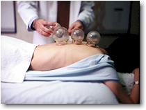
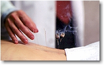
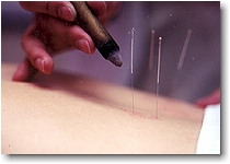


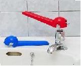
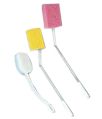
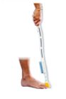

 A raised toilet seat increases the height of the toilet pan. It makes it easier for you to sit down on the toilet and stand up. Raised toilet seats of different heights are available commercially. The adequate height should allow the user to place his/her foot flat on the floor while seated on the toilet. If you live with other people who use the same bathroom, they will need to know how to remove the raised toilet seat. NOTE: It is important to make sure that it is securely attached to the toilet each time you use it. Incorrect fitting of the raised toilet seat increases risks of falls since it can move. It can also damage the seat if it is placed incorrectly.
 If using toilet paper is hard for you because you have difficulty moving one arm, you can also try to use a bidet, which is similar in appearance to a toilet bowl, however it projects water for personal cleaning. In addition to being expensive, bidets are not recommended if you have difficulty standing safely.

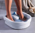
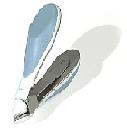
 If you have difficulty squeezing the toothpaste out of the tube, you can use a tube dispenser that will allow you to put toothpaste on your toothbrush while using only one hand.While brushing your teeth, you may find that using your strong hand to hold the toothbrush is easier. However, it is sometimes recommended that you still try to use your weak hand in daily tasks if regaining function is one of your rehabilitation goals. If you have dentures, you can use a one-handed technique to soak them. For example, you can open the container and put them in water or in a denture cleansing liquid using only your strong hand.
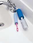
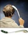
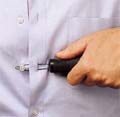

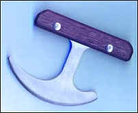

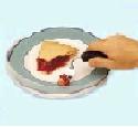 These can help you use utensils without pushing food off of your plate. This is helpful for people who cannot use their second hand to hold the knife.