Agnosia

Evidence Reviewed as of before: 15-02-2023
Author(s)*: Tamara Lefranc; Audrey-Pascaline Segla
Editor(s): Annie Rochette
Table of contents

Introduction

Agnosia is defined as the inability to recognize, identify and name familiar objects using one or more senses, or the inability to recognize one’s own deficits (anosognosia). This inability is not associated with a sensory impairment, but may be expressed specifically in one or more senses, such as sight (visual agnosia), hearing (auditory agnosia) or touch (tactile agnosia or astereognosia). Agnosia can also be characterized according to the nature of the object rather than the modality; for example, prosopagnosia is a form of visual agnosia where the person is unable to recognize faces. Agnosia affects less than 1% of the neurologically impaired population.

Patient/Family Information

What is agnosia?

Agnosia is defined as the inability to recognize, identify and name familiar objects using one or more of the senses, or the inability to recognize physical, cognitive and/or affective impairments (anosognosia). Agnosias are rare deficits, with less than 1% of people with neurological disorders suffering from agnosia.

Are there different types of agnosia?

Agnosia can be described as specific to the stimulus modality, such as sight (visual agnosia), hearing (auditory agnosia), smell (olfactory agnosia) or touch (tactile agnosia or astereognosia).

Visual agnosias are the most common and best-understood forms, divided into two main classes: aperceptive and associative visual agnosias.

Visual aperceptive agnosias are characterized by an inability to perceive the primary characteristics of objects. Sufferers are unable to copy shapes or objects. As for people with associative visual agnosia, they are unable to recognize the object despite perceiving all its features. So, even if they are able to copy or describe an object, they remain incapable of recognizing it, naming it, describing its function or using it.

Agnosia can also be characterized according to the nature of the object or stimulus rather than the modality, for example, prosopagnosia is a form of visual agnosia specific to the inability to recognize faces.

Another example of stimulus-specific agnosia is anosognosia, which is the inability to recognize the presence or severity of cognitive, sensory, motor or affective deficits.

The following table provides a non-exhaustive list of several forms of visual agnosia documented in the literature.

Table 1: Different forms of visual agnosia found in the literature
Stimulus-specific visual agnosia Description
Achromatopsia Inability to recognize colors.
Shape agnosia Inability to perceive the shape, orientation, length and contours of an object.
Integrative agnosia Inability to integrate features such as contours, shape, color and orientation as a whole to form an object.
Visuospatial agnosia Difficulty perceiving the spatial relationship between objects or between the object and oneself.
Akinetopsia Inability to perceive movement.
Agnostic alexia Language disorder consisting of the inability to recognize a word visually.
Topographical disorientation/ landmark agnosia Inability to orientate oneself in familiar surroundings due to inability to recognize landmarks once known.
Prosopagnosia Inability to recognize a familiar face or even one’s own face. In some cases, the person can deduce information such as age, gender and emotion, while others cannot recognize a face as one.
Simultagnosia Inability to perceive more than one object or object component at a time.
Balint syndrome Triad of symptoms including simultagnosia, ocular apraxia and ataxia.

Why do people become agnostic after a stroke?

The information captured by our senses is interpreted in different places in the brain according to its auditory, tactile, visual, olfactory or gustatory modality. In the event of a stroke, a brain lesion may occur in the regions linking the different primary sensory areas that interpret information from a specific modality, resulting in an inability to interpret the information that is perceived and, therefore, to recognize it. Depending on the location of the lesion, different forms of agnosia may occur.

For example, visual information captured by the eyes, such as colors, shapes, contours or movement, is interpreted in the brain by the primary visual cortex located in the occipital lobe. A lesion in the temporal, occipital or parietal lobes can result in visual agnosia. A lesion in the right temporal lobe could result in auditory agnosia.

What impact does agnosia have on my daily life?

All people carry out their activities of daily living by interacting with different elements in their environment. People with post-stroke visual agnosia may perceive the characteristics of familiar objects and environments differently from before the stroke. This can lead to feelings of confusion and insecurity when interacting with their environment on a daily basis, as they may perceive objects as obstacles rather than tools. Agnosia can also present a safety issue. For example, if a person with visual agnosia fails to recognize the sharp edge of a knife or road signs (making it impossible to drive safely), or if a person with olfactory agnosia fails to recognize the smell of a gas leak, smoke or burnt food.

Here are some other possible examples:

  • Difficulty recognizing familiar objects: can make simple daily activities difficult. For example, feeding oneself, choosing food at the grocery store, using tools at work, or getting dressed if the person doesn’t recognize his or her clothes.
  • Difficulties in social interaction: difficulty recognizing familiar faces (prosopagnosia) or understanding language due to difficulty recognizing words (auditory agnosia). This can lead to social isolation if not addressed by a professional.
  • Emotional impact: Living with agnosia can be stressful for the person, due to the constant challenges encountered. This can eventually lead to anxiety, depression and loss of self-confidence.

In short, agnosia can reduce a person’s autonomy, as they are unable to analyze their environment and interact with it adequately. Impacts vary according to the specific type of agnosia, and can have a significant impact on an individual’s quality of life.

Who diagnoses and treats agnosia?

The diagnosis of agnosia is made following medical imaging and neurological examination by a physician, most often a specialist in neurology. Agnosias can also be diagnosed following a neuropsychological examination, in which case the diagnosis is made by a neuropsychologist.

The resulting difficulties can be addressed in rehabilitation by the occupational therapist and speech language therapist.

Will my agnosia improve?

Few people recover their perceptual abilities. However, significant improvement may occur in the first 3 months post-stroke, and may continue to progress up to a year later. Recovery depends on a number of factors, including age, extent of disability, type, severity and location of stroke, and the effectiveness of therapies.

What therapies are available for agnosia?

Agnosia is a perceptual deficit for which the literature on interventions is scarce compared with other deficits such as hemineglect.

There are two types of approach, namely remedial and compensatory. The remedial approach consists in training the person’s cognitive abilities through exercises. The effectiveness of this approach has not been demonstrated in the literature.

There is currently no cure for agnosia. However, there are compensatory strategies that can help limit the impact on daily life. These strategies mainly involve using the other senses to compensate for modality-specific agnosia. Occupational therapists and speech therapists can help sufferers to adapt their environment and use compensatory strategies to assist recognition of environmental elements. In general, the use of compensatory strategies is accompanied by teaching about them, and training in the task to use them effectively.

Visual agnosias

These strategies include modifying the environment to facilitate recognition of objects relevant to the task, and to reduce risk. The strategies used must be adapted to the individual’s needs.

The organization of the environment is also a strategy that can help the person interact with his or her surroundings by purifying the space and organizing it in a way that assists recognition. Here are a few examples:

  • Lock rooms considered to be at risk, such as the garage;
  • Adding tactile cues to help the person recognize certain elements, such as a rough texture, can help identify dangerous objects by touch;
  • Organize the refrigerator so that fruits and vegetables can be found in an easily accessible place;
  • etc.

Auditory agnosia

In the case of auditory agnosias, compensatory strategies to improve the ability to communicate aim to compensate via the visual modality. This may involve using non-verbal cues such as intonation, facial expression or gestures to deduce the meaning of the conversation. They can also learn to lip-read. Reducing ambient noise can help the person to better understand their interlocutor. Adapting the environment can also help the person with auditory agnosia to identify risks in their environment, for example, by replacing an audible alarm with a flashing one.

Tactile agnosia

For tactile agnosias, it is generally recommended to compensate for object recognition using vision.

Anosognosia

For anosognosia, self-awareness training is the most common type of intervention. Training can take the form of a formal intensive program, the modalities of which are indicated by the therapist, or recurrent follow-up with the therapist, in which the teaching and application of compensatory strategies are prioritized (e.g., presenting a stimulus in the attained direction and then in an unattained direction, splitting the task into small steps, etc.). The use of video self-observation can serve as a relevant tool for precipitating the patient’s awareness. In both cases, the aim is to improve the person’s ability to become aware of his or her difficulties in order to better compensate for them (e.g., using visual scanning methods to encourage the person to become aware of objects to his or her left in the case of hemineglect). In addition to training, some interventions combine personal training with education for the patient and those around him/her.

What can I expect from agnosia therapies?

Studies on the effectiveness of agnosia-specific interventions are few and far between. Thus, the level of scientific evidence is insufficient to date. What’s more, interventions are mainly aimed at compensating for agnosia on a day-to-day basis, rather than recovering perceptual skills.

How does agnosia affect my stroke recovery?

Anosognosia can limit a person’s ability to benefit from rehabilitation. Indeed, since they do not recognize the presence of their disabilities, they cannot devote their efforts to compensatory strategies, or see the relevance of therapy. However, there are strategies that can be used to help the person become aware of their difficulties.

A member of my family is agnostic. How can I help them?

Educate yourself: it’s beneficial for family members to learn about agnosia, its symptoms and its impact on daily life. Understanding the condition can help them better support their loved one.

Emotional support: Offering emotional support and encouragement is important. Listen to their frustrations and reassure your loved one. Being patient, understanding and empathetic is essential to overcoming the challenges associated with agnosia. In the case of anosognosia, it’s important to remain patient and avoid rushing your loved one and confronting them with their difficulties. This is even more likely to upset him/her.

Don’t forget to seek support for yourself, as a loved one.

Help, but not too much!

Depending on the severity of the agnosia, help may be needed with everyday tasks such as meal preparation, grocery shopping, personal care and leisure activities. Be careful not to do everything for your loved one.

It is possible to help the person with agnosia by adapting the environment to his or her needs to promote independence. This may involve organizing the refrigerator in a logical way, while helping the person to understand how his or her environment is organized. As a loved one, you have an important role to play in the person’s rehabilitation, and can help integrate the strategies learned in home therapy into everyday life. Therapists can guide you in the optimal strategies to use.

When working with a person with auditory agnosia, you can facilitate communication by adapting your approach. You could use gestures, writing, or make sure the environment is free of noises that interfere with communication.

Participate in rehabilitation: Interact with therapists and apply the strategies they teach to adapt the environment and/or activities in the community (e.g., reduce clutter, label objects, maintain good lighting to facilitate daily living). Keep them informed of your needs and the strategies that work with your loved one in their environment.

References

Besharati, S., Kopelman, M., Avesani, R., Moro, V. and Fotopoulou, A. (2015, 2015/05/04). Another perspective on anosognosia: Self-observation in video replay improves motor awareness. Neuropsychological Rehabilitation, 25(3), 319-352. https://doi.org/10.1080/09602011.2014.923319

Bouwmeester, L., van de Wege, A., Haaxma, R. and Snoek, J. W. (2015, 2015/01/02). Rehabilitation in a complex case of topographical disorientation. Neuropsychological Rehabilitation, 25(1), 1-14. https://doi.org/10.1080/09602011.2014.923318

Buchmann, I., Finkel, L., Dangel, M., Erz, D., Maren Harscher, K., Kaupp-Merkle, M., Liepert, J., Rockstroh, B. and Randerath, J. (2020). A combined therapy for limb apraxia and related anosognosia. Neuropsychological Rehabilitation, 30(10), 2016-2034. https://doi.org/https://dx.doi.org/10.1080/09602011.2019.1628075

Burns, M. S. (2004, 2004/01/01). Clinical Management of Agnosia. Topics in Stroke Rehabilitation, 11(1), 1-9. https://doi.org/10.1310/N13K-YKYQ-3XX1-NFAV

Cappa, S., Sterzi, R., Vallar, G. and Bisiach, E. (1987, 1987/01/01/). Remission of hemineglect and anosognosia during vestibular stimulation. Neuropsychologia, 25(5), 775-782. https://doi.org/https://doi.org/10.1016/0028-3932(87)90115-1

Coslett, H. B. (2011). Frontiers in Neuroscience Sensory Agnosias. In J. A. Gottfried (ed.), Neurobiology of Sensation and Reward. CRC Press/Taylor & Francis Copyright © 2011 by Taylor and Francis Group, LLC.

Coslett, H. B. (2018, Jun). Apraxia, Neglect, and Agnosia. Continuum (Minneap Minn), 24(3, behavioral neurology and psychiatry), 768-782. https://doi.org/10.1212/con.0000000000000606

Cuomo, J., Flaster, M. and Biller, J. (2012). Right Brain: A descriptive account of two patients’ experience with and adaptations to Bálint syndrome. Neurology, 79(11), e95-e96. https://doi.org/10.1212/WNL.0b013e3182698d28

Denis, M. (2016). Fragilités. In Petit traité de l’espace (pp. 123-136). Mardaga. https://www.cairn.info/petit-traite-de-l-espace–9782804703226-page-123.htm

Dirette, D. (2010, 2010/07/01). Self-Awareness Enhancement through Learning and Function (SELF): A Theoretically Based Guideline for Practice. British Journal of Occupational Therapy, 73(7), 309-318. https://doi.org/10.4276/030802210X12759925544344

Fotopoulou, A., Rudd, A., Holmes, P. and Kopelman, M. (2009, Apr). Self-observation reinstates motor awareness in anosognosia for hemiplegia. Neuropsychologia, 47(5), 1256-1260. https://doi.org/10.1016/j.neuropsychologia.2009.01.018

Gazzaniga, M. S., Ivry, R. B., Mangun, G. R., Coquery, J. M. and Macar, F. (2000). Cognitive neuroscience: The biology of the mind. De Boeck Supérieur. https://books.google.ca/books?id=P__aswEACAAJ 

Gillen, G. (2009). Managing agnosia to optimize function. In Cognitive and Perceptual Rehabilitation: Optimizing Function. Mosby Elsevier.

Hazelton, C., Thomson, K., Todhunter-Brown, A., Campbell, P., Chung, C. S. Y., Dorris, L., Gillespie, D. C., Hunter, S. M., McGill, K., Nicolson, D. J. et al. (2022). Interventions for perceptual disorders following stroke. Cochrane Database of Systematic Reviews, (11). https://doi.org/10.1002/14651858.CD007039.pub3

Heutink, J., Indorf, D. L., & Cordes, C. (2019, 2019/11/26). The neuropsychological rehabilitation of visual agnosia and Balint’s syndrome. Neuropsychological Rehabilitation, 29(10), 1489-1508. https://doi.org/10.1080/09602011.2017.1422272

Huang, J. (2023, August). Agnosia. https://www.merckmanuals.com/fr-ca/professional/troubles-neurologiques/fonction-et-dysfonctionnement-des-lobes-c%C3%A9r%C3%A9braux/agnosie?query=agnosie

Kumar, A. and Wroten, M. (2023). Agnosia. StatPearls Publishing LLC. https://www.ncbi.nlm.nih.gov/books/NBK493156/

Lampinen, J. and Tham, K. (2003, 2003/12/01). Interaction with the Physical Environment in Everyday Occupation after Stroke: A Phenomenological Study of Persons with Visuospatial Agnosia. Scandinavian Journal of Occupational Therapy, 10(4), 147-156. https://doi.org/10.1080/11038120310016580

Martinaud, O. (2012). Prosopagnosia and other visual agnosias. Revue de neuropsychologie, 4, 277. https://doi.org/10.3917/rne.044.0277

Martinaud, O. (2017). Visual agnosia and focal brain injury. Revue Neurologique, 173(7), 451-460. https://doi.org/https://doi.org/10.1016/j.neurol.2017.07.009

Olson, E. (1991). Perceptual deficits affecting the stroke patient. Rehabil Nurs, 16(4), 212-213. https://doi.org/10.1002/j.2048-7940.1991.tb01215.x

Reid, L. and Edmans, J. (2010). Management of Perceptual Impairments. In Occupational Therapy and Stroke (pp. 158-172). https://doi.org/https://doi.org/10.1002/9781444323801.ch8

Roberts, S. P. (1992). Visual disorders of higher cortical function. Journal of the American Optometric Association, 63 10.

Rosselli, M., Ardila, A. and Beltran, C. (2001). Rehabilitation of Balint’s syndrome: a single case report. Appl Neuropsychol, 8(4), 242-247. https://doi.org/10.1207/s15324826an0804_7

Sohlberg, M. M. (2000). Assessing and managing unawareness of self. Semin Speech Lang, 21(2), 135-150; quiz 150-131. https://doi.org/10.1055/s-2000-7561

Sohlberg, M. M., Glang, A. and Todis, B. (1998, Apr). Improvement during baseline: three case studies encouraging collaborative research when evaluating caregiver training. Brain Inj, 12(4), 333-346. https://doi.org/10.1080/026990598122638

Ward, J. (2020). The Seeing Brain. In Routledge (ed.), The Student’s Guide to Cognitive Neuroscience (4e  ed.).

Ward, J. (2020). The student’s guide to cognitive neuroscience (Fourth editione  ed.). Routledge Abingdon, Oxon. https://www.taylorfrancis.com/books/9781351035187 

Wilkinson, D., Ko, P., Kilduff, P., McGlinchey, R. and Milberg, W. (2005). Improvement of a face perception deficit via subsensory galvanic vestibular stimulation. Journal of the International Neuropsychological Society, 11(7), 925-929.https://doi.org/10.1017/S1355617705051076

Apraxia

Evidence Reviewed as of before: 28-08-2020
Author(s)*: Annabel McDermott, OT; Annie Rochette, erg./OT (c) Ph.D.
Content consistency: Gabriel Plumier
Patient/Family Information Table of contents

Introduction

Apraxia is a neuropsychological deficit that interrupts an individual’s ability to perform purposeful movement, in the absence of basic sensorimotor difficulties such as lack of sensation or muscle weakness. Approximately 30% of individuals display apraxia or partial signs of apraxia (i.e. dyspraxia) following stroke, with a greater incidence among individuals with left hemisphere damage. Apraxia effects an individual’s autonomy for work and daily activities.

Patient/Family Information

What is apraxia?

Apraxia is a cognitive disorder that can occur after stroke. Apraxia is the inability to make purposeful movement, but is not due to sensory or motor disturbances (e.g. loss of sensation, muscle weakness). Apraxia effects the ability to perform movements and gestures.

Why do people get apraxia?

Approximately 30% of people who have had a stroke will display apraxia or partial signs of apraxia (i.e. dyspraxia). Apraxia is more common among people with damage to the left hemisphere of the brain. However, apraxia can also result from damage to other parts of the brain.

Are there different types of apraxia?

There are many different types of apraxia. The most common type of apraxia is buccofacial (or orofacial) apraxia:

  • Buccofacial apraxia: difficulty making movements of the mouth, eyes or face.

The most common forms of limb apraxia (i.e. affecting use of the arms/legs) are ideational apraxia and ideomotor apraxia:

  • Ideational apraxia: difficulty organizing actions to achieve a goal.
  • Ideomotor apraxia: difficulty selecting, sequencing and using objects.

Different forms of apraxia can also affect speech, touch, writing/drawing skills, eye movements, and body movements.

How can I recognize limb apraxia?

Limb apraxia affects a person’s ability to perform simple movements. This may be seen as difficulty imitating an action, performing an action in response to a spoken command, or understanding an action. Limb apraxia can affect the person’s arm movements for communication (e.g. using gestures) and daily activities (e.g. using familiar objects for everyday tasks).

Who diagnoses and treats apraxia?

Apraxia is difficult to diagnose because of the many different types of apraxia, the different definitions used to describe apraxia, and a lack of suitable assessments. Medical/health professionals can assess for apraxia in several different ways including using formal tests, and by observing the patient’s movements when imitating gestures, following spoken commands (e.g. “pretend to drink from a cup”), or using common objects.

Treatment will depend on the type of apraxia.

  • A Speech Language Pathologist can help the person who is experiencing difficulties with speech, language, communication/gestures, feeding, swallowing and mouth movements.
  • A Physiotherapist can help the person who is experiencing difficulties moving their body and limbs to make intended movements.
  • An Occupational Therapist can help the person who is having difficulty doing activities around the home and at work.

How does apraxia affect my recovery?

Apraxia impacts on a person’s ability to perform movements and gestures. Apraxia can impact on the person’s ability to do rehab activities (e.g. walking), communicate with others (e.g. using gestures) and complete common tasks (e.g. self-care tasks). This can affect their ability to relearn movements or learn new skills after stroke, which can impact on the person’s recovery, as well as their ability to perform daily activities and work tasks.

Will my apraxia get better?

Apraxia typically spontaneously recovers in the first few months post-stroke and is responsive to rehabilitation. The recovery process and rate of recovery will be different for each individual.

What can I expect from apraxia therapies?

Intervention can be customized to suit the person’s difficulties. Interventions for apraxia include:

  • Strategy training for daily activities (i.e. teaching specific strategies to overcome the difficulties to patient experiences)
  • Gesture training (i.e. relearning gestures)
  • Direct ADL training (i.e. relearning – or learning new ways to perform – daily tasks)
  • Using assistive technology to compensate for difficulties.

Will apraxia therapies work?

A small number of studies have investigated apraxia treatment. Results from these studies show benefits immediately after the treatment, but benefits may not last several months later. The lack of research regarding apraxia interventions impacts on the ability to draw strong conclusions regarding their effectiveness at this time.

Are there any side effects?

There are no significant side-effects from apraxia treatments.

My family member has apraxia. How can I help?

Stroke recovery requires patience and persistence from the person who had a stroke and their family/caregivers. If you or your loved one is experiencing apraxia after a stroke, the recovery process might be frustrating and stressful. It is important to continue with therapies, even if apraxia makes it challenging.

Follow this link to the Stroke Association (https://www.stroke.org.uk) for useful tips for communicating with a person who has had a stroke.

Where can I find more information about apraxia?

  • American Stroke Association (https://www.stroke.org)

Clinician Information

When reviewing the findings, it is important to note that they are always made according to randomized clinical trial (RCT) criteria – specifically as compared to a control group. To clarify, if a treatment is “effective” it implies that it is more effective than the control treatment to which it was compared. Non-randomized studies are no longer included when there is sufficient research to indicate strong evidence (level 1a) for an outcome.

Apraxia is a neuropsychological deficit that disrupts an individual’s ability to perform purposeful movement, in the absence of basic sensorimotor difficulties such as lack of sensation or muscle weakness (Koski, Iacoboni & Mazziotta, 2002; Landry & Spaulding, 1999; West et al., 2008). Approximately 30% of individuals display apraxia or dyspraxia (i.e. partial signs of apraxia) following stroke, with a greater incidence among individuals with left hemisphere damage (Koski, Iacoboni & Mazziotta, 2002; Pazzaglia & Galli, 2019). Apraxia effects an individual’s independence for work and daily activities (Cantagallo, Maini & Rumiati, 2012; Dovern, Fink & Weiss, 2012; Koski, Iacoboni & Mazziotta, 2002; van Heugten, 2001).

Apraxia is most common among individuals with damage to the left parietal lobe, however can also result from damage to the right parietal lobe, temporal lobe, frontal lobe or subcortical regions (Koski, Iacoboni & Mazziotta, 2002). Apraxia impacts on an individual’s mental representation of an action, which in turn affects his/her ability to organize and imitate actions to achieve a goal (Bowen et al., 2009). Accordingly, apraxia limits an individual’s participation in rehabilitation, use of gestures for non-verbal communication, and performance of daily activities (Koski, Iacoboni & Mazziotta, 2002).

Numerous forms of apraxia have been defined (Koski, Iacoboni & Mazziotta, 2002). Ideational apraxia and ideomotor apraxia are the most common forms of the disorder, and are defined as follows:

Ideational apraxia: Difficulty in the ability to organize actions required to achieve a goal.

Ideomotor apraxia: Difficulty in the ability to select, sequence and use objects (West et al., 2008).

The heterogeneity of apraxia, as well as inconsistent definitions and the absence of a gold standard for assessment contribute to difficulty diagnosing the disorder (Dovern, Fink & Weiss, 2012; Lindsten-McQueen et al., 2014; West et al., 2008). For research purposes diagnosis is based on (i) neuropsychological testing to determine the presence/absence of apraxia; and (ii) standardized assessment of activities of daily living (ADLs) to determine the degree of impairment (van Heugten, 2001).

Apraxia typically spontaneously recovers in the first few months post-stroke (Cantagallo, Maini & Rumiati, 2012) and is responsive to rehabilitation (Buxbaum et al., 2008). Intervention can be customized to the presenting difficulties (Landry & Spaulding, 1999). Accordingly, interventions used in the treatment of apraxia include strategy training for ADLs using internal/external compensatory strategies; sensory stimulation using proprioceptive/deep pressure and sharp/soft touch; cueing using verbal or physical prompts; error reduction through chaining (forward/backward strategies); gesture training; conductive education; and normal movement approaches (Buxbaum et al., 2008; West et al., 2008).

A number of systematic reviews of interventions for apraxia have been conducted (Bowen et al., 2009; Lindsten-McQueen et al., 2014; Pazzaglia & Galli, 2019; Saikaley et al., n.d.; Worthington, 2016; van Heugten, 2001). A Cochrane Review of apraxia interventions following stroke by West et al. (2008) included three randomized controlled trials (two of which were considered suitable for inclusion in this review) that used strategy training, gesture training and transfer of training. The review showed a significant treatment effect immediately following apraxia intervention, but results were not sustained at 6 months post-stroke. Conclusive evidence of the benefit of apraxia therapies was not attained.

This review of interventions for apraxia following stroke includes one high quality RCT, three fair quality RCTs and six non-randomized studies. The majority of studies were conducted with individuals in the subacute phase of stroke recovery. Interventions include gesture training (three studies), strategy training (five studies) and direct training of ADLs (two studies). While strategy training and gesture training were both shown to benefit some outcomes, the lack of research regarding apraxia interventions impacts on the ability to draw strong conclusions regarding their effectiveness at this time.

Results Table

View results table

Outcomes

Acute phase

No studies have been conducted in the acute phase of stroke recovery.

Subacute phase: Direct training of Activities of Daily Living for apraxia

Activities of Daily Living (ADLs)
Effective
2b

One non-randomised study (Goldenberg & Hagmann, 1998) investigated the use of direct training for apraxia on activities of daily living (ADLs) in the subacute phase of stroke recovery. This study assigned patients with left hemisphere stroke and apraxia to receive direct training and explorative training of daily tasks. Performance of ADLs was measured according to the number of fatal and reparable errors made during three trained/untrained tasks, assessed weekly over the intervention period (2-5 weeks) and at follow-up (6-30 months). At end of treatment 10 participants were able to complete all three ADL tasks without fatal errors; the remaining 5 participants made one fatal error. There was no generalisation of training effects from trained to untrained tasks. Participants who continued to practice activities at home showed fewer fatal errors at follow-up.

Conclusion: There is limited evidence (level 2b) from one non-randomised study that direct training of activities of daily living is effective in improving performance of trained activities of daily living among individuals with apraxia in the subacute phase of stroke recovery.
Note: Between-group comparisons were not made.

Subacute phase: Gesture training for apraxia

Gestural expression
Effective
2b

One non-randomised study (Daumuller & Goldenberg, 2010) investigated the effect of gesture training on gestural expression among individuals with apraxia in the subacute phase of stroke recovery. The non-randomised study assigned patients with left hemisphere stroke and severe aphasia (number of patients with apraxia not specified) to receive gestural therapy for 3 weeks or no gestural therapy. Gestural expression of participants who received gesture training was measured at week 1, week 2 and week 3 (practised gestures, unpractised gestures). A significant improvement in practised gestures was found at all timepoints, and a significant improvement in unpractised gestures was found at week 1 and week 2. A significant between-group difference in expression of unpractised gestures was found at week 2, in favour of gestural therapy vs. no therapy.

Conclusion: There is limited evidence (level 2b) from one non-randomised study that gesture training is more effective than no training for improving gestural expression in the subacute phase of stroke recovery. The study also reported a significant improvement in gestural expression following gesture training.

Chronic phase: Direct training of Activities of Daily Living for apraxia

Activities of Daily Living (ADLs)
Effective
2b

One non-randomised study (Goldenberg, Daumuller & Hagmann, 2001) investigated the use of direct training for apraxia on activities of daily living (ADLs) in the chronic phase of stroke recovery. This non-randomized crossover trial assigned patients with left hemisphere stroke and severe apraxia to receive direct training or explorative training of four activities. ADLs were measured according to number of errors and assistance provided during performance of trained and untrained tasks at 2-weekly intervals. A significant reduction in errors and assistance for ADL tasks was found at post-treatment (4 weeks) following direct training only. Results remained significant for assistance (but not errors) at follow-up (3 months).

Conclusion: There is limited evidence (level 2b) from one non-randomised study that direct training of activities of daily living is effective in improving performance of trained activities of daily living among patients with apraxia in the chronic phase of stroke recovery.
Note: Between-group comparisons were not made.

Phase not specific to one period: Gesture training for apraxia

Activities of Daily Living (ADLs) – carers’ perception
Effective
2a

One fair quality RCT (Smania et al., 2006) investigated the effect of gesture training for apraxia on carers’ self-perception of the patient’s ability to perform activities of daily living (ADLs) following stroke. The fair quality RCT randomized patients with subacute/chronic left hemisphere stroke and apraxia and aphasia to receive gesture training or conventional aphasia rehabilitation. ADLs were measured by caregiver questionnaire at post-treatment (30 sessions) and follow-up (2 months post-treatment). A significant between-group difference was found at post-treatment, in favour of gesture training vs. aphasia rehabilitation. Results did not remain significant at follow-up.

Conclusion: There is limited evidence (level 2a) from one fair quality RCT that gesture training is more effective than a comparison intervention (aphasia rehabilitation) for improving carers’ perception of the patient’s ability to perform activities of daily living immediately following treatment.

Constructional apraxia
Not effective
2a

Two fair quality RCTs (Smania et al., 2000; Smania et al., 2006) investigated the effect of gesture training for apraxia on constructional apraxia following stroke.

The first fair quality RCT (Smania et al., 2000) randomized patients with subacute/chronic left hemispheric stroke and apraxia to receive gesture-production training or conventional aphasia rehabilitation. Constructional apraxia was measured at post-treatment (35 sessions). No significant improvement was found.
Note: Between-group differences were not reported.

The second fair quality RCT (Smania et al., 2006) randomized patients with subacute/chronic left hemisphere stroke and apraxia and aphasia to receive gesture training or conventional aphasia rehabilitation. Constructional apraxia was measured at post-treatment (30 sessions). No significant between-group difference was found.

Conclusion: There is limited evidence (level 2a) from one fair quality RCT that gesture training is not more effective than a comparison intervention (aphasia rehabilitation) for improving constructional apraxia following stroke. A second fair quality RCT found no significant improvement in constructional apraxia following gesture-production training.

Gesture comprehension
Effective
2a

Two fair quality RCTs (Smania et al., 2000; Smania et al., 2006) investigated the effect of gesture training for apraxia on gesture comprehension following stroke.

The first fair quality RCT (Smania et al., 2000) randomized patients with subacute/chronic left hemispheric stroke and apraxia to receive gesture-production training or conventional aphasia rehabilitation. Gesture comprehension was measured by the gesture comprehension test at post-treatment (35 sessions). No significant improvement was found.

Note: Between-group differences were not reported.

The second fair quality RCT (Smania et al., 2006) randomized patients with subacute/chronic left hemisphere stroke and apraxia and aphasia to receive gesture training or conventional aphasia rehabilitation. Gesture comprehension was measured at post-treatment (30 sessions) and follow-up (2 months post-treatment). A significant between-group difference was found at post-treatment, in favour of gesture training therapy vs. aphasia rehabilitation. Results did not remain significant at follow-up.

Conclusion: There is limited evidence (level 2a) from one fair quality RCT that gesture training is more effective than a comparison intervention (aphasia rehabilitation) for improving gesture comprehension among patients with apraxia following stroke.
Note: However, a second fair quality RCT found no significant improvement in gesture comprehension following gesture production training.

Ideational apraxia
Not effective
2a

Two fair quality RCTs (Smania et al., 2000; Smania et al., 2006) investigated the effect of gesture training for apraxia on ideational apraxia following stroke.

The first fair quality RCT (Smania et al., 2000) randomized patients with subacute/chronic left hemispheric stroke and apraxia to receive gesture-production training or conventional aphasia rehabilitation. Ideational apraxia was measured at post-treatment (35 sessions) according to the use of real objects and errors made (inadequate utilisation, sequence error, substitution, perplexity, localisation error, awkwardness, omission, total errors). A significant improvement in ideational apraxia was found following gesture-production training, but not conventional aphasia rehabilitation. Significant reduction in some errors (awkwardness, omissions, total errors) was also seen following gesture-production training.
Note: Between-group differences were not reported.

The second fair quality RCT (Smania et al., 2006) randomized patients with subacute/chronic left hemisphere stroke and apraxia and aphasia to receive gesture training or conventional aphasia rehabilitation. Ideational apraxia was measured at post-treatment (30 sessions) and follow-up (2 months post-treatment). No significant between-group difference was found at either timepoint.
Note: There was a significant improvement in ideational apraxia at post-treatment, within the gesture training group only.

Conclusion: There is limited evidence (level 2a) from one fair quality RCT that gesture training is not more effective than a comparison intervention (aphasia rehabilitation) for improving ideational apraxia following stroke.
Note:
However, both fair quality RCTs reported a significant improvement in ideational apraxia among participants who received gesture training.

Ideomotor apraxia
Effective
2a

Two fair quality RCTs (Smania et al., 2000; Smania et al., 2006) investigated the effect of gesture training for apraxia on ideomotor apraxia following stroke.

The first fair quality RCT (Smania et al., 2000) randomized patients with subacute/chronic left hemispheric stroke and apraxia to receive gesture-production training or conventional aphasia rehabilitation. Ideomotor apraxia was measured at post-treatment (35 sessions) using De Renzi’s test of ideomotor apraxia and errors made (unrecognisable, intrusion, position, perseveration, omission, inappropriate sequence, conduit d’approche, substitution, total errors). A significant improvement was found following gesture-production training, but not conventional aphasia rehabilitation. Significant reduction in some errors (unrecognisable, intrusion, position, total errors) was also seen following gesture-production training.
Note: Between-group differences were not reported.

The second fair quality RCT (Smania et al., 2006) randomized patients with subacute/chronic left hemisphere stroke and apraxia and aphasia to receive gesture training or conventional aphasia rehabilitation. Ideomotor apraxia was measured at post-treatment (30 sessions) and follow-up (2 months post-treatment). A significant between-group difference was found at post-treatment, in favour of gesture training therapy vs. aphasia rehabilitation. Results did not remain significant at follow-up.

Conclusion: There is limited evidence (level 2a) from one fair quality RCT that gesture training is more effective than a comparison intervention (aphasia rehabilitation) for improving ideomotor apraxia following stroke.
Note: The other fair quality RCT also reported a significant improvement in ideomotor apraxia following gesture training but not aphasia rehabilitation.

Intelligence
Not effective
2a

Two fair quality RCTs (Smania et al., 2000; Smania et al., 2006) investigated the effect of gesture training for apraxia on intelligence following stroke.

The first fair quality RCT (Smania et al., 2000) randomized patients with subacute/chronic left hemispheric stroke and apraxia to receive gesture-production training or conventional aphasia rehabilitation. Intelligence was measured using Raven’s Progressive Matrices at post-treatment (35 sessions). No significant improvement was found.
Note: Between-group differences were not reported.

The second fair quality RCT (Smania et al., 2006) randomized patients with subacute/chronic left hemisphere stroke and apraxia and aphasia to receive gesture training or conventional aphasia rehabilitation. Intelligence was measured using Raven’s Progressive Matrices at post-treatment (30 sessions). No significant between-group difference was found.
Note: There was a significant improvement in intelligence within the aphasia rehabilitation group only.

Conclusion: There is limited evidence (level 2a) from one fair quality RCT that gesture training is not more effective than a comparison intervention (aphasia rehabilitation) for improving performance on a test of intelligence following stroke.

Verbal comprehension
Not effective
2a

Two fair quality RCTs (Smania et al., 2000; Smania et al., 2006) investigated the effect of gesture training for apraxia on verbal comprehension following stroke.

The first fair quality RCT (Smania et al., 2000) randomized patients with subacute/chronic left hemispheric stroke and apraxia to receive gesture-production training or conventional aphasia rehabilitation. Verbal comprehension was measured using the Token Test at post-treatment (35 sessions). No significant improvement was found.
Note: Between-group differences were not reported.

The second fair quality RCT (Smania et al., 2006) randomized patients with subacute/chronic left hemisphere stroke and apraxia and aphasia to receive gesture training or conventional aphasia rehabilitation. Verbal comprehension was measured by the Token Test at post-treatment (30 sessions). No significant between-group difference was found.
Note: There was a significant improvement in verbal comprehension within the aphasia rehabilitation group only.

Conclusion: There is limited evidence (level 2a) from one fair quality RCT that gesture training for apraxia is not more effective than a comparison intervention (aphasia training) for improving verbal comprehension following stroke.

Phase not specific to one period: Strategy training for apraxia

Activities of Daily Living (ADLs)
Effective
1b

One high quality RCT (Donkervoort et al., 2001), one fair quality RCT (Geusgens et al., 2006), and two non-randomised studies (van Heugten et al., 1998; Geusgens et al., 2007) have investigated the effect of strategy training for apraxia on activities of daily living (ADLs) following stroke.

The high quality RCT (Donkervoort et al., 2001) randomised patients with subacute/chronic left hemisphere stroke and apraxia to receive strategy training integrated with occupational therapy or occupational therapy alone. ADLs were measured using (i) the Barthel Index, (ii) standardised ADL observations for apraxia, and (iii) an ADL judgement list scored by the occupational therapist at post-treatment (8 weeks) and follow-up (3 months post-treatment). Significant between-group differences were found on two measures (Barthel Index, standardised ADL observations) at post-treatment, in favour of strategy training vs. occupational therapy alone; results did not remain significant at follow-up.

Further to the study by Donkervoort et al. (2001), a fair quality study (Geusgens et al., 2006) measured transfer of skills during trained and untrained ADLs on standardised observation of 4 tasks (washing face and upper body, putting on a shirt/blouse, preparing and eating a sandwich, preparing a cup of hot chocolate) at post-treatment (8 weeks) and follow-up (3 months). A significant between-group difference in non-trained ADLs was seen at post-treatment*, in favour of strategy training vs. occupational therapy alone; results did not remain significant at follow-up.
* Note: Results reflect change scores from baseline to post-treatment.

The first non-randomised study (van Heugten et al., 1998) assigned patients with acute/subacute left hemisphere stroke and apraxia to receive strategy training. ADLs were measured using (i) the Barthel Index, (ii) standardized observations of ADL performance (independence, initiation, execution and control) when completing 4 tasks (washing face and upper body, putting on a shirt, preparing and eating a sandwich, preparing coffee or tea), and (iii) a 16-item ADL questionnaire derived from the Rivermead ADL index, completed by the OT at post-treatment (12 weeks). Significant improvements were found on all measures at post-treatment.
Note: Further, van Heugten et al. (2000) noted a ceiling effect on ADL observations, whereby patients who were competent with ADLs prior to intervention demonstrated minimal improvement over time.

The second non-randomised study (Geusgens et al., 2007) assigned patients with subacute/chronic left hemisphere stroke and apraxia to receive strategy training of ADLs. ADLs were measured by (i) the Barthel Index at post-treatment (8 weeks), and (ii) standardised ADL observations (trained tasks, untrained tasks, total) at post-treatment and follow-up (20 weeks). A significant improvement in all measures of ADLs was found at post-treatment; results did not remain significant at follow-up.
Note: Lasting transfer effects from trained to non-trained tasks was seen at follow-up.

Conclusion: There is moderate evidence (level 1b) from one high quality RCT and one fair quality RCT that strategy training for apraxia is more effective, in the short term, than a comparison intervention (occupational therapy alone) for improving performance of activities of daily living following stroke. Further, two non-randomised studies reported significant improvements in activities of daily living following strategy training (significant improvements were not maintained at follow-up in one of these non-randomised studies).

Activities of Daily Living (ADLs) – patient perception
Not effective
1b

One high quality RCT (Donkervoort et al., 2001) investigated the effect of strategy training for apraxia on self-perception of activities of daily living (ADLs) following stroke. The high quality RCT randomised patients with subacute/chronic left hemisphere stroke and apraxia to receive strategy training integrated with occupational therapy or occupational therapy alone. Self-perception of ADLs was measured using an ADL judgement list scored by the patient at post-treatment (8 weeks) and follow-up (3 months post-treatment). No significant between-group difference was found at either timepoint.

Conclusion: There is moderate evidence (level 1b) from one high quality RCT that strategy training for apraxia is not more effective than a comparison intervention (occupational therapy alone) for improving individuals’ perception of their ability to perform activities of daily living following stroke.

Apraxia
Not effective
1b

One high quality RCT (Donkervoort et al., 2001) and two non-randomised studies (van Heugten et al., 1998; Geusgens et al., 2007) investigated the effect of strategy training for apraxia on apraxia following stroke.

The high quality RCT (Donkervoort et al., 2001) randomised patients with subacute/chronic left hemisphere stroke and apraxia to receive strategy training integrated with occupational therapy or occupational therapy alone. Apraxia was measured using the Apraxia Test (object use, gesture imitation) at post-treatment (8 weeks) and follow-up (3 months post-treatment). No significant between-group difference was found at either time point.

The first non-randomised study (van Heugten et al., 1998) assigned patients with acute/subacute left hemisphere stroke and apraxia to receive strategy training. Apraxia was measured at post-treatment (12 weeks) using a 2-item assessment adapted from De Renzi evaluating use of objects and imitation of gestures. A significant improvement in was found.

The second non-randomised study (Geusgens et al., 2007) assigned patients with subacute/chronic left hemisphere stroke and apraxia to receive strategy training of activities of daily living. Apraxia was measured by the Apraxia Test at post-treatment (8 weeks). A significant improvement was found.

Conclusion: There is moderate evidence (level 1b) from one high quality RCT that strategy training for apraxia is not more effective than a comparison intervention (occupational therapy alone) for improving apraxia following stroke.
Note: However, two non-randomised studies found a significant improvement in apraxia following strategy training. Results suggest that strategy training is as effective as occupational therapy alone for improving apraxia following stroke.

Motor function
Not effective
1b

One high quality RCT (Donkervoort et al., 2001) and two non-randomised studies (van Heugten et al., 1998; Geusgens et al., 2007) investigated the effect of strategy training for apraxia on motor function following stroke.

The high quality RCT (Donkervoort et al., 2001) randomised patients with subacute/chronic left hemisphere stroke and apraxia to receive strategy training integrated with occupational therapy or occupational therapy alone. Motor function was measured using the Functional Motor Test at post-treatment (8 weeks) and follow-up (3 months post-treatment). No significant between-group difference was found at either time point.

The first non-randomised study (van Heugten et al., 1998) assigned patients with acute/subacute left hemisphere stroke and apraxia to receive strategy training. Motor functioning was measured at post-treatment (12 weeks) using an 8-item assessment of contralateral function (trunk balance, shoulder movement, arm movement, grasp and release of a cylinder, grasp and release a dice, tactile sensitivity). A significant improvement in motor functioning was found.

The second non-randomised study (Geusgens et al., 2007) assigned patients with subacute/chronic left hemisphere stroke and apraxia to receive strategy training of activities of daily living. Motor function was assessed by the Functional Motor Test at post-treatment (8 weeks). No significant improvement was found.

Conclusion: There is moderate evidence (level 1b) from one high quality RCT that strategy training for apraxia is not more effective than a comparison intervention (occupational therapy alone) for improving motor function following stroke.
Note: Further, a non-randomized study found no significant improvement in motor function following strategy training. However, another non-randomized study found a significant improvement in motor function following strategy training.

Motor impairment
Not effective
1b

One high quality RCT (Donkervoort et al., 2001) investigated the effect of strategy training for apraxia on motor impairment following stroke. The high quality RCT randomised patients with subacute/chronic left hemisphere stroke and apraxia to receive strategy training integrated with occupational therapy or occupational therapy alone. Voluntary movement of the affected limbs was measured using the Motricity Index at post-treatment (8 weeks) and follow-up (3 months post-treatment). No significant between-group difference was found at either time point.

Conclusion: There is moderate evidence (level 1b) from one high quality RCT that strategy training for apraxia is not more effective than a comparison intervention (occupational therapy alone) for reducing motor impairment following stroke.

References

Daumuller, M. & Goldenberg, G. (2010). Therapy to improve gestural expression in aphasia: a controlled clinical trial. Clinical Rehabilitation, 24, 55-65. https://doi.org/10.1177/2F0269215509343327

Donkervoort, M., Dekker, J., Stehmann-Saris, F.C., Deelman, B.G. (2001). Efficacy of strategy training in left hemisphere stroke patients with apraxia: a randomised clinical trial. Neuropsychological Rehabilitation, 11(5), 549-566. https://doi.org/10.1080/09602010143000093

Geusgens, C., van Heugten, C., Donkervoort, M., van den Ende, E., Jolles, J., & van den Heuvel, W. (2006). Transfer of training effects in stroke patients with apraxia: An exploratory study. Neuropsychological Rehabilitation, 16(2), 213-29. https://doi.org/10.1080/09602010500172350

Geusgens, C., van Heugten, C., Cooijmans, J.P.J., Jolles, J., & van de Heuvel, W. (2007). Transfer effects of a cognitive strategy training for stroke patients with apraxia. Journal of Clinical and Experimental Neuropsychology, 29(8), 831-41. https://doi.org/10.1080/13803390601125971

Goldenberg, G. & Hagmann, S. (1998). Therapy of activities of daily living in patients with apraxia. Neuropsychological Rehabilitation, 8(2), 123-41. https://doi.org/10.1080/713755559

Goldenberg, G., Daumuller, M., & Hagmann, S. (2001). Assessment and therapy of complex activities of daily living in apraxia. Neuropsychological Rehabilitation: An International Journal, 11(2), 147-69. https://doi.org/10.1080/09602010042000204

Smania, N., Girardi, F., Domenicali, C., Lora, E., & Aglioti, S. (2000). The rehabilitation of limb apraxia: a study in left-brain-damaged patients. Archives of Physical Medicine and Rehabilitation, 81, 379-88. https://doi.org/10.1053/mr.2000.6921

Smania, N., Aglioti, S.M., Girardi, F., Tinazzi, M., Fiaschi, A., Cosentino, A., & Corato, E. (2006). Rehabilitation of limb apraxia improves daily life activities in patients with stroke. Neurology, 67, 2050-2. https://doi.org/10.1212/01.wnl.0000247279.63483.1f

van Heugten, C.M., Dekker, J., Deelman, B.G., van Dijk, A.J., Stehmann-Saris, J.C., & Kinebanian, A. (1998). Outcome of strategy training in stroke patients with apraxia: a phase II study. Clinical Rehabilitation, 12, 294-303. https://doi.org/10.1191/2F026921598674468328

Van Heugten, C.M., Dekker, J., Deelman, B.G., Stehmann-Saris, J.C., & Kinebanian, A. (2000). Rehabilitation of stroke patients with apraxia: the role of additional cognitive and motor impairments. Disability and Rehabilitation, 22(12), 547-54. https://doi.org/10.1080/096382800416797

Excluded studies:

Bolognini, N., Convento, S., Banco, E., Mattioli, F., Tesio, L., & Vallar, G. (2015). Improving ideomotor limb apraxia by electrical stimulation of the left posterior parietal cortex. Brain, 138, 428-39.
Reason for exclusion: Study compared anodal transcranial direct current stimulation with sham stimulation.

Buchmann, I., Finkel, L., Dangel, M., Erz, D., Harscher, K.M., Kaupp-Merkle, M., Liepert, J., Rockstroh, B., & Randerath, J. (2019). A combined therapy for limb apraxia and related anosognosia. Neuropsychological Rehabilitationhttps://doi.org/10.1080/09602011.2019.1628075
Reason for exclusion: Case study (n=2)

Edmans, J.A., Webster, J., & Lincoln, N.B. (2000). A comparison of two approaches in the treatment of perceptual problems after strokeClinical Rehabilitation, 14, 230-243.
Reason for exclusion: The majority of participants did not present with limb dyspraxia.

Additional references:

Bowen, A., West, C., Hesketh, A., & Vail, A. (2009). Rehabilitation for apraxia: evidence for short-term improvements in activities of daily living. Stroke, 40:e396-e397. https://doi.org/10.1161/STROKEAHA.108.536946

Buxbaum, L.J., Haaland, K.Y., Hallett, M., Wheaton, L., Heilman, K.M., Rodriguez, A., & Gonzalez Rothi, L.J. (2008). Treatment of limb apraxia: moving forward to improved action. American Journal of Physical Medicine & Rehabilitation, 87(2), 149-61. https://europepmc.org/article/med/18209511

Cantagallo, A., Maini, M., & Rumiati, R.I. (2012). The cognitive rehabilitation of limb apraxia in patients with stroke. Neuropsychological Rehabilitation, 22(3), 473-88. http://dx.doi.org/10.1080/09602011.2012.658317

Dovern, A., Fink, G.R., & Weiss, P.H. (2012). Diagnosis and treatment of upper limb apraxia. Journal of Neurology, 259, 1269-83. https://link.springer.com/article/10.1007/s00415-011-6336-y

Koski, L., Iacoboni, M., & Mazziotta, J.C. (2002). Deconstructing apraxia: understanding disorders of intentional movement after stroke. Current Opinion in Neurology, 15, 71-7. https://europepmc.org/article/med/11796953

Landry, J. & Spalding, S. (1999). Assessment and intervention with clients with apraxia: contributions from the literature. Canadian Journal of Occupational Therapy, 66(1), 52-61. https://doi.org/10.1177%2F000841749906600106

Lindsten-McQueen, K., Williamson Weiner, N., Wang, H.-Y., Josman, N., & Tabor Connor, L. (2014). Systematic review of apraxia treatments to improve occupational performance outcomes. OTJR: Occupation, Participation and Health, 34(4), 183-92. https://doi.org/10.3928/2F15394492-20141006-02

Pazzaglia, M. & Galli, G. (2019). Action observation for neurorehabilitation in apraxia. Frontiers in Neurology, 10:309. https://doi.org/10.3389/fneur.2019.00309

Saikaley, M., Iruthayarajah, J., Orange, J., Welch-West, P., Salter, K., Macaluso, S., & Teasell, R. (n.d.) Chapter 14: Aphasia and apraxia rehabilitation (19th edition). Evidence-Based Review of Stroke Rehabilitation. Retrieved from http://www.ebrsr.com/sites/default/files/ch%2014_version19.pdf

van Heugten, C. (2001). Rehabilitation and management of apraxia after stroke. Reviews in Clinical Gerontology, 11, 177-84.

West, C., Bowen, A., Hesketh, A., & Vail, A. (2008). Interventions for motor apraxia following stroke. Cochrane Database of Systematic Reviews, 2008(1), 1-17. https://www.cochranelibrary.com/cdsr/doi/10.1002/14651858.CD004132.pub2/abstract

Worthington, A. (2016). Treatment and technologies in the rehabilitation of apraxia and action disorganization syndrome: a review. NeuroRehabilitation, 39, 163-74 https://pubmed.ncbi.nlm.nih.gov/27314872/

Mirror Therapy – Upper Extremity

Evidence Reviewed as of before: 26-10-2018
Author(s)*: Annabel McDermott, OT; Adam Kagan, B.Sc.; Samuel Harvey-Vaillancourt, PT U3; Shahin Tavakol, PT U3; Dan Moldoveanu, PT U3; Phonesavanh Cheang, PT U3; Elissa Sitcoff, BA BSc; Nicol Korner-Bitensky, PhD OT
Content consistency: Gabriel Plumier
Patient/Family Information Table of contents

Introduction

Mirror therapy is a type of motor imagery whereby the patient moves his unaffected limb while watching the movement in a mirror; this in turn sends a visual stimulus to the brain to promote movement in the affected limb. Some of the effects of mirror therapy on the brain have already been demonstrated. A crossover study on healthy individuals by Garry, Loftus & Summers (2004) showed that viewing the mirror image of an individual’s active hand increased the excitability of neurons in the ipsilateral primary motor cortex significantly more than viewing the inactive hand directly (no mirror). The study also found a trend toward significance in favour of viewing a mirror image of the active hand compared to viewing the active hand directly (no mirror).

There is a growing body of evidence regarding the use of mirror therapy on the upper extremity following stroke. Please also see our Mirror Therapy – Lower Extremity module for studies that have investigated the use of mirror therapy with the lower limbs.

Patient/Family Information

What is mirror therapy?

Mirror therapy is a specific therapy designed to strengthen arms and hands weakened by a stroke. In mirror therapy, we use movements of the stronger hand and arm to “trick our brain” into thinking that the weaker arm is moving. Researchers have shown that this “tricking of the brain” actually works – the brain areas responsible for making the weaker arm move become stimulated. There is also some new work being done using mirror therapy on the leg (see photographs – under section – How often do I need to practice?)

How do I set up mirror therapy at home?

You start by placing a solid stand-alone mirror on a table lengthwise in front of you. NOTE: You should sit in a sturdy chair while doing this activity. Place both your arms on the table one on either side of the mirror. The mirror side (where you can see the reflection of your arm) is placed so that you see your stronger arm. It is important that the mirror is large enough so that you can see your whole arm and hand in it. You should not look at your weaker hand and arm – only focus on looking into the mirror. Now move your stronger hand while you watch the mirror. The image that you see in the mirror will make it seem like your weak hand is moving. This information on arm movement is sent to your brain that is then “tricked” into thinking that your weaker arm is moving.

Will it be of benefit to me?

Mirror therapy is especially useful for people who have very little movement of their arm and hand after a stroke. The research on how well this intervention works is still quite new. There is some encouraging evidence that suggests that by using mirror therapy, the part of your brain that is damaged is stimulated, encouraging recovery. In fact, research has shown that some patients experience greater improvements in movement when they participate in mirror therapy in addition to their regular therapy, instead of just regular therapy alone. More research in the future will give us more information on just how beneficial mirror therapy is after a stroke.

Are there any risks to me?

There are no specific risks involved in participating in mirror therapy. It is important to use a non-breakable mirror just in case it falls over. It is also important to work in a seated position so that you can focus on your arm and hand without having to think about your balance and standing safety.

Mirror therapy is actually quite easy to do at home and many people find it a fun way of having additional therapy for their hand and arm.

Do I need any special equipment?

While specialized mirror boxes are available for purchase, using a sturdy table-sized mirror with a good solid stand works just as well.

How often do I need to practice?

There is no standardized protocol for mirror therapy. You should start with whatever amount of time you can tolerate and enjoy, and then gradually work up to a full series of movements and activities.

How do I begin?

Your rehabilitation therapist should be able to provide you with a program that will meet your individual needs. She or he can guide you as to:

  • how many times a week you should do mirror therapy,
  • what specific activities and movements you should do,
  • what activities you should not do,
  • how long each mirror therapy session should be,
  • how to change activities as your hand and arm get stronger.

Clinician Information

Note: When reviewing the findings, it is important to note that they are always made according to randomized clinical trial (RCT) criteria – specifically as compared to a control group. To clarify, if a treatment is “effective” it implies that it is more effective than the control treatment to which it was compared. Non-randomized studies are no longer included when there is sufficient research to indicate strong evidence (level 1a) for an outcome.

Thirty-five studies (18 high quality RCTs, 14 fair quality RCTs, two poor quality RCTs and one non-randomized study) have investigated the effect of mirror therapy post-stroke. Of these, just three studies (one high quality RCT, one fair quality RCT and one non-randomized study) were conducted specifically with patients in the acute phase of stroke recovery, whereas the majority of studies were conducted with patients in the subacute or chronic phases of recovery. Across studies, outcomes included functional independence, dexterity, grip strength and hand function, upper extremity kinematics, sensory function, motor function and activity, pain, range of motion, and unilateral spatial neglect.

Results from this StrokEngine review showed strong evidence (level 1a) to support the use of mirror therapy to improve unilateral spatial neglect in the subacute phase of stroke recovery, and to improve upper extremity kinematics and motor function in the chronic phase of stroke recovery. Mirror therapy was comparable with other interventions for other outcomes. No adverse effects were reported.

Results Table

View results table

Outcomes

Acute Phase

Functional independence
Effective
2A

One fair quality RCT (Invernizzi et al., 2013) and one non-randomized study (Yeldan et al., 2015) investigated the effect of mirror therapy on functional independence in patients with acute stroke.

The fair quality RCT (Invernizzi et al., 2013) randomized patients to receive mirror therapy or sham mirror therapy; both groups received conventional rehabilitation. Functional independence was measured by the Functional Independence Measure at post-treatment (4 weeks). There was a significant between-group difference, in favour of mirror therapy vs. sham mirror therapy.

The non-randomized study (Yeldan et al., 2015) assigned patients to receive mirror therapy or no mirror therapy; both groups received neurodevelopmental treatment. Functional independence was measured by the Barthel Index at post-treatment (3 weeks). No significant between-group difference was found.

Conclusion: There is limited evidence (Level 2a) from one fair quality RCT that mirror therapy is more effective than a comparison intervention (sham mirror therapy) for improving functional independence in patients with acute stroke.

Note: A non-randomized study found no difference between mirror therapy and no mirror therapy, when both patient groups also received neurodevelopmental treatment.

Motor function
Effective
2A

One fair quality RCT (Invernizzi et al., 2013) and one non-randomized study (Yeldan et al., 2015) investigated the effect of mirror therapy on upper extremity motor function in patients with acute stroke.

The fair quality RCT (Invernizzi et al., 2013) randomized patients to receive mirror therapy or sham mirror therapy; both groups received conventional rehabilitation. Upper extremity motor function was measured by the Action Research Arm Test at post-treatment (4 weeks). There was a significant between-group difference, in favour of mirror therapy vs. sham mirror therapy.

The non-randomized study (Yeldan et al., 2015) assigned patients to receive mirror therapy or no mirror therapy; both groups received neurodevelopmental treatment. Upper extremity motor function was measured by the Fugl-Meyer Assessment – Upper Extremity, and Stroke Upper Limb Capacity Scale at post-treatment (3 weeks). No significant between-group differences on any of the measures were found.

Conclusion: There is limited evidence (Level 2a) from one fair quality RCT that mirror therapy is more effective than a comparison intervention (sham mirror therapy) for improving upper extremity motor function in patients with acute stroke.

Note: A non-randomized study found no difference between mirror therapy and no mirror therapy, when both patient groups also received neurodevelopmental treatment.

Sensory integration
Not effective
2b

One non-randomized study (Yeldan et al., 2015) investigated the effect of mirror therapy on upper extremity sensory integration in patients with acute stroke. This study assigned patients to receive mirror therapy or no mirror therapy; both groups received neurodevelopmental treatment. Somatosensory perception was measured by the Ayres Southern Californian Sensory Integration Tests (Finger identification, Right-left discrimination items) at post-treatment (3 weeks). No significant between-group differences on any of the measures were found.

Conclusion: There is limited evidence (Level 2b) from one non-randomized study that mirror therapy is not more effective than no mirror therapy for improving somatosensory perception in patients with acute stroke.

Strength
Effective
2a

One fair quality RCT (Invernizzi et al., 2013) and one non-randomized study (Yeldan et al., 2015) investigated the effect of mirror therapy on upper extremity strength in patients with acute stroke.

The fair quality RCT (Invernizzi et al., 2013) randomized patients to receive mirror therapy or sham mirror therapy; both groups received conventional rehabilitation. Upper extremity strength was measured by the Motricity Index at post-treatment (4 weeks). There was a significant between-group difference, in favour of mirror therapy vs. sham mirror therapy.

The non-randomized study (Yeldan et al., 2015) assigned patients to receive mirror therapy or no mirror therapy; both groups received neurodevelopmental treatment. Upper extremity strength was measured by the Motricity Index at post-treatment (3 weeks). No significant between-group difference was found.

Conclusion: There is limited evidence (Level 2a) from one fair quality RCT that mirror therapy is more effective than a comparison intervention (sham mirror therapy) for improving upper extremity strength in patients with acute stroke.

Note: A non-randomized study found no difference between mirror therapy and no mirror therapy, when both patient groups also received neurodevelopmental treatment.

Unilateral spatial neglect
Effective
1B

One high quality RCT (Pandian et al., 2014) investigated the effect of mirror therapy on unilateral spatial neglect in patients with acute stroke. This high quality RCT randomized patients to receive mirror therapy or sham mirror therapy. Unilateral spatial neglect was measured by the Star Cancellation Test, Line Bisection Test and Picture Identification Task at post-treatment (1 month) and follow-up (3 months, 6 months). There were significant between-group differences in all measures at all time points, in favour of mirror therapy vs. sham mirror therapy.

Conclusion: There is moderate evidence (Level 1b) from one high quality RCT that mirror therapy is more effective than a comparison intervention (sham mirror therapy) for improving unilateral spatial neglect in patients with acute stroke.

Subacute Phase

Dexterity
Conflicting
4

Two high quality RCTs (Kim, Lee & Song, 2014; Samuelkamaleshkumar et al., 2014) investigated the effect of mirror therapy on dexterity in patients with subacute stroke.

The first high quality RCT (Kim, Lee & Song, 2014) randomized patients to receive mirror therapy and functional electrical stimulation (FES) or sham mirror therapy and FES; both groups received conventional rehabilitation. Dexterity was measured by the Box and Block Test at post-treatment (4 weeks). No significant between-group difference was found.

The second high quality RCT (Samuelkamaleshkumar et al., 2014) randomized patients to receive mirror therapy or no mirror therapy; both groups received conventional rehabilitation. Dexterity was measured by the Box and Block Test at post-treatment (3 weeks). A significant between-group difference was found, in favour of mirror therapy vs. no mirror therapy.

Conclusion: Conflicting evidence (Level 4) between two high quality RCTs was found regarding the effect of mirror therapy on dexterity in patients with subacute stroke . These evidence indicate that mirror therapy is not more effective than simulated mirror therapy and functional electrical stimulation, but more effective than no mirror therapy.

Functional independence
Not effective
1A

Three high quality RCTs (Dohle et al., 2009; Thieme et al., 2012; Lim et al., 2016) and two fair quality RCTs (Radajewska et al., 2013, 2017; Gurbuz et al., 2016) investigated the effect of mirror therapy on functional independence in patients with subacute stroke.

The first high quality RCT (Dohle et al., 2009) randomized patients to receive mirror therapy or upper extremity training while watching the affected limb. Functional independence was measured by the Functional Independence Measure (FIM – Motor score) at post-treatment (6 weeks). No significant between-group difference was found.

The second high quality RCT (Thieme et al., 2012) randomized patients to receive individual mirror therapy, group mirror therapy, or sham group mirror therapy. Functional independence was measured by the Barthel Index (BI) at post-treatment (5 weeks). No significant between-group differences were found.

The third high quality RCT (Lim et al., 2016) randomized patients to receive mirror therapy or sham mirror therapy. Functional independence was measured by the modified BI at post-treatment (4 weeks). A significant between-group difference was found, in favour of mirror therapy vs. sham mirror therapy.

The first fair quality RCT (Radajewska et al., 2013, 2017) randomized patients to receive mirror therapy or no mirror therapy; both groups received conventional stroke rehabilitation. Functional independence was measured by the Functional Index ‘Repty’ at post-treatment (3 weeks). No significant between-group difference was found.

The second fair quality RCT (Gurbuz et al., 2016) randomized patients to receive mirror therapy or sham mirror therapy. Functional independence was measured by the FIM at post-treatment (4 weeks). No significant between-group difference was found.

Conclusion: There is strong evidence (Level 1a) from two high quality RCTs and two fair quality RCTs that mirror therapy is not more effective than comparison interventions (upper extremity training while watching the affected limb, sham group mirror therapy, no mirror therapy) in improving functional independence in patients with subacute stroke.

Note: However, a third high quality RCT found that mirror therapy was more effective than sham mirror therapy.

Motor activity
Effective
1b

One high quality RCT (Cacchio et al., 2009a) investigated the effect of mirror therapy on upper limb motor activity in patients with subacute stroke. This high quality RCT randomized patients with subacute stroke and Complex Regional Pain Syndrome type 1 to receive mirror therapy or sham mirror therapy; both groups received conventional rehabilitation. Upper extremity motor activity was measured by the Motor Activity Log – Quality of Movement score at post-treatment (4 weeks) and follow-up (6 months). A significant between-group difference was found at both time points, in favour of mirror therapy vs. sham mirror therapy.

Conclusion: There is moderate evidence (Level 1b) from one high quality RCT that mirror therapy is more effective than a comparison intervention (sham mirror therapy) for improving upper extremity motor activity in patients with subacute stroke.

Motor function
Conflicting
4

Six high quality RCTs (Cacchio et al., 2009a; Dohle et al., 2009; Thieme et al., 2012; Kim, Lee & Song, 2014; Samuelkamaleshkumar et al., 2014; Lim et al., 2016) and eight fair quality RCTs (Yun et al., 2011; Lee, Cho & Song, 2012; Bae, Jeong & Kim, 2012; Radajewska et al., 2013, 2017; Mirela et al., 2015; Nagapattinam et al., 2015; Rehani, kumari & Midha, 2015; Gurbuz et al., 2016) investigated the effect of mirror therapy on upper extremity motor function in patients with subacute stroke.

The first high quality RCT (Cacchio et al., 2009a) randomized patients with subacute stroke and Complex Regional Pain Syndrome type 1 to receive mirror therapy or sham mirror therapy; both groups received conventional rehabilitation. Upper extremity motor function was measured by the Wolf Motor Function Test – Functional Ability and Performance Time (WMFT-FA; WMFT-PT) at post-treatment (4 weeks) and follow-up (6 months). Significant between-group differences were found on both measures and at both time points, in favour of mirror therapy vs. sham mirror therapy.

The second high quality RCT (Dohle et al., 2009) randomized patients to receive mirror therapy or upper extremity training while watching the affected limb. Upper extremity motor function was measured by the Action Research Arm Test (ARAT – Grasp, Grip, Pinch, Gross movement scores) and the Fugl-Meyer Assessment (FMA – Proximal arm, Hand, Finger scores) at post-treatment (6 weeks). No significant between-group differences were found.

Note: However, in a subgroup of patients with distal plegia, a significant difference was seen in distal function (FMA – Finger score), in favour of mirror therapy vs. sham mirror therapy.

The third high quality RCT (Thieme et al., 2012) randomized patients to receive individual mirror therapy, group mirror therapy, or sham group mirror therapy. Upper extremity motor function was measured by the ARAT and the FMA (Motor score) at post-treatment (5 weeks). No significant between-group differences on any of the measures were found.

The fourth high quality RCT (Kim, Lee & Song, 2014) randomized patients to receive mirror therapy and functional electrical stimulation (FES) or sham mirror therapy and FES; both groups received conventional rehabilitation. Upper extremity motor function was measured by the FMA (Shoulder/elbow/forearm, Wrist, Hand, Coordination subtests) and the Manual Function Test (MFT – Shoulder, Hand subtests) at post-treatment (4 weeks). Significant between-group differences in measures of distal function (FMA – Wrist, Hand subtests; MFT – Hand subtest) were seen, in favour of mirror therapy + FES vs. sham mirror therapy + FES.

The fifth high quality RCT (Samuelkamaleshkumar et al., 2014) randomized patients to receive mirror therapy or no mirror therapy; both groups received conventional rehabilitation. Upper extremity motor function was measured by the FMA – Upper Extremity (FMA-UE) at post-treatment (3 weeks). A significant between-group difference was found, in favour of mirror therapy vs. no mirror therapy.

The sixth high quality RCT (Lim et al., 2016) randomized patients to receive mirror therapy or sham mirror therapy. Upper extremity motor function was measured by the FMA at post-treatment (4 weeks). A significant between-group difference was found, in favour of mirror therapy vs. sham mirror therapy.

The first fair quality RCT (Yun et al., 2011) randomized patients to receive mirror therapy + neuromuscular electrical stimulation (NMES), mirror therapy, or NMES. Upper extremity motor function was measured by the FMA (Wrist, Hand, Coordination, combined scores) at post-treatment (3 weeks). There was no significant difference between mirror therapy vs. NMES.

Note: There were significant between-group differences in favour of mirror therapy + NMES vs. mirror therapy alone or NMES alone.

The second fair quality RCT (Lee, Cho & Song, 2012) randomized patients to receive mirror therapy or no mirror therapy; both groups received conventional rehabilitation. Upper extremity motor function was measured by the FMA (Shoulder/elbow/forearm, Wrist, Hand, Coordination subtests) and the MFT (Upper limb, Hand subtests) at post-treatment (4 weeks). Significant between-group differences were found for most measures (FMA – Shoulder/elbow/forearm, Wrist, Hand subtests; MFT – Upper limb, Hand subtests), in favour of mirror therapy vs. no mirror therapy.

The third fair quality RCT (Bae, Jeong & Kim, 2012) randomized patients to receive mirror therapy or unilateral upper limb exercises while watching the non-paretic limb; both groups received conventional rehabilitation. Upper extremity motor function was measured by the MFT at post-treatment (4 weeks). A significant between-group difference was found, in favour of mirror therapy vs. unilateral upper limb exercises.

The fourth fair quality RCT (Radajewska et al., 2013, 2017) randomized patients to receive mirror therapy or no mirror therapy; both groups received conventional stroke rehabilitation. Upper extremity motor function was measured by the Frenchay Arm Test and Motor Status Score at post-treatment (3 weeks). A significant between-group difference in one measure of upper extremity motor function (Frenchay Arm Test) was found, in favour of mirror therapy vs. no mirror therapy.

The fifth fair quality RCT (Mirela et al., 2015) randomized patients to receive mirror therapy or no mirror therapy; both groups received conventional rehabilitation. Upper extremity motor function was measured by the FMA-UE at post-treatment (6 weeks). A significant between-group difference was found, in favour of mirror therapy vs. no mirror therapy.

The sixth fair quality RCT (Nagapattinam et al., 2015) randomized patients to receive mirror therapy, FES, or mirror therapy + FES. Upper extremity motor function was measured by the ARAT (Grasp, Grip, Pinch, Gross movement, Total scores) at post-treatment (2 weeks). No significant between-group differences were found.

The seventh fair quality RCT (Rehani, kumari & Midha, 2015) randomized patients to receive mirror therapy or a Motor Relearning Principles exercise program; both groups received conventional physiotherapy. Upper extremity motor function was measured using the Chedoke Arm and Hand Activity Inventory at post-treatment (4 weeks). No significant between-group difference was found.

The eight fair quality RCT (Gurbuz et al., 2016) randomized patients to receive mirror therapy or sham mirror therapy. Upper extremity motor function was measured by the FMA-UE at post-treatment (4 weeks). A significant between-group difference was found, in favour of mirror therapy vs. sham mirror therapy.

Conclusion: There is conflicting evidence (level 4) regarding the effect of mirror therapy on upper extremity motor function in patients with subacute stroke. Three high quality RCTs and five fair quality RCTs found that mirror therapy was more effective than no mirror therapy or comparison interventions (sham mirror therapy and unilateral upper limb exercises); however, two high quality RCTs and three fair quality RCTs found no difference between mirror therapy and comparison interventions (upper extremity training, sham group mirror therapy, neuromuscular electrical stimulation, functional electrical stimulation or Motor Relearning Principles exercise program).

Note: A high quality RCT found benefits were localised to distal function when mirror therapy was combined with FES. Another high quality RCT saw a significant difference in distal function of patients with distal plegia, in favour of mirror therapy vs. sham mirror therapy.

Motor recovery
Conflicting
4

Three high quality RCTs (Kim, Lee & Song, 2014; Samuelkamaleshkumar et al., 2014; Lim et al., 2016) and three fair quality RCTs (Lee, Cho & Song, 2012; Mirela et al., 2015; Gurbuz et al., 2016) investigated the effect of mirror therapy on upper extremity motor recovery in patients with subacute stroke.

The first high quality RCT (Kim, Lee & Song, 2014) randomized patients to receive mirror therapy and functional electrical stimulation (FES) or sham mirror therapy and FES; both groups received conventional rehabilitation. Upper extremity motor recovery was measured by Brunnstrom stages of motor recovery (Upper extremity, Hand scores) at post-treatment (4 weeks). A significant between-group difference in distal recovery (Hand score) was seen, in favour of mirror therapy + FES vs. sham mirror therapy + FES.

The second high quality RCT (Samuelkamaleshkumar et al., 2014) randomized patients to receive mirror therapy or no mirror therapy; both groups received conventional rehabilitation. Upper extremity motor recovery was measured by Brunnstrom stages of motor recovery (Upper extremity, Hand scores) at post-treatment (3 weeks). Significant between-group differences were found in proximal and distal recovery, in favour of mirror therapy vs. no mirror therapy.

The third high quality RCT (Lim et al., 2016) randomized patients to receive mirror therapy or sham mirror therapy. Upper extremity motor recovery was measured by Brunnstrom stages of motor recovery (Upper extremity, Hand scores) at post-treatment (4 weeks No significant between-group differences on any of the measures were found.

The first fair quality RCT (Lee, Cho & Song, 2012) randomized patients to receive mirror therapy or no mirror therapy; both groups received conventional rehabilitation. Upper extremity motor recovery was measured by Brunnstrom stages of motor recovery (Upper extremity, Hand scores) at post-treatment (4 weeks). Significant between-group differences were found in proximal and distal recovery, in favour of mirror therapy vs. no mirror therapy.

The second fair quality RCT (Mirela et al., 2015) randomized patients to receive mirror therapy or no mirror therapy; both groups received conventional rehabilitation. Upper extremity motor recovery was measured by Brunnstrom stages of motor recovery at post-treatment (6 weeks). No significant between-group difference was found.

The third fair quality RCT (Gurbuz et al., 2016) randomized patients to receive mirror therapy or sham mirror therapy; both groups received conventional rehabilitation. Motor recovery was measured by Brunnstrom stages of motor recovery (Upper extremity, Hand scores) at post-treatment (4 weeks). No significant between-group differences on any of the measures were found.

Conclusion: There is conflicting evidence (Level 4) regarding the effect of mirror therapy on upper extremity motor recovery in patients with subacute stroke: One high quality RCT and one fair quality RCT found that mirror therapy was more effective than no mirror therapy, whereas one high quality RCT and two fair quality RCTs found no difference in outcomes between mirror therapy and sham/no mirror therapy.

Note: A third high quality RCT found that benefits were localised to distal motor recovery when mirror therapy was combined with FES.

Muscle power
Not effective
2a

One fair quality RCT (Yun et al., 2011) investigated the effect of mirror therapy on upper extremity muscle power in patients with subacute stroke. This fair quality RCT randomized patients to receive mirror therapy + neuromuscular electrical stimulation (NMES), mirror therapy, or NMES. Upper extremity muscle power (hand flexion/extension, wrist flexion/extension) was measured by manual muscle testing at post-treatment (3 weeks). There was no significant difference between mirror therapy and NMES.

Note: There were significant between-group differences in hand extension power only, in favour of mirror therapy + NMES vs. mirror therapy alone or NMES alone.

Conclusion: There is limited evidence (Level 2a) from one fair quality RCT that mirror therapy is not more effective than a comparison intervention (neuromuscular electrical stimulation) for improving muscle power in patients with subacute stroke.

Pain
Not effective
1a

Three high quality RCTs (Cacchio et al., 2009a; Dohle et al., 2009; Thieme et al., 2012) investigated the effect of mirror therapy on upper limb pain in patients with subacute stroke.

The first high quality RCT (Cacchio et al., 2009a) randomized patients with subacute stroke and Complex Regional Pain Syndrome type 1 to receive mirror therapy or sham mirror therapy; both groups received conventional rehabilitation. Upper extremity pain (at rest, on movement) and tactile allodynia were measured by visual analogue scale at post-treatment (4 weeks) and follow-up (6 months). Significant between-group differences in all measures were found at both time points, in favour of mirror therapy vs. sham mirror therapy.

The second high quality RCT (Dohle et al., 2009) randomized patients to receive mirror therapy or upper extremity training while watching the affected limb. Pain was measured by the Fugl-Meyer Assessment of Sensorimotor Recovery After Stroke (FMA – Pain score) at post-treatment (6 weeks). No significant between-group difference was found.

The third high quality RCT (Thieme et al., 2012) randomized patients to receive individual mirror therapy, group mirror therapy, or sham group mirror therapy. Upper extremity pain was measured by the FMA (Pain score) at post-treatment (5 weeks). No significant between-group difference was found.

Conclusion: There is strong evidence (Level 1a) from two high quality RCTs that mirror therapy is not more effective than comparison interventions (upper extremity training while watching the affected limb, sham group mirror therapy) for reducing upper limb pain in patients with subacute stroke.
Note:
However, one high quality RCT found that mirror therapy was more beneficial than sham mirror therapy for reducing pain and tactile allodynia in patients with subacute stroke and Complex Regional Pain Syndrome type 1, when measured using a visual analogue scale.

Range of motion
Not effective
1A

Two high quality RCTs (Dohle et al., 2009; Thieme et al., 2012) investigated the effect of mirror therapy on upper extremity range of motion (ROM) in patients with subacute stroke.

The first high quality RCT (Dohle et al., 2009) randomized patients to receive mirror therapy or upper extremity training while watching the affected limb. Upper extremity ROM was measured by the Fugl-Meyer Assessment of Sensorimotor Recovery After Stroke (FMA – ROM score) at post-treatment (6 weeks). No significant between-group difference was found.

The second high quality RCT (Thieme et al., 2012) randomized patients to receive individual mirror therapy, group mirror therapy, or sham group mirror therapy. Upper extremity ROM was measured by the FMA (ROM score) at post-treatment (5 weeks). No significant between-group difference was found.

Conclusion: There is strong evidence (Level 1a) from two high quality RCTs that mirror therapy is not more effective than comparison interventions (upper extremity training while watching the affected limb or sham group mirror therapy) for improving upper extremity range of motion in patients with subacute stroke.

Sensory function
Not effective
1A

Two high quality RCTs (Dohle et al., 2009; Thieme et al., 2012) investigated the effect of mirror therapy on upper limb sensorimotor function in patients with subacute stroke.

The first high quality RCT (Dohle et al., 2009) randomized patients to receive mirror therapy or upper extremity training while watching the affected limb. Upper extremity sensorimotor function was measured by the Fugl-Meyer Assessment of Sensorimotor Recovery After Stroke (FMA – Light touch, Proprioception scores) at post-treatment (6 weeks). A significant between-group difference in surface sensibility (FMA – Light touch) was found in favour of mirror therapy group vs. upper extremity training.

The second high quality RCT (Thieme et al., 2012) randomized patients with subacute stroke to receive individual mirror therapy, group mirror therapy, or sham group mirror therapy. Upper extremity sensorimotor function was measured by the FMA (Sensory score) at post-treatment (5 weeks). No significant between-group difference was found.

Conclusion: There is strong evidence (Level 1a) from two high quality RCTs that mirror therapy is not more effective than comparison interventions (upper extremity training, sham group mirror therapy) for improving upper extremity sensory function (proprioception only) in patients with subacute stroke.

Note: There was conflicting evidence between the two studies regarding the effect of mirror therapy on light touch – mirror therapy was more effective than upper extremity training but was no more effective than sham group mirror therapy.

Spasticity
Not effective
1A

Two high quality RCTs (Thieme et al., 2012; Samuelkamaleshkumar et al., 2014) and two fair quality RCTs (Yun et al., 2011; Mirela et al., 2015) investigated the effect of mirror therapy on upper extremity spasticity in patients with subacute stroke.

The first high quality RCT (Thieme et al., 2012) randomized patients to receive individual mirror therapy, group mirror therapy, or sham group mirror therapy. Upper extremity spasticity was measured by the Modified Ashworth Scale (MAS – Finger flexors, Wrist flexors) at post-treatment (5 weeks). No significant differences between mirror therapy vs. sham group mirror therapy were found.

Note: A significant between-group difference in resistance to passive movement of finger flexors was found, in favour of individual mirror therapy vs. group mirror therapy.

The second high quality RCT (Samuelkamaleshkumar et al., 2014) randomized patients to receive mirror therapy or no mirror therapy; both groups received conventional rehabilitation. Upper extremity spasticity was measured by the MAS at post-treatment (3 weeks). No significant between-group difference was found.

The first fair quality RCT (Yun et al., 2011) randomized patients to receive mirror therapy + neuromuscular electrical stimulation (NMES), mirror therapy, or NMES. Upper extremity spasticity was measured by the MAS at post-treatment (3 weeks). No significant between-group difference was found.

The second fair quality RCT (Mirela et al., 2015) randomized patients to receive mirror therapy or no mirror therapy; both groups received conventional rehabilitation. Upper extremity spasticity was measured by the MAS (Shoulder, Elbow, Wrist scores) and the Bhakta Test (Finger flexion scale) at post-treatment (6 weeks). Significant between-group differences in distal spasticity (MAS – Wrist; Bhakta Test – Finger flexion scale) were found, in favour of mirror therapy vs. no mirror therapy.

Conclusion: There is strong evidence (Level 1a) from two high quality RCTs and one fair quality RCT that mirror therapy is not more effective than comparison interventions (group sham mirror therapy, no mirror therapy, neuromuscular electrical stimulation) for reducing upper extremity spasticity in patients with subacute stroke.

Note: One fair quality RCT found that mirror therapy was more effective than no mirror therapy for reducing distal spasticity.

Stroke outcomes
Not effective
1B

One high quality RCT (Thieme et al., 2012) investigated the effect of mirror therapy on stroke outcomes in patients with subacute stroke. This high quality RCT randomized patients to receive individual mirror therapy, group mirror therapy, or sham group mirror therapy. Stroke outcomes were measured by the Stroke Impact Scale at post-treatment (5 weeks). There was no significant difference between groups.

Conclusion: There is moderate evidence (Level 1b) from one high quality RCT that mirror therapy is not more effective than a comparison intervention (sham group mirror therapy) for improving stroke outcomes in patients with subacute stroke.

Unilateral spatial neglect
Effective
1A

Two high quality RCTs (Dohle et al., 2009; Thieme et al., 2012) investigated the effect of mirror therapy on unilateral spatial neglect in patients with subacute stroke.

The first high quality RCT (Dohle et al., 2009) randomized patients to receive mirror therapy or upper extremity training while watching the affected limb. Unilateral spatial neglect was measured by a non-validated 5-point rating scale derived from the Behavioural Inattention Test and the Tests of Attentional Performance at post-treatment (6 weeks). A significant between-group difference was found, in favour of mirror therapy vs. upper extremity training watching the affected limb.

The second high quality RCT (Thieme et al., 2012) randomized patients to receive individual mirror therapy, group mirror therapy, or sham group mirror therapy. Unilateral spatial neglect was measured by the Star Cancellation Test at post-treatment (5 weeks). A significant between-group difference was found, in favour of individual mirror therapy vs. sham group mirror therapy.

Note: There were no significant differences between individual vs. group mirror therapy, nor between group mirror therapy vs. sham group mirror therapy.

Conclusion: There is strong evidence (Level 1a) from two high quality RCTs that mirror therapy is more effective than comparison interventions (upper extremity training while watching the affected limb, group sham mirror therapy) for improving unilateral spatial neglect in patients with subacute stroke.

Chronic phase

Dexterity
Conflicting
4

Two high quality RCTs (Ji, Cha & Kim, 2014; Lin et al., 2014), two fair quality RCTs (Cho & Cha, 2015; Kim et al., 2016) and one poor quality RCT (Park et al., 2015a) examined the effect of mirror therapy on dexterity in patients with chronic stroke.

The first high quality RCT (Ji, Cha & Kim, 2014) randomized patients to receive mirror therapy, mirror therapy + repetitive Transcranial Magnetic Stimulation (rTMS), or sham mirror therapy. Dexterity was measured by the Box and Block Test (BBT) at post-treatment (6 weeks). A significant between-group difference was found, in favour of mirror therapy vs. sham mirror therapy.
Note: A significant between-group difference was also found in favour of mirror therapy + rTMS vs. mirror therapy.

The second high quality RCT (Lin et al., 2014) randomized patients to receive mirror therapy, mirror therapy + electrical stimulation, or conventional rehabilitation (task-oriented training). Manual dexterity was measured by the BBT at post-treatment (4 weeks). A significant between-group difference was found, in favour of task-oriented training vs. mirror therapy.
Note: A significant between-group difference was found in favour of mirror therapy + electrical stimulation vs. mirror therapy. There were no differences between mirror therapy + electrical stimulation vs. task-oriented training.

The first fair quality RCT (Cho & Cha, 2015) randomized patients to receive mirror therapy or sham mirror therapy; both groups received transcranial direct current stimulation. Dexterity was measured by the BBT at post-treatment (6 weeks). There was a significant between-group difference in favour of mirror therapy vs. sham mirror therapy.

The second fair quality RCT (Kim et al., 2016) randomized patients to receive mirror therapy or conventional rehabilitation. Dexterity was measured by the BBT at post-treatment (4 weeks). There was a significant between-group difference, favouring mirror therapy vs. conventional rehabilitation.

The poor quality RCT (Park et al., 2015a) randomized patients to receive mirror therapy or sham mirror therapy; both groups received conventional rehabilitation. Dexterity was measured by the BBT at post-treatment (4 weeks). A significant between-group difference was found, in favour of mirror therapy vs. sham mirror therapy.

Conclusion: There is conflicting evidence (Level 4) regarding the effect of mirror therapy on dexterity in the chronic phase of stroke recovery. While one high quality RCT, two fair quality RCTs and one poor quality RCT found that mirror therapy was more effective than comparison interventions (sham mirror therapy, conventional rehabilitation), a second high quality RCTfound that mirror therapy was not more effective than task-oriented training.

Note: Two high quality RCTs found that mirror therapy and repetitive Transcranial Magnetic Stimulation / electrical stimulation are more effective than mirror therapy alone for improving dexterity in the chronic phase of stroke recovery.

Functional independence
Effective
2a

Two fair quality RCTs (Park et al., 2015b; Kim et al., 2016) and one poor quality RCT (Park et al., 2015a) investigated the effect of mirror therapy on functional independence in patients with chronic stroke.

The first fair quality RCT (Park et al., 2015b) randomized patients to receive mirror therapy or sham mirror therapy. Functional independence was measured by the Functional Independence Measure (FIM) at baseline and at post-treatment (6 weeks). A significant between-group difference in change scores from baseline to post-treatment was found, in favour of mirror therapy vs. sham mirror therapy.

The second fair quality RCT (Kim et al., 2016) randomized patients to receive mirror therapy or conventional rehabilitation. Functional independence was measured by the FIM at post-treatment (4 weeks). A significant between-group difference was found, in favour of mirror therapy vs. conventional rehabilitation.

The poor quality RCT (Park et al., 2015a) randomized patients to receive mirror therapy or sham mirror therapy; both groups received conventional rehabilitation. Functional independence was measured by the Functional Independence Measure (Total, Self-care, Sphincter control, Transfer, Locomotion, Communication, Social cognition scores) at post-treatment (4 weeks). Significant between-group differences were found (FIM: Total, Self-care scores), in favour of mirror therapy vs. sham mirror therapy.

Conclusion: There is limited evidence (Level 2a) from two fair quality RCTs and one poor quality RCT that mirror therapy is more effective than comparison interventions (sham mirror therapy, conventional rehabilitation) for improving functional independence in patients with chronic stroke.

Grip strength
Not effective
1b

One high quality RCT (Michielsen et al., 2010) and one fair quality RCT (Cho & Cha, 2015) investigated the effect of mirror therapy on grip strength in patients with chronic stroke.

The high quality RCT (Michielsen et al., 2010) randomized patients to receive mirror therapy or bimanual exercises with sight of both hands. Grip force was measured by Jamar handheld dynamometer at post-treatment (6 weeks) and follow-up (6 months). No significant between-group difference was found at either time point.

The fair quality RCT (Cho & Cha, 2015) randomized patients to receive mirror therapy or sham mirror therapy; both groups received transcranial direct current stimulation. Grip strength was measured by Jamar handheld dynamometer at post-treatment (6 weeks). There was a significant between-group difference, in favour of mirror therapy vs. sham mirror therapy.

Conclusion: There is moderate evidence (Level 1b) from one high quality RCT that mirror therapy is not more effective than a comparison intervention (bimanual exercises with sight of both hands) in improving grip strength among patients with chronic stroke.

Note: However, a fair quality RCT found that mirror therapy was more effective than sham mirror therapy. In this study, participants in the comparison group performed bilateral exercises without vision of the non-paretic arm; in the high quality RCT participants completed bilateral exercises with sight of both hands. Differences in treatment regime and intensity may also account for discrepancies between studies.

Hand function
Not effective
2a

One fair quality RCT (Cho & Cha, 2015) examined the effect of mirror therapy on hand function in patients with chronic stroke. This fair quality RCT randomized patients to receive mirror therapy or sham mirror therapy; both groups received transcranial direct current stimulation. Hand function was measured by the Jebsen Taylor Test of Hand Function at post-treatment (6 weeks). No significant between-group difference was found.

Conclusion: There is limited evidence (Level 2a) from one fair quality RCT that mirror therapy is not more effective than a comparison therapy (sham mirror therapy) for improving hand function among patients with chronic stroke.

Kinematics
Effective
1a

Two high quality RCTs (Wu et al., 2013; Lin et al., 2014) investigated the effect of mirror therapy on upper extremity kinematics in patients with chronic stroke.

The first high quality RCT (Wu et al., 2013) randomized patients to receive mirror therapy or task-oriented training. Upper extremity kinematics (reaction time, normalized movement time, normalized total displacement, normalized shoulder flexion, normalized elbow extension, maximum shoulder abduction, maximum shoulder/elbow cross-correlation) were measured at post-treatment (4 weeks). Significant between-group differences in some kinematic variables (reaction time, normalized total displacement, maximum shoulder-elbow cross-correlation) were found, in favour of mirror therapy vs. task-oriented training.

The second high quality RCT (Lin et al., 2014) randomized patients to receive mirror therapy, mirror therapy + electrical stimulation, or conventional rehabilitation (task-oriented training). Upper extremity kinematics (wrist normalized movement time, wrist normalized movement units, normalized shoulder flexion, normalized elbow extension, maximum shoulder abduction) were measured at post-treatment (4 weeks). A significant between-group difference in maximum shoulder abduction was found in favour of mirror therapy vs. task-oriented training. Conversely, a significant difference in normalized shoulder flexion was found in favour of task-oriented training vs. mirror therapy.
Note: A significant between-group difference in maximum shoulder abduction was found in favour of mirror therapy + electrical stimulation vs. task-oriented training.

Conclusion: There is strong evidence (Level 1a) from two high quality RCTs that mirror therapy is more effective than a comparison intervention (task-oriented training) for improving some kinematic variables among patients with chronic stroke.

Mobility
Not effective
1b

One high quality RCT (Lin et al., 2014) investigated the effect of mirror therapy on mobility in patients with chronic stroke. This high quality RCT randomized patients to receive mirror therapy, mirror therapy + electrical stimulation, or conventional rehabilitation (task-oriented training). Mobility was measured by the 10-Minute Walk Test (velocity, stride length) performed at two speeds (self-paced, as quick as possible) at post-treatment (4 weeks). Significant between-group differences in most measures of mobility (self-paced – velocity, stride length; as quick as possible – velocity) were found, in favour of task-oriented training vs. mirror therapy.

Note: There were significant between-group differences in mobility (self-paced – velocity, stride length; as quick as possible – velocity), in favour of mirror therapy + electrical stimulation vs. mirror therapy. There were no differences between mirror therapy + electrical stimulation vs. task-oriented training.

Conclusion: There is moderate evidence (Level 1b) from one high quality RCT that upper extremity mirror therapy is not more effective than a comparison intervention (task-oriented training) for improving mobility in patients with chronic stroke.

Motor activity
Not effective
1A

Three high quality RCTs (Wu et al., 2013; Lin et al., 2014; Rodrigues et al., 2016) investigated the effect of mirror therapy on upper extremity motor activity in patients with chronic stroke.

The first high quality RCT (Wu et al., 2013) randomized patients to receive mirror therapy or task-oriented training. Upper extremity motor activity was measured by the Motor Activity Log – Amount of Use (MAL-AOU) and – Quality of Movement (MAL-QOM) subtests at post-treatment (4 weeks) and follow-up (6 months). There were no significant between-group differences at either time point.

The second high quality RCT (Lin et al., 2014) randomized patients to receive mirror therapy, mirror therapy + electrical stimulation, or conventional rehabilitation (task-oriented training). Upper extremity motor activity was measured by the MAL-AOU and MAL-QOM at post-treatment (4 weeks). There were no significant differences between groups.

The third high quality RCT (Rodrigues et al., 2016) randomized patients to receive mirror therapy or sham mirror therapy watching the paretic upper limb. Upper extremity motor activity was measured by the Brazilian version of the TEMPA (Total, Unilateral, Bilateral scores) at post-treatment (4 weeks). No significant between-group difference was found.

Conclusion: There is strong evidence (Level 1a) from three high quality RCTs that mirror therapy is not more effective than comparison interventions (task-oriented training, sham mirror therapy watching the paretic upper limb) for improving upper extremity motor activity in patients with chronic stroke.

Motor function
Effective
1A

Eight high quality RCTs (Michielsen et al., 2010; Wu et al., 2013; Ji, Cha & Kim, 2014; Lin et al., 2014; Arya et al., 2015; Colomer, Noe & Llorens, 2016; Rodrigues et al., 2016; Arya et al., 2018), four fair quality RCTs (Altschuler et al., 1999; Cho & Cha, 2015; Park et al., 2015b; Kim et al., 2016) and one poor quality RCT (Park et al., 2015a) investigated the effect of mirror therapy on upper extremity motor function in the chronic phase of stroke recovery.

The first high quality RCT (Michielsen et al., 2010) randomized patients to receive mirror therapy or bimanual exercise training with sight of both hands. Upper extremity motor function was measured by the Action Research Arm Test (ARAT) and the Fugl-Meyer Assessment (FMA) at post-treatment (6 weeks) and follow-up (6 months). A significant between-group difference in one measure (FMA) was found at post-treatment, in favour of mirror therapy vs. bimanual exercises. Results did not remain significant at follow-up.

The second high quality RCT (Wu et al., 2013) randomized patients to receive mirror therapy or task-oriented training. Upper extremity motor function was measured by the Fugl-Meyer Assessment – Upper Extremity (FMA-UE – Total, Proximal, Distal scores) at post-treatment (4 weeks). Significant between-group differences (FMA-UE – Total, Distal scores) were found, in favour of mirror therapy vs. task-oriented training.

The third high quality RCT (Ji, Cha & Kim, 2014) randomized patients to receive mirror therapy, mirror therapy + repetitive Transcranial Magnetic Stimulation (rTMS), or sham mirror therapy. Upper extremity motor function was measured by the FMA at post-treatment (6 weeks). A significant between-group difference was found, in favour of mirror therapy vs. sham mirror therapy.

Note: A significant between-group difference was also found in favour of mirror therapy + rTMS vs. mirror therapy.

The fourth high quality RCT (Lin et al., 2014) randomized patients to receive mirror therapy, mirror therapy + electrical stimulation, or conventional rehabilitation (task-oriented training). Upper extremity motor function was measured by the FMA at post-treatment (4 weeks). A significant between-group difference was found, in favour of mirror therapy vs. task-oriented training.

Note: There was also a significant between-group difference in favour of mirror therapy + electrical stimulation vs. task-oriented training. There were no significant differences between mirror therapy vs. mirror therapy + electrical stimulation.

The fifth high quality RCT (Arya et al., 2015) randomized patients to receive mirror therapy or conventional occupational therapy. Upper extremity motor function was measured using the FMA-UE, Upper arm (FMA-UA) and Wrist/hand (FMA-W/H) scores at post-treatment (8 weeks). Significant between-group differences in two measures (FMA-UE, FMA-W/H) were found, in favour of mirror therapy vs. conventional occupational therapy.

The sixth high quality RCT (Colomer, Noe & Llorens, 2016) randomized patients to receive mirror therapy or passive mobilisation of the upper extremity. Upper extremity motor function was measured by the Wolf Motor Function Test – Performance time (WMFT-PT) and Functional ability (WMFT-FA) scores and the FMA-UE at post-treatment (8 weeks). No significant between-group differences on any of the measures were found.

The seventh high quality RCT (Rodrigues et al., 2016) randomized patients to receive mirror therapy or sham mirror therapy watching the paretic upper limb. Upper extremity motor function was measured by the FMA-UE (Total, Proximal, Distal scores) at post-treatment (4 weeks). No significant between-group differences on any of the measures were found.

The eighth high quality RCT (Arya et al., 2018) randomized patients to receive mirror therapy or time-matched standard motor and sensory rehabilitation. Upper extremity motor function was measured using the FMA/WH subscore at post-treatment (6 weeks). A significant between-group difference was found, in favour of mirror therapy vs. standard motor and sensory rehabilitation.

The first fair quality crossover study (Altschuler et al., 1999) randomized patients to receive mirror therapy or bilateral exercises with view of the affected arm. Upper extremity motor function (speed, accuracy of cardinal movement) was measured using a 7-point Likert scale at mid-treatment (2 weeks), post-treatment (4 weeks) and follow-up (6 weeks, 8 weeks). Patients demonstrated better outcomes following mirror therapy than the comparison intervention at all time points.

Note: Statistical data were not provided.

The second fair quality RCT (Cho & Cha, 2015) randomized patients to receive mirror therapy or sham mirror therapy; both groups received transcranial direct current stimulation. Upper extremity motor function was measured by the FMA at post-treatment (6 weeks). No significant between-group difference was found.

The third fair quality RCT (Park et al., 2015b) randomized patients to receive mirror therapy or sham mirror therapy. Upper extremity motor function was measured by the Manual Function Test at post-treatment (6 weeks). A significant between-group difference was found, in favour of mirror therapy vs. sham mirror therapy.

The fourth fair quality RCT (Kim et al., 2016) randomized patients to receive mirror therapy or conventional rehabilitation. Upper extremity motor function was measured by the ARAT and the FMA at post-treatment (4 weeks). Significant between-group differences were found on both measures, favouring mirror therapy vs. conventional rehabilitation.

The poor quality RCT (Park et al., 2015a) randomized patients to receive mirror therapy or sham mirror therapy; both groups received conventional rehabilitation. Upper extremity motor function was measured by the FMA at post-treatment (4 weeks). A significant between-group difference was found, in favour of mirror therapy vs. sham mirror therapy.

Conclusion: There is strong evidence (Level 1a) from six high quality RCTs, two fair quality RCTs and one poor quality RCT that mirror therapy is more effective than comparison interventions (bimanual exercises with sight of both hands, task-oriented training, sham mirror therapy, conventional occupational therapy, standard motor and sensory rehabilitation or conventional rehabilitation) for improving upper extremity motor function in patients with chronic stroke. A third fair quality RCT also reported improved motor function following mirror therapy.

Note: However, two high quality RCTs and one fair quality RCT reported no significant difference between mirror therapy and comparison interventions (passive mobilisation, sham mirror therapy). Differences in outcome measures used, as well as the intensity and duration of interventions, may account for discrepancies in results among studies.

Pain
Conflicting
4

Two high quality RCTs (Cacchio et al., 2009b; Michielsen et al., 2010) have investigated the effect of mirror therapy on pain in patients with chronic stroke.

The first high quality RCT (Cacchio et al., 2009b) randomized patients with chronic stroke and Complex Regional Pain Syndrome type 1 to receive mirror therapy, sham mirror therapy or mental imagery. Pain was measured by visual analogue scale (VAS) at post-treatment (4 weeks). Significant between-group differences in pain on movement were found, in favour of mirror therapy vs. sham mirror therapy and mental imagery.

The second high quality RCT (Michielsen et al., 2010) randomized patients to receive mirror therapy or bimanual exercises with sight of both hands. Pain was measured by VAS at post-treatment (6 weeks) and follow-up (6 months). No significant between-group difference was found at either time point.

Conclusion: There is conflicting evidence (Level 4) between two high quality RCTs regarding the effect of mirror therapy on pain in patients with chronic stroke. One high quality RCT found that mirror therapy was more effective than comparison interventions (sham mirror therapy, mental imagery) for improving pain in patients with chronic stroke and Complex Regional Pain Syndrome type 1, whereas a second high quality RCT found that mirror therapy is not more effective than a comparison therapy (bimanual exercises with sight of both hands).

Quality of life
Not effective
1B

One high quality RCT (Michielsen et al., 2010) investigated the effect of mirror therapy on quality of life in patients with chronic stroke. This high quality RCT randomized patients to receive mirror therapy or bimanual exercises with sight of both hands. Quality of life was measured by the EuroQol-5D at post-treatment (6 weeks) and follow-up (6 months). No significant difference was found at either time point.

Conclusion: There is moderate evidence (Level 1b) from one high quality RCT that mirror therapy is not more effective than a comparison intervention (bimanual exercises with sight of both hands) for improving quality of life in patients with chronic stroke.

Range of motion
Insufficient evidence
5

One fair quality (Altschuler et al., 1999) investigated the effect of mirror therapy on upper extremity range of motion in patients with chronic stroke. This fair quality crossover study randomized patients to receive mirror therapy or bilateral exercises with view of the affected arm. Upper extremity range of motion was measured using a 7-point Likert scale at mid-treatment (2 weeks), post-treatment (4 weeks) and follow-up (6 weeks, 8 weeks). Patients demonstrated better range of motion following mirror therapy than the comparison intervention at all time points.

Note: Statistical data were not provided.

Conclusion: There is insufficient evidence (Level 5) regarding the effect of mirror therapy on range of motion in patients with chronic stroke. One fair quality RCT found improvements in range of motion following mirror therapy in comparison to bilateral exercises with view of the affected arm.

Self-perceived upper extremity function
Not effective
1a

Three high quality RCTs (Michielsen et al., 2010; Wu et al., 2013; Lin et al., 2014) investigated the effect of mirror therapy on self-perceived upper extremity function in patients with chronic stroke.

The first high quality RCT (Michielsen et al., 2010) randomized patients to receive mirror therapy or bimanual exercise training with sight of both hands. Self-perceived upper extremity function was measured by the ABILHAND at post-treatment (6 weeks) and follow-up (6 months). No significant between-group difference was found at either time point.

The second high quality RCT (Wu et al., 2013) randomized patients to receive mirror therapy or task-oriented training. Self-perceived upper extremity motor function was measured by the ABILHAND at post-treatment (4 weeks) and follow-up (6 months). No significant between-group difference was found at either time point.

The third high quality RCT (Lin et al., 2014) randomized patients to receive mirror therapy, mirror therapy + electrical stimulation, or conventional rehabilitation (task-oriented training). Self-report of upper extremity motor function was measured by the ABILHAND at post-treatment (4 weeks). No significant between-group difference was found.

Conclusion: There is strong evidence (Level 1a) from three high quality RCTs that mirror therapy is not more effective than comparison interventions (bimanual exercises with sight of both hands, task-oriented training, mirror therapy + electrical stimulation) in improving self-perceived upper extremity function in patients with chronic stroke.

Sensory function
Not effective
1A

Three high quality RCTs (Wu et al., 2013; Colomer, Noe & Llorens, 2016; Arya et al., 2018) investigated the effect of mirror therapy on upper extremity sensory function in patients with chronic stroke.

The first high quality RCT (Wu et al., 2013) randomized patients to receive mirror therapy or task-oriented training. Upper extremity sensory function was measured by the revised Nottingham Sensory Assessment – Tactile subtest (Light touch, Temperature, Pinprick, Pressure, Tactile localization, Bilateral simultaneous touch, Tactile total score) at post-treatment (4 weeks). A significant between-group difference was found on only one measure of sensory function (Temperature), in favour of mirror therapy vs. task-oriented training.

The second high quality RCT (Colomer, Noe & Llorens, 2016) randomized patients to receive mirror therapy or passive mobilisation of the upper extremity. Upper extremity sensation was measured by the Nottingham Sensory Assessment – Tactile (Light touch, Pressure, Pinprick, Temperature, Tactile localisation, Bilateral simultaneous touch), Kinaesthetic and Stereognosis scores at post-treatment (8 weeks). A significant between-group difference was found on one measure of upper extremity sensation (Light touch), in favour of mirror therapy vs. passive mobilisation.

The third high quality RCT (Arya et al., 2018) randomized patients to receive mirror therapy or time-matched standard motor and sensory rehabilitation. Sensory function was measured at post-treatment (6 weeks) using the Semmes-Weinstein Monofilaments to assess cutaneous thresholds of the palm and fingers, and the 2-Point Discrimination Test to measure touch discrimination. No significant between-group difference in mean change in cutaneous thresholds for the affected fingers and palm were found. An increase in the number of positive responses for the finger quadrants and palm was found, in favour of mirror therapy vs. motor and sensory rehabilitation.

Note: A reliable assessment of touch discrimination was not achieved as only 26% of participants (n=17, 4 respectively) responded to touch discrimination testing on the affected side.

Conclusion: There is strong evidence (Level 1a) from three high quality RCTs that mirror therapy is not more effective than comparison interventions (task-oriented training, passive mobilisation or motor and sensory rehabilitation) for improving upper extremity sensory function in patients with chronic stroke.

Spasticity/tone
Not effective
1A

Two high quality RCTs (Michielsen et al., 2010; Lin et al., 2014) investigated the effect of mirror therapy on upper extremity spasticity in patients with chronic stroke.

The first high quality RCT (Michielsen et al., 2010) randomized patients to receive mirror therapy or bimanual exercise training with sight of both hands. Upper extremity spasticity was measured by the Tardieu Scale at post-treatment (6 weeks) and follow-up (6 months). There was no significant between-group difference at either time point.

The second high quality RCT (Lin et al., 2014) randomized patients to receive mirror therapy, mirror therapy + electrical stimulation, or conventional rehabilitation (task-oriented training). Upper extremity tone in the biceps, flexor carpi radialis and flexor carpi ulnaris was measured by Myoton-3 myometer at post-treatment (4 weeks). There were no significant between-group differences.

Conclusion: There is strong evidence (Level 1a) from two high quality RCTs that mirror therapy is not more effective than comparison interventions (bimanual exercises with sight of both hands, task-oriented training, mirror therapy + electrical stimulation) in reducing upper extremity spasticity/tone in patients with chronic stroke.

Upper extremity usage
Not effective
1B

One high quality RCT (Michielsen et al., 2010) investigated the effect of mirror therapy on amount of upper extremity use in patients with chronic stroke. This high quality RCT randomized patients to receive mirror therapy or bimanual exercises with sight of both hands. Upper extremity use was measured by the Stroke Upper Limb Activity Monitor at post-treatment (6 weeks). No significant between-group difference was found at either time point.

Conclusion: There is moderate evidence (Level 1b) from one high quality RCT that mirror therapy is not more effective than a comparison therapy (bimanual exercises with sight of both hands) in improving upper extremity usage in patients with chronic stroke.

Phase not specific to one period

Dexterity
Ineffective
2A

One fair quality RCT (Amasyali & Yaliman, 2016) investigated the effect of mirror therapy on dexterity in patients with stroke. This fair quality RCT randomized patients with subacute / chronic stroke to receive mirror therapy, electrostimulation or no additional treatment; all participants received conventional rehabilitation. Dexterity was measured by the Box and Block Test at post-treatment (3 weeks) and follow-up (3 months). Significant between-group differences were found at follow-up only, in favour of mirror therapy vs. electrostimulation and no additional treatment.

Note: There was no significant difference between electrostimulation and no additional treatment.

Conclusion: There is limited evidence (Level 2a) from one fair quality RCT that mirror therapy is not more effective than comparison interventions (electrostimulation, no additional treatment) for improving dexterity in patients with stroke.

Note: However, significant between-group differences were found at follow up, in favour of mirror therapy vs. electrostimulation and no additional treatment.

Functional independence
Conflicting
4

Two high quality RCTs (Yavuzer et al., 2008; Purvane Vural et al., 2016) investigated the effect of mirror therapy on functional independence in patients with stroke.

The first high quality RCT (Yavuzer et al., 2008) randomized patients with subacute / chronic stroke to receive mirror therapy or sham mirror therapy; both groups received conventional rehabilitation. Functional independence was measured by the Functional Independence Measure (FIM – Self-care score) at post-treatment (4 weeks) and follow-up (6 months). A significant between-group difference was found at both time points, in favour of mirror therapy vs. sham mirror therapy.

The second high quality RCT (Purvane Vural et al., 2016) randomized patients with subacute/chronic stroke to receive mirror therapy or no mirror therapy; both groups received conventional rehabilitation. Functional independence was measured by the FIM – Motor score at post-treatment (4 weeks). No significant between-group difference was found.

Conclusion: There is conflicting evidence (Level 4) regarding the effect of mirror therapy on functional independence following stroke. While one high quality RCT found that mirror therapy was more effective than sham mirror therapy, a second high quality RCT reported no significant difference between mirror therapy vs. no mirror therapy.

Note: The two studies used different measures of functional independence (FIM Self Care items vs. FIM Motor score), which may account for discrepancies in results.

Grip strength
Not effective
2A

One fair quality RCT (Amasyali & Yaliman, 2016) investigated the effect of mirror therapy on grip strength in patients with stroke. This fair quality RCT randomized patients with subacute / chronic stroke to receive mirror therapy, electrostimulation or no additional treatment; all participants received conventional rehabilitation. Grip strength was measured by handheld dynamometer at post-treatment (3 weeks) and follow-up (3 months). No significant between-group differences were found at either time point.

Conclusion: There is limited evidence (Level 2a) from one fair quality RCT that mirror therapy is not more effective than a comparison intervention (electrostimulation) or no treatment for improving grip strength in patients with stroke.

Motor function
Effective
1B

One high quality RCT (Purvane Vural et al., 2016), one fair quality RCT (Amasyali & Yaliman, 2016) and one poor quality RCT (Rajappan et al., 2015) investigated the effect of mirror therapy on upper extremity motor function in patients with stroke.

The high quality RCT (Purvane Vural et al., 2016) randomized patients with subacute / chronic stroke to receive mirror therapy or no mirror therapy; both groups received conventional rehabilitation. Upper extremity motor function was measured by the Fugl-Meyer Assessment – Upper Extremity (FMA-UE – Wrist, Hand scores) at post-treatment (4 weeks). Significant between-group differences were found on both scores, in favour of mirror therapy vs. no mirror therapy.

The fair quality RCT (Amasyali & Yaliman, 2016) randomized patients with subacute / chronic stroke to receive mirror therapy, electrostimulation or no additional treatment; all participants received conventional rehabilitation. Upper extremity motor function was measured by the FMA-UE at post-treatment (3 weeks) and follow-up (3 months). A significant between-group difference was found at post-treatment only, in favour of mirror therapy vs. no additional treatment.

Note: There were no significant differences between mirror therapy vs. electrostimulation, or between electrostimulation vs. no additional treatment at either time point.

The poor quality RCT (Rajappan et al., 2015) randomized patients with subacute / chronic stroke to receive mirror therapy or sham mirror therapy; all participants received conventional rehabilitation. Upper extremity motor function was measured by the FMA-UE (Total, Wrist, Hand, Speed scores) and the Upper Extremity Functional Index at post-treatment (4 weeks). Significant between-group differences were found on all scores, in favour of mirror therapy vs. sham mirror therapy.

Conclusion: There is moderate evidence (Level 1b) from one high quality RCT, one fair quality RCT and one poor quality RCT that mirror therapy is more effective than comparison interventions (no mirror therapy, sham mirror therapy) for improving upper extremity motor function in patients with stroke.

Motor recovery
Conflicting
4

Two high quality RCTs (Yavuzer et al., 2008; Purvane Vural et al., 2016) investigated the effect of mirror therapy on upper extremity motor recovery in patients with stroke.

The first high quality RCT (Yavuzer et al., 2008) randomized patients with subacute / chronic stroke to receive mirror therapy or sham mirror therapy; both groups received conventional rehabilitation. Upper extremity motor recovery was measured by the Brunnstrom stages of motor recovery (Upper extremity, Hand change scores) at post-treatment (4 weeks) and follow-up (6 months). Significant between-group differences were found at both time points, in favour of mirror therapy vs. sham mirror therapy.

The second high quality RCT (Purvane Vural et al., 2016) randomized patients with subacute/chronic stroke to receive mirror therapy or no mirror therapy; both groups received conventional rehabilitation. Upper extremity motor recovery was measured by the Brunnstrom stages of motor recovery (Upper Extremity, Hand scores) at post-treatment (4 weeks). No significant between-group differences were found.

Conclusion: There is conflicting evidence (Level 4) regarding the effect of mirror therapy on upper extremity motor recovery following stroke. While one high quality RCT found that mirror therapy was more effective than sham mirror therapy, a second high quality RCT reported no significant difference between mirror therapy vs. no mirror therapy.

Pain
Effective
1b

One high quality RCT (Purvane Vural et al., 2016) investigated the effect of mirror therapy on pain in patients with stroke. This high quality RCT randomized patients with subacute / chronic stroke to receive mirror therapy or no mirror therapy; both groups received conventional rehabilitation. Pain was measured by a visual analogue scale at post-treatment (4 weeks). A significant between-group difference was found, in favour of mirror therapy vs. no mirror therapy.

Conclusion: There is moderate evidence (Level 1b) from one high quality RCT that mirror therapy is more effective than no mirror therapy for reducing pain in patients with stroke.

Range of motion
Not effective
2A

One fair quality RCT (Amasyali & Yaliman, 2016) investigated the effect of mirror therapy on range of motion in patients with stroke. This fair quality RCT randomized patients with subacute/chronic stroke to receive mirror therapy, electrostimulation or no additional treatment; all participants received conventional rehabilitation. Wrist range of motion was measured by goniometer at post-treatment (3 weeks) and follow-up (3 months). A significant between-group difference was found at follow-up only, in favour of mirror therapy vs. no additional treatment.

Note: A significant between-group difference was found at follow-up in favour of electrostimulation vs. no additional therapy.

Conclusion: There is limited evidence (Level 2a) from one fair quality RCT that mirror therapy is not more effective than a comparison intervention (electrostimulation) or no additional treatment for improving range of motion (wrist) in patients with stroke.

Spasticity
Not effective
1A

Two high quality RCTs (Yavuzer et al., 2008; Purvane Vural et al., 2016) investigated the effect of mirror therapy on upper extremity spasticity in patients with stroke.

The first high quality RCT (Yavuzer et al., 2008) randomized patients with subacute / chronic stroke to receive mirror therapy or sham mirror therapy; both groups received conventional rehabilitation. Upper extremity spasticity was measured by the Modified Ashworth Scale (MAS) at post-treatment (4 weeks) and follow-up (6 months). No significant between-group difference was found at either time point.

The second high quality RCT (Purvane Vural et al., 2016) randomized patients with subacute / chronic stroke to receive mirror therapy or no mirror therapy; both groups received conventional rehabilitation. Upper extremity spasticity was measured by the MAS at post-treatment (4 weeks). No significant between-group difference was found.

Conclusion: There is strong evidence (Level 1a) from two high quality RCTs that mirror therapy is not more effective than a comparison intervention (sham mirror therapy) or no mirror therapy for reducing upper extrremity spasticity in patients with stroke.

Clinician How-To

Pocket card

Pocket_Card_mirror_therapy

What is mirror therapy?

In mirror therapy, a mirror is placed in the client’s sagittal plane so the client cannot see the affected upper limb. The client watches in the mirror the movements made with the healthy limb and simultaneously tries to move the affected limb on the other side of the mirror. To explain why the reflection of the healthy limb in the mirror helps with the motor recovery of the affected limb, the current hypothesis is that the mirror neurons in the brain are activated during the imitation movements and interact simultaneously with the motor neurons.

Who should have mirror therapy?

Regarding the affective dimension , one of the main elements to consider is the client’s motivation to commit to the treatment process since it requires an almost daily investment of time. Mirror therapy is not recommended for clients who have a recent history of alcohol or drug abuse, severe depression or claustrophobia because these elements could interfere with the treatment.

Regarding the cognitive dimension , the key element is that the client must be able to follow instructions. Clients with cognitive disorders, aphasia, dementia, a mental health problem or attention deficit could have the therapy as long as these problems do not interfere with their understanding of the instructions and thus with the treatment for 30 minutes each day.

It is even more important to consider cognitive difficulties for clients who wish to have the therapy at home since they must be able to participate in self-directed treatment. This means they must not only be able to administer the therapy themselves, they must also be able to manage the treatment schedule, manage the material and how it is set up, keep their attention on the mirror without being reminded, self-correct, etc.

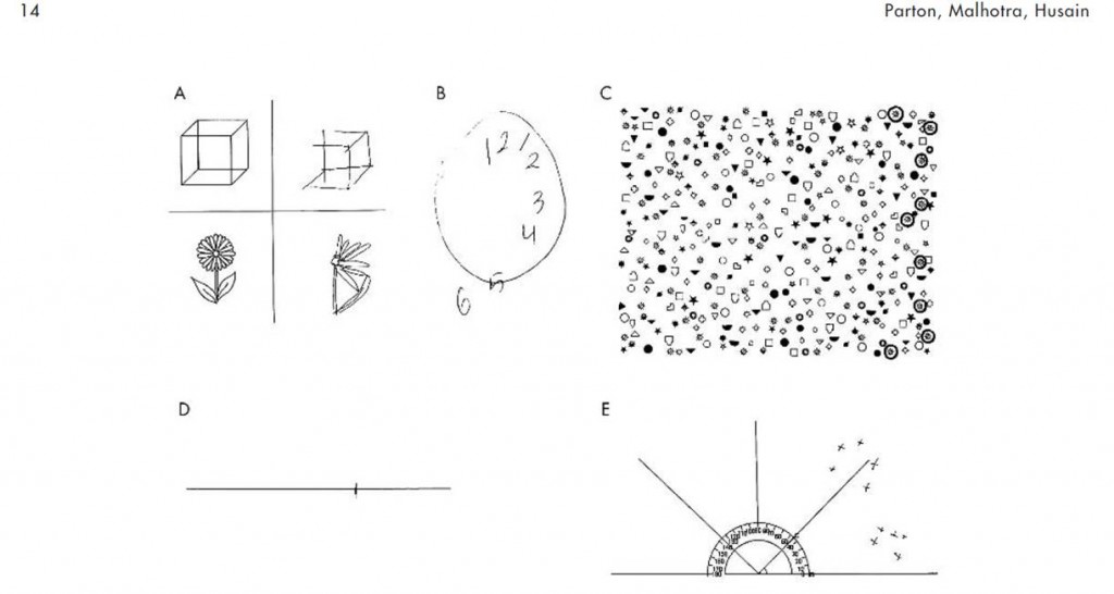
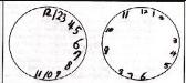
Clients with hemispatial neglect can have mirror therapy. Some studies exclude them from their research protocol but this is because their research objectives focus more specifically on the motor or functional recovery of the affected upper limb. Hemispatial neglect is excluded from their sample so that it cannot interfere with the expected results. Furthermore, clients who present severe hemispatial neglect and cannot turn their head on the contralateral side of the lesion upon request cannot have the therapy since they would not be able to keep their attention on the mirror.

Regarding the physical dimension , studies include both men and women, right- or left-handed, regardless of whether the affected side is the dominant side or not. The stroke may be ischemic or hemorrhagic with a cortical or subcortical lesion. Clients may present mild to severe hemiparesis as well as sensory deficits (paresthesia). They must be medically stable and able to maintain a sitting position throughout the treatment. Clients with vision impairments, apraxia and neurological disorders are usually excluded from studies for research purposes. Therefore, it is not known if mirror therapy is effective in the presence of these problems.

Who can give the therapy?

Mirror therapy is often given by occupational therapists and physiotherapists but any health professional can administer it.

It can also be given by a family caregiver or be self-administered following a brief explanation of how to do it and after receiving tools to do the therapy at home (user guide, written explanations, photos or videos of the movements to be done). It is important to do a regular follow-up (once a week) with clients doing the therapy at home, with or without the help of a family caregiver. The aim is to verify that the client is following instructions, understands the exercises and applies them correctly, is not making any compensating movements or taking the wrong position. The aim is also to vary the degree of difficulty, answer the client’s questions, etc. The follow-up can be done on the phone, at personal meetings, in a journal, etc. If the mirror therapy is done at home, it is important to consider the motivation of carers and the client to commit to the treatment.

If the hemiparesis is too severe and the client cannot reproduce with the affected side, simultaneously and as accurately as possible, the movements made with the healthy side, a therapist may be needed to guide or control the movements on the affected side passively. Hence it would be more difficult for such clients to do the therapy at home.

How is mirror therapy administered?

The person who administers the mirror therapy can stand in front of the client on the other side of the table. This person supervises the movements made on each side of the mirror and ensures that the client is really looking at the reflection of his/her healthy limb in the mirror.

How many weeks should mirror therapy last?

Between 3 and 6 weeks but most studies have a 4-week protocol.

How many times per week?

A minimum of 5 days per week.

How long does a mirror therapy session last?

A minimum of 30 minutes per day. This can be split into two shorter periods during the day.

What can be done in front of the mirror?

What voluntary movements are done in front of the mirror?

  • Flexion and extension of the shoulder
  • Flexion and extension of the elbow
  • Flexion and extension of the wrist
  • Flexion and extension of the fingers
  • Abduction and adduction of the shoulder
  • Abduction and adduction of the fingers
  • Internal and external rotation of the shoulder
  • Pronation and supination of the forearm
  • Ulnar and radial deviation of the wrist
  • Circumduction of the wrist

What actions can be done in front of the mirror?

Some examples of actions:

  • Squeeze and release the fist
  • Open and close the hand
  • Tap the fingers on the table
  • Oppose (touch) each finger to the thumb one by one
  • With the hand closed, try to lift each finger, including the thumb, one at a time

What tasks can be done in front of the mirror?

Some examples of tasks:

  • Handle objects using different types of grips, for example, make small balls of theraplast or modeling clay with the fingers, turn a cylindrical object in the hand (complex rotation), pick up beads or paper clips, put clothes pegs on the lip of a mug, insert pegs in a board, etc.
  • Grasp and release objects with different textures (balls, sponges, etc.)
  • Pick up and move various objects (balls, sticks, cubes, mug, glass, etc.) in different directions, for example, move an object following a sequence of movements forming a square or an ‘X’, put a ball in a glass and take it out, lift a glass, lift a rectangular object, place beads or pegs in a container with a small opening, insert pegs in holes in a piece of wood, transfer grains of rice from one pot to another, manipulate rings, etc.
  • Turn over playing cards
  • Color, connect the dots to make a drawing, copy shapes on a piece of paper
  • Use different shaped stamps with an ink pad
  • Handle utensils
  • Wipe, clean and dust the table with cloths with different textures (scouring pad, soft sponge, silk cloth, etc.)

How must the movements be done?

The movements in front of the mirror must be done simultaneouly on the affected and the non-affected side. This encourages bilateral use of the upper limbs. If the mirror therapy involves use of an object by the healthy upper limb, the affected upper limb must try to reproduce the movement as accurately as possible but without the object.

At what speed must the movements be done?

  • The client can choose the speed of the movements.
  • Some studies combining the mirror with electrical stimuli suggest that the voluntary movements are done in 5 or 10 seconds. The time the movements take must match the duration of the electrical stimuli.

What assistance is offered during the sessions?

  • The emphasis should be on active movements in front of the mirror. Clients must try their best to make the same movements with their affected upper limb. According to some studies, assistance can be offered to make the movements with the affected upper limb passively, especially if the hemiparesis is severe. To date, there is no consensus regarding whether physical assistance interferes with the mirror therapy treatment.
  • The desired movement may be demonstrated.
  • The client can be given an instruction booklet containing written instructions, photos or videos of the movements, actions and tasks to be done, especially if the mirror therapy is done at home. A toolbox containing the material needed to use the mirror may also be offered or loaned to the client.
  • The client can use a journal, especially if the therapy is done at home, as a tool to help with follow-up and to inform the therapists of the progress of the therapy (mirror therapy schedule, client’s experiences, exercises done, etc.).

What assessments and other intervention can be done with mirror therapy?

Studies exploring mirror therapy have used different assessments to measure participants’ progress, including:

  • Fugl-Meyer Assessment to measure motor recovery of the upper limb
  • Brunnstrom Scale to measure the type of movement done
  • Demeurisse Motricity Index for the upper limb to assess motor function
  • Action Research Arm Test (ARAT) to measure specific changes in upper limb activity
  • Functional Independence Measure (FIM)

There are no contraindications to using mirror therapy at the same time as other therapies. In fact, some studies combine training on specific tasks, bilateral use of the upper limbs and electrical stimuli with the use of mirror therapy.

What are the side effects of mirror therapy?

Study participants have not reported any side effects. However, one study reported that some participants got bored during the therapy.

What important features must the mirror have?

  • The mirror is placed in the client’s midsagittal plane. The client must watch the reflection of his/her healthy limb in the mirror in order to simulate that the image is actually of the affected upper limb.
  • Different materials can be used to build the structure of the mirror (wood, corrugated cardboard, plastic, etc.).
  • The size of the mirror can vary between 12 in x 12 in and 28 in x 48 in. Its size depends on the types of movements to be done in front of the mirror.
  • To ensure that the client cannot see it behind the mirror, the affected upper limb can be inserted in an enclosed box or the affected hand can be covered by a screen.
  • One study designed a folding mirror that is easy to carry. The angle of the mirror is supported by velcro strips.

What else is important to remember before using the mirror?

All jewelry and watches must be removed so that the illusion of the reflection is as credible as possible.

When and in what settings can mirror therapy be used?

When is the best time to have mirror therapy?

Mirror therapy has positive results in the acute, subacute and chronic phase post-stroke. Most studies advocate early intervention (as soon as possible) up to 14 months post-stroke.

In what settings can mirror therapy be used?

Mirror therapy can be used in different settings, including in the hospital (acute care), in-patient rehabilitation, out-patient rehabilitation and at home. The treatment setting varies with the client’s level of functional and socio-residential autonomy.

Mirror therapy can be done at home, self-administered or supervised by a family caregiver. However, clinical reasoning must be used before determining if a client is eligible.

Can mirror therapy be given in a group?

Mirror therapy can be given individually or in a group. However, including clients with attention deficits or hemispatial neglect in a group is not recommended because there is less progress in a group with respect to reducing hemispatial neglect.

References used in this section

Altschuler, E. L., et al. (1999). “Rehabilitation of hemiparesis after stroke with a mirror.” The Lancet 353(9169): 2035-2036.

Arya, K. N. and S. Pandian (2013). “Effect of task-based mirror therapy on motor recovery of the upper extremity in chronic stroke patients: a pilot study.” Topics in Stroke Rehabilitation 20(3): 210-217.

Cacchio, A., et al. (2009). “Mirror therapy for chronic complex regional pain syndrome type 1 and stroke.” New England Journal of Medicine 361(6): 634-636.

Cacchio, A., et al. (2009). “Mirror therapy in complex regional pain syndrome type 1 of the upper limb in stroke patients.” Neurorehabilitation & Neural Repair 23(8): 792-799.

de Almeida Oliveira, R., et al. (2014). “Mental practice and mirror therapy associated with conventional physical therapy training on the hemiparetic upper limb in poststroke rehabilitation: a preliminary study.” Topics in Stroke Rehabilitation 21(6): 484-494.

Dohle, C., et al. (2009). “Mirror therapy promotes recovery from severe hemiparesis: a randomized controlled trial.” Neurorehabilitation & Neural Repair 23(3): 209-217.

Ezendam, D., et al. (2009). “Systematic review of the effectiveness of mirror therapy in upper extremity function.” Disability & Rehabilitation 31(26): 2135-2149.

Faralli, A., et al. (2013). “Noninvasive strategies to promote functional recovery after stroke.” Neural Plasticity 2013: 854597.

Invernizzi, M., et al. (2013). “The value of adding mirror therapy for upper limb motor recovery of subacute stroke patients: a randomized controlled trial.” European journal of physical & rehabilitation medicine. 49(3): 311-317.

Kim, H., et al. (2014). “Effect of functional electrical stimulation with mirror therapy on upper extremity motor function in poststroke patients.” Journal of Stroke & Cerebrovascular Diseases 23(4): 655-661.

Kojima, K., et al. (2014). “Feasibility study of a combined treatment of electromyography-triggered neuromuscular stimulation and mirror therapy in stroke patients: a randomized crossover trial.” Neurorehabilitation 34(2): 235-244.

Lee, M. M., et al. (2012). “The mirror therapy program enhances upper-limb motor recovery and motor function in acute stroke patients.” American Journal of Physical Medicine & Rehabilitation 91(8): 689-696, quiz 697-700.

Lin, K. C., et al. (2014). “Combining afferent stimulation and mirror therapy for rehabilitating motor function, motor control, ambulation, and daily functions after stroke.” Neurorehabilitation & Neural Repair 28(2): 153-162.

Lisa, L. P., et al. (2013). “The effectiveness of different treatment modalities for the rehabilitation of unilateral neglect in stroke patients: a systematic review.” Neurorehabilitation 33(4): 611-620.

Michielsen, M. E., et al. (2011). “Motor recovery and cortical reorganization after mirror therapy in chronic stroke patients: a phase II randomized controlled trial.” Neurorehabilitation & Neural Repair 25(3): 223-233.

Nilsen, D. M. and T. DiRusso (2014). “Using mirror therapy in the home environment: a case report.” American Journal of Occupational Therapy 68(3): e84-89.

Pandian, J. D., et al. (2014). “Mirror therapy in unilateral neglect after stroke (MUST trial): a randomized controlled trial.” Neurology 83(11): 1012-1017.

Pollock, A., et al. (2014). “Interventions for improving upper limb function after stroke.” Cochrane Database of Systematic Reviews 11: CD010820.

Radajewska, A., et al. (2013). “The effects of mirror therapy on arm and hand function in subacute stroke in patients.” International Journal of Rehabilitation Research 36(3): 268-274.

Rothgangel, A. S., et al. (2011). “The clinical aspects of mirror therapy in rehabilitation: a systematic review of the literature.” International Journal of Rehabilitation Research 34(1): 1-13.

Samuelkamaleshkumar, S., et al. (2014). “Mirror therapy enhances motor performance in the paretic upper limb after stroke: a pilot randomized controlled trial.” Archives of Physical Medicine & Rehabilitation 95(11): 2000-2005.

Sathian, K., et al. (2000). “Doing it with mirrors: a case study of a novel approach to neurorehabilitation.” Neurorehabilitation & Neural Repair 14(1): 73-76.

Shinoura, N., et al. (2008). “Mirror therapy activates outside of cerebellum and ipsilateral M1.” Neurorehabilitation 23(3): 245-252.

Stevens, J. A. and M. E. P. Stoykov (2003). “Using motor imagery in the rehabilitation of hemiparesis.” Archives of physical medicine and rehabilitation 84(7): 1090-1092.

Thieme, H., et al. (2012). “Mirror therapy for improving motor function after stroke.” Cochrane Database of Systematic Reviews 3: CD008449.

Thieme, H., et al. (2013). “Mirror therapy for patients with severe arm paresis after stroke–a randomized controlled trial.” Clinical Rehabilitation 27(4): 314-324.

Wang, J., et al. (2013). “Cerebral activation evoked by the mirror illusion of the hand in stroke patients compared to normal subjects.” Neurorehabilitation 33(4): 593-603.

Wu, C. Y., et al. (2013). “Effects of mirror therapy on motor and sensory recovery in chronic stroke: a randomized controlled trial.” Archives of Physical Medicine & Rehabilitation 94(6): 1023-1030.

Yavuzer, G., et al. (2008). “Mirror therapy improves hand function in subacute stroke: a randomized controlled trial.” Archives of Physical Medicine & Rehabilitation 89(3): 393-398.

Yun, G. J., et al. (2011). “The synergic effects of mirror therapy and neuromuscular electrical stimulation for hand function in stroke patients.” Annals of Rehabilitation Medicine 35(3): 316-321.

Info Pocket Booklet

Pocket Card for mirror therapy

References

Altschuler, E.L., Wisdom, S.B., Stone, L., Foster, C., Galasko, D., Llewellyn, M.E., & Ramachandran, V.S. (1999). Rehabilitation of hemiparesis after stroke with a mirror. The Lancet, 353, 2035-2036. doi: 10.1016/S0140-6736(99)00920-4

Amasyali, S.Y. & Yaliman, A. (2016). Comparison of the effects of mirror therapy and electromyography-triggered neuromuscular stimulation on hand functions in stroke patients: a pilot study. International Journal of Rehabilitation Research, 39, 302-7. doi: 10.1097/MRR.0000000000000186

Arya, K.N., Pandian, S., Kumar, D., & Puri, V. (2015). Task-based mirror therapy augmenting motor recovery in poststroke hemiparesis: a randomized controlled trial. Journal of Stroke & Cerebrovascular Diseases, 24(8), 1738-48. doi: 10.1016/j.jstrokecerebrovasdis.2015.03.026

Bae, S.H., Jeong, W.S., & Kim, K.Y. (2012). Effects of mirror therapy on subacute stroke patients’ brain waves and upper extremity functions. Journal of Physical Therapy Science, 24(11), 1119-22. doi: 10.1589/jpts.24.1119

Cacchio, A., De Blasis, E., De Blasis, V., Santilli, V., & Spacca, G. (2009a). Mirror therapy in Complex Regional Pain Syndrome type 1 of the upper limb in stroke patients. Neurorehabilitation and Neural Repair, 23, 792-799. doi: 10.1177/1545968309335977

Cacchio, A., De Blasis, E., Necozione, S., di Orio, F., & Santilli, V. (2009b). Mirror therapy for Chronic Complex Regional Pain Syndrome type 1 and stroke. New England Journal of Medicine, 361(6), 634-636. doi: 10.1056/NEJMc0902799

Cho, H.-S. & Cha, H.-G. (2015). Effect of mirror therapy with tDCS on functional recovery of the upper extremity of stroke patients. Journal of Physical Therapy Science, 27, 1045-7. doi: 10.1589/jpts.27.1045

Colomer, C., Noe, E., & Llorens, R. (2016). Mirror therapy in chronic stroke survivors with severely impaired upper limb function: a randomized controlled trial. European Journal of Physical and Rehabilitation Medicine, 52(3), 271-8. Retrieved from: http://www.minervamedica.it/en/journals/europa-medicophysica/article.php?cod=R33Y2016N03A0271

Costa, V.S, Silveira, J.C.C., Clementino, T.C.A., Borges, L.R.D.M., & Melo, L.P. (2016). Effects of mirror therapy on the motor and functional recovery of post-stroke paretic upper limbs: a systematic review. Fisioterapia e Pesquisa, 23(4), 431-8. doi: 10.1590/1809-2950/15809523042016

Dohle, C., Püllen, J., Nakaten, A., Küst, J., Rietz, C., & Karbe, H. (2009). Mirror therapy promotes recovery from severe hemiparesis: a randomized controlled trial. Neurorehabilitation and Neural Repair, 23, 209-217. doi: 10.1177/1545968308324786

Ezendam, D., Bongers, R. M., Jannink, M. J. A. (2009). Systematic review of the effectiveness of mirror therapy in upper extremity function. Disability and Rehabilitation, 31, 2135-2149. doi: 10.3109/09638280902887768

Gurbuz, N., Afsar, S.I., Ayas, S., & Cosar, S.N.S. (2016). Effect of mirror therapy on upper extremity motor function in stroke patients: a randomized controlled trial. The Journal of Physical Therapy Science, 28(9), 2501-6. DOI: 10.1589/jpts.28.2501

Invernizzi, M., Negrini, S., Cara, S., Lanzotti, L., Cisari, C., & Baricich, A. (2013). The value of adding mirror therapy for upper limb motor recovery of subacute stroke patients: a randomized controlled trial. European Journal of Physical and Rehabilitation Medicine, 49(3), 311-7. Retrieved from: https://www.minervamedica.it/en/journals/europa-medicophysica/article.php?cod=R33Y2013N03A0311

Ji, S.-G., Cha, H.-G., & Kim, M.-K. (2014). Stroke recovery can be enhanced by using repetitive transcranial magnetic stimulation combined with mirror therapy. Journal of Magnetics, 19(1), 28-31. doi: 10.4283/JMAG.2014.19.1.028

Kim, K., Lee, S., Kim, D., Lee, K., & Kim, Y. (2016). Effects of mirror therapy combined with motor tasks on upper extremity function and activities daily living of stroke patients. Journal of Physical Therapy Science, 28(2), 483-7. doi: 10.1589/jpts.28.483

Kim, H.J., Lee, G.C., & Song, C.H. (2014). Effect of functional electrical stimulation with mirror therapy on upper extremity motor function in poststroke patients. Journal of Stroke and Cerebrovascular Diseases, 23(4), 655-61. doi: 10.1016/j.jstrokecerebrovasdis.2013.06.017

Lee, M.M., Cho, H., & Song, C.H. (2012). The mirror therapy program enhances upper-limb motor recovery and motor function in acute stroke patients. American Journal of Physical Medicine & Rehabilitation, 91(8), 689-700. doi: 10.1097/PHM.0b013e31824fa86d

Lim, K.-B., Lee, H.-J., Yoo, J., Yun, H.-J., & Hwang, H.-J. (2016). Efficacy of mirror therapy containing functional tasks in poststroke patients. Annals of Rehabilitation Medicine, 40(4), 629-36. doi: 10.5535/arm.2016.40.4.629

Lin, K.-C., Huang, P.-C., Chen, Y.-T., Wu, C.-Y., & Huang, W.-L. (2014). Combining afferent stimulation and mirror therapy for rehabilitating motor function, motor control, ambulation, and daily functions after stroke. Neurorehabilitation and Neural Repair, 28(2), 153-62. doi: 10.1177/1545968313508468

Michielsen, M. E., Selles, R. W., van der Geest, J. N., Eckhardt, M., Yavuzer, G., Stam, H. J., Smits, M., Ribbers, G. M., & Bussmann, J. B. J. (2010). Motor recovery and cortical reorganization after mirror therapy in chronic stroke patients: a phase II randomized controlled trial. Neurorehabilitaiton and Neural Repair, 4, 1-11. doi: 10.1177/1545968310385127

Mirela, C.L., Matei, D., Ignat, B., & Popescu, C.D. (2015). Mirror therapy enhances upper extremity motor recovery in stroke patients. Acta Neurologica Belgica, 115, 597-603. doi: 10.1007/s13760-015-0465-5

Nagapattinam, S., Vinod Babu, K., Sai Kumar, N., & Ayyappan, V.R. (2015). Effect of task specific mirror therapy with functional electrical stimulation on upper limb function for subacute hemiplegia. International Journal of Physiotherapy, 2(5), 840-9. doi: 10.15621/ijphy/2015/v2i5/78243

Pandian, J.D., Arora, R., Kaur, P., Sharma, D., Vishwambaran, D.K., & Arima, H. (2014). Mirror therapy in unilateral neglect after stroke (MUST trial): a randomized controlled trial. Neurology, 83, 1012-7. doi: 10.1212/WNL.0000000000000773

Park, J.-Y., Chang, M., Kim, K.-M., & Kim, H.J. (2015a). The effect of mirror therapy on upper-extremity function and activities of daily living in stroke patients. Journal of Physical Therapy Science, 27, 1681-3. doi: 10.1589/jpts.27.1681

Park, Y., Chang, M., Kim, K.-M., & An, D.-H. (2015b). The effects of mirror therapy with tasks on upper extremity function and self-care in stroke patients. Journal of Physical Therapy Science, 27, 1499-501. doi: 10.1589/jpts.27.1499

Purvane Vural, S., Yuzer, G.F.N., Ozcan, D.S., Ozbudak, S.D., & Ozgirgin, N. (2016). Effects of mirror therapy in stroke patients with complex regional pain syndrome type 1: a randomized controlled study. Archives of Physical Medicine and Rehabilitation, 97, 575-81. doi: 10.1016/j.apmr.2015.12.008

Radajewska, A., Opara, J.A., Kucio, C., Blaszczyszyn, M., Mehlich, K., Szczygiel, J. (2013). The effects of mirror therapy on arm and hand function in subacute stroke in patients. International Journal of Rehabilitation Research, 36(3), 268-74. doi: 10.1097/MRR.0b013e3283606218

Radajewska, A., Opara, J., Bilinski, G., Kaczorowska, A., Nawrat-Szoltysik, A., Kucinsak, A., &. Lepsy, E. (2017). Effectiveness of mirror therapy for subacute stroke in relation to chosen factors. Rehabilitation Nursing, 42(4), 223-9. doi: 10.1002/rnj.275

Rajappan, R., Abudaheer, S., Selvaganapathy, K., & Gokanadason, D. (2015). Effect of mirror therapy on hemiparetic upper extremity in subacute stroke patients. International Journal of Physiotherapy, 2(6), 1041-6. doi: 10.15621/ijphy/2015/v2i6/80766

Rehani, P., Kumari, R., & Midha, D. (2015). Effectiveness of motor relearning programme and mirror therapy on hand functions in patients with stroke – a randomized clinical trial. International Journal of Therapies and Rehabilitation Research, 4(3), 20-4. doi: 10.5455/ijtrr.00000058

Rodrigues, L.C., Farias, N.C., Gomes, R.P., & Michaelsen, S.M. (2016). Feasibility and effectiveness of adding object-related bilateral symmetrical training to mirror therapy in chronic stroke: a randomized controlled pilot study. Physiotherapy Theory and Practice, 32(2), 83-91. doi: 10.3109/09593985.2015.1091872

Rothgangel, A. S., Braun, S. M., Beurskens, A. J., Seitz, R. J., & Wade, D. T. (2011). The clinical aspects of mirror therapy in rehabilitation: A systematic review of the literature. International Journal of Rehabilitation Research, 34, 1-13. doi: 10.1097/MRR.0b013e3283441e98

Samuelkamaleshkumar, S., Reethajanetsureka, S., Pauljebaraj, P., Benshamir, B., Padankatti, S.M., David, J.A. (2014). Mirror therapy enhances motor performance in the paretic upper limb after stroke: a pilot randomized controlled trial. Archives of Physical Medicine and Rehabilitation, 95(11), 2000-5. doi: 10.1016/j.apmr.2014.06.020

Thieme, H., Bayn, M., Wurg, M., Zange, C., Pohl, M., & Behrens, J. (2012). Mirror therapy for patients with severe arm paresis after stroke – a randomized controlled trial. Clinical Rehabilitation, 27(4), 314-24. doi: 10.1177/0269215512455651

Wu, C.-Y., Huang, P.-C., Chen, Y.-T., Lin, K.-C., & Yang, H.-W. (2013). Effects of mirror therapy on motor and sensory recovery in chronic stroke: a randomized controlled trial. Archives of Physical Medicine and Rehabilitation, 94, 1023-30. doi: 10.1016/j.apmr.2013.02.007

Yavuzer G., Selles R., Sezer N., Sütbeyaz S., Bussmann J.B., Köseoglu F., Atay M.B., Stam H.J.(2008). Mirror Therapy Improves Hand Function in Subacute Stroke: A Randomized Controlled Trial. Archive of Physical Medicine, 89, 393-398. doi: 10.1016/j.apmr.2007.08.162

Yeldan, I., Huseyinsinoglu, B.E., Akinci, B., Tarakci, E., Baybas, S., & Ozdincler, A.R. (2015). The effects of very early mirror therapy on functional improvement of the upper extremity in acute stroke patients. Journal of Physical Therapy Science, 27, 3519-24. doi: 10.1589/jpts.27.3519

Yun, G.J., Chun, M.H., Park, J.Y., & Kim, B.R. (2011). The synergic effects of mirror therapy and neuromuscular electrical stimulation for hand function in stroke patients. Annals of Rehabilitation Medicine, 35, 316-21. doi: 10.5535/arm.2011.35.3.316

Excluded Studies

Arya, K.N. & Pandian, S. (2013). Effect of task-based mirror therapy on motor recovery of the upper extremity in chronic stroke patients: a pilot study. Topics in Stroke Rehabilitation, 20(3), 210-7. doi: 10.1310/tsr2003-210
Reason for exclusion: Not a randomized controlled trial.

Dalla Libera, D., Regazzi, S., Fasoletti, C., Dinacci Ruggieri, D., & Rossi Hildebrand, P. (2015). Beneficial effect of transcranial magnetic stimulation combined with mirror therapy in stroke patients: a pilot study in neurorehabilitative setting. Brain Stimulation, 8, 360-77. doi: 10.1016/j.brs.2015.01.206
Reason for exclusion: Abstract only – statistical data of clinical outcome measures not provided.

Geller, D., Nilsen, D., Van Lew, S., Gillen, G., & Bernardo, M. (2016). Home mirror therapy: a randomized controlled pilot study comparing unimanual and bimanual mirror therapy for improved upper limb function post-stroke. Archives of Physical Medicine and Rehabilitation, 97(10), e4. doi: 10.1016/j.apmr.2016.08.008
Reason for exclusion: Oral presentation – no statistical data provided for clinical outcome measures.

Harmsen, W., Bussmann, J.B.J., Selles, R.W., Hurkmans, H.L.P., & Ribbers, G.M. (2015). A mirror therapy-based action observation protocol to improve motor learning after strokeNeurorehabilitation and Neural Repair, 29(6), 509-16. doi: 10.1177/1545968314558598
Reason for exclusion: Observational study; intervention provided over one session.

Ju, Y. & Yoon, I.-J. (2018). The effects of modified constraint-induced movement therapy and mirror therapy on upper extremity function and its influence on activities of daily living. The Journal of Physical Therapy Science, 30, 77-81. doi: 10.1589/jpts.30.77
Reason for exclusion: No between-group comparisons.

Kim, H. & Shim, J. (2015). Investigation of the effects of mirror therapy on the upper extremity functions of stroke patients using the manual function test. The Journal of Physical Therapy Science, 27, 227-9. doi: 10.1589/jpts.27.227
Reason for exclusion: Not a randomized controlled trial; results do not add strength to current evidence.

Kojima, K., Ikuno, K., Morii, Y., Tokuhisa, K., Morimoto, S., & Shomoto, K. (2014). Feasibility study of a combined treatment of electromyography-triggered neuromuscular stimulation and mirror therapy in stroke patients: a randomized crossover trial. NeuroRehabilitation, 34, 235-44. doi: 10.3233/NRE-131038
Reason for exclusion: Combined therapy (mirror therapy + electromyography-triggered neuromuscular stimulation) impacts on ability to determine effects of mirror therapy alone.

Lee, D., Lee, G. & Jeong, J. (2016). Mirror therapy with neuromuscular electrical stimulation for improving motor function of stroke survivors: a pilot randomized clinical study. Technology and Health Care, 24(4), 503-11. doi: 10.3233/THC-161144
Reason for exclusion: Combined therapy (mirror therapy + neuromuscular electrical stimulation) impacts on ability to determine effects of mirror therapy alone.

Medeiros, C.S., Fernandes, S.G., Lopes, J.M., Cacho, E.N., & Cacho, R.O. (2014). Effects of mirror therapy through functional activities and motor standards in motor function of the upper limb after strokeFisioterapia & Pesquisa, 21(3), 264-70. doi: 10.590/1809-2950/87821032014.
Reason for exclusion: Both groups received mirror therapy (mirror therapy using functional activities vs. mirror therapy using isolated motor patterns).

Moustapha, A. & Rousseaux, M. (2012). Immediate effects of mirror therapy on spatial neglect. Annals of Physical and Rehabilitation Medicine, 55(S1), e197. doi: 10.1016/j.rehab.2012.07.501
Reason for exclusion: Abstract, insufficient information.

Paik, Y.-R., Kim, S.-K., Lee, J.-S., & Jeon, B.-Y. (2014). Simple and task-oriented mirror therapy for upper extremity function in stroke patients: a pilot study. Hong Kong Journal of Occupational Therapy, 24, 6-12. doi: 1016/j.hkjot.2014.01.002
Reason for exclusion: Non-randomized study with no between-group comparisons; results do not add strength to current evidence.

Rothgangel, A.S., Morton, A.R., van den Hout, J.W.E., Beurkens, A.J.H.M. (2004). Phantoms in the brain: spiegeltherapie bij chronische CVA-patienten; een pilot-study. Nederlands Tijdschrift voor Fysiotherapie, 114, 36-40. Accessed from: https://www.researchgate.net/profile/Andreas_Rothgangel/publication/290130135_Phantoms_in_the_brain_Mirror_therapy_in_chronic_stroke_patients_a_pilot_study/links/56a0b3ec08aee4d26ad74c6a/Phantoms-in-the-brain-Mirror-therapy-in-chronic-stroke-patients-a-pi
Reason for exclusion: Language other than English/French.

Selles, R.W., Michielsen, M.E., Bussmann, J.B.J., Stam, H.J., Hurkmans, H.L., Heijnen, I., Groot, D.d., Ribbers, G.M. (2014). Effects of a mirror-induced visual illusion on a reachgin task in stroke patients: implications for mirror therapy training. Neurorehabilitation and Neural Repair, 28(7), 652-9. doi: 10.1177/1545968314521005
Reason for exclusion: Not an intervention.

Salhab, G., Sarraj, A.R., & Saleh, S. (2016). Mirror therapy combined with functional electrical stimulation for rehabilitation of stroke survivors’ ankle dorsiflexion. IEEE explore digital library, 38th Annual International Conference of the IEEE Engineering in Medicine and Biology Society (EMBC). doi: 10.1109/EMBC.2016.7591776
Reason for exclusion: Combined therapy (mirror therapy + electrical stimulation) impacts on ability to determine effect of mirror therapy alone.

Sathian K., Greenspan A.I. & Wolf S.L. (2000). Doing It with Mirrors: A Case Study of a Novel Approach to Neurorehabilitation. Neurorehabilitation and Neural Repair, 14(1), 73-76. doi: 10.1177/154596830001400109
Reason for exclusion: Not a randomized controlled trial; results do not add to strength to current evidence.

Seok, H., Kim, S.H., Jang, Y.W., Lee, J.B., & Kim, S.W. (2010). Effect of mirror therapy on recovery of upper limb function and strength in subacute hemiplegia after strokeJournal of Korean Academy of Rehabilitation Medicine, 34, 508-12. Accessed from: http://www.koreascience.or.kr/article/ArticleFullRecord.jsp?cn=DJHOB7_2010_v34n5_508
Reason for exclusion: Language other than English/French.

Stevens J.A., Stoykov P.M.E. (2003). Using motor imagery in the rehabilitation of hemiparesis. Archives of Physical Medicine, 84(7), 1090-2. doi: 10.1016/S0003-9993(03)00042-X
Reason for exclusion: Not a randomized controlled trial; results do not add to strength to current evidence.

Yoon, J.A., Koo, B.I., Shin, M.J., Shin, Y.B., Ko, H.-Y., & Shin, Y.-I. (2014). Effect of constraint-induced movement therapy and mirror therapy for patients with subacute strokeAnnals of Rehabilitation Medicine, 38(4), 458-66. doi: 10.5535/arm.2014.38.4.458
Reason for exclusion: Combined therapy (mirror therapy + CIMT) impacts on ability to determine effects of mirror therapy alone.

Zacharis, D., Moumtzi, E., Terzis, N., Roussos, N., & Patatoukas, D. (2014). The use of mirror therapy in stroke patients with hemiplegic upper limb: a randomized controlled trial. Annals of Physical and Rehabilitation Medicine, 57S, e27. doi: 10.1016/j.rehab.2014.03.101
Reason for exclusion: Abstract, insufficient information.

Motor Imagery / Mental Practice

Evidence Reviewed as of before: 01-06-2017
Author(s)*: Tatiana Ogourtsova, MSc BSc OT; Annabel McDermott, OT; Angela Kim, B.Sc.; Adam Kagan, B.Sc.; Emilie Belley B.A. Psychology, B.Sc PT; Mathilde Parent-Vachon Bsc PT; Josee-Anne Filion; Alison Nutter; Marie Saulnier; Stephanie Shedleur, Bsc PT; Tsz Ting Wan, BSc PT; Elissa Sitcoff, BA BSc; Nicol Korner-Bitensky, PhD OT
Expert Reviewer: Stephen Page, PhD (C)
Patient/Family Information Table of contents

Introduction

Motor imagery or mental practice/mental imagery/mental rehearsal involves activation of the neural system while a person imagines performing a task or body movement without actually physically performing the movement. Motor imagery has been used after a stroke to attempt to treat loss of arm, hand and lower extremity movement, to help improve performance in activities of daily living, to help improve gait, and to minimize the effects of unilateral spatial neglect. Motor imagery can be used in the acute phase, subacute phase or chronic phase of rehabilitation. It has been shown that while motor imagery is beneficial by itself, it is most effective when used in addition to physical practice. In fact, many of the first studies on motor imagery were designed to investigate whether motor imagery improved motor performance in athletes. Brain scanning techniques have shown that similar areas of the brain are activated during motor imagery and physical movement. In addition, motor imagery has been shown in one study to help the brain reorganize its neural pathways, which may help promote learning of motor tasks after a stroke.

Patient/Family Information

Authors: Tatiana Ogourtsova, MSc BSc OT, Annabel McDermott, OT, Erica Kader; Emilie Belley, BA Psychology, BSc PT; Josee-Anne Filion; Alison Nutter; Mathilde Parent-Vachon; Marie Saulnier; Stephanie Shedleur, Bsc PT; Tsz Ting Wan, BSc PT; Elissa Sitcoff, BA BSc; Nicol Korner-Bitensky, PhD OT

What is motor imagery?

Motor imagery is a form of therapy that can be used to strengthen the arms, hands, feet and legs which may be weakened by stroke. In motor imagery, we mentally rehearse the movement of the affected body parts, without ever actually attempting to perform the movement. In other words, you imagine doing the motion in your mind. For example, you may imagine hitting a golf ball or drinking a cup of tea. Researchers have shown that this “mental rehearsal” actually works, as it stimulates the brain areas responsible for making the weaker arm or leg move.

Courtesy of Dr. Stephen Page and his team at Drake Center and University of Cincinnati

What is motor imagery used for?

It has been used to improve strength, increase hip movements, and improve postural control in the elderly, as well as treat people who have health problems, including injury to the spinal cord, Parkinson’s disease, or fibromyalgia (general muscle pain). It is especially useful for people with problems with the arms, legs, and hands.

Are there different types of motor imagery?

There are two distinct types of motor imagery:

  • Kinaesthetic motor imagery – imagining the feeling associated with performing a movement.
  • Visual motor imagery – imagining the movement itself.

What can I expect from a motor imagery session?

An example of a motor imagery session for a person with a weakened arm might include:

  • 5 minutes of listening to a tape recording of relaxation techniques
  • 20 minutes of exercises related to motor imagery. In week one the mental imagery training involves using computer images and movies to analyze steps and sequences required to successfully complete a task ie. reaching for a cup or turning a page in a book. In week two, patients are trained to identify problems they are having with the tasks and correct them using mental imagery. In the third week, they practice the corrected tasks mentally as well as perform the actual tasks.
  • The session concludes with time given to the individual to refocus on the room around them.

Does it Work for Stroke?

Experts have done experiments to compare mental imagery with other treatments, to see if mental imagery helps people who have had a stroke.

In individuals with ACUTE stroke (up to 1 month after stroke), 1 high quality study and one fair quality study found that mental imagery:

  • Was more helpful than the usual treatment alone for improving self-care skills (e.g. dressing and shopping);
  • Was as helpful as other treatments for improving thinking skills (e.g. attention) and motor function of the arms and legs.

In individuals with SUBACUTE stroke (1 month to 6 months after stroke), 2 high quality studies and 1 fair quality study found that mental imagery:

  • Was more helpful than the usual treatment alone for improving walking speed;
  • Was as helpful as other treatments for improving self-care skills (e.g. dressing) and physical skills of the arms and legs, including mobility, dexterity and grip strength.

In individuals with CHRONIC stroke (more than 6 months after stroke), 10 high quality studies, 6 fair quality studies in 1 poor quality study found that mental imagery:

  • Was more helpful than the usual treatment alone for improving balance, walking speed, and motor function of the arms and legs;
  • Was as helpful as other treatments for improving self-care skills (e.g. dressing and shopping) and spasticity.

When can motor imagery be used after stroke?

Motor imagery techniques can be started at any time following a stroke. However, it is believed that the treatments would be most useful in the first 6 to 18 months after a stroke when the majority of post-stroke recovery occurs.

Are there any risks to me?

There are no specific risks involved in participating in motor imagery. Motor imagery is actually quite easy to do at home, and many people find it a fun and relaxing way of having additional therapy.

How do I begin?

Your rehabilitation therapist should be able to provide you with a program to meet your individual needs. She/He can guide you as to:

  • how many times a week you should do motor imagery exercises,
  • what specific activities and movements you should do,
  • what activities you should not do,
  • how long each motor imagery session should be,
  • how to change activities as you improve.

How much does it cost? Do I need special equipment?

Motor imagery is inexpensive and accessible. Insurance will cover the services that you will receive in the hospital or rehabilitation centre. Once you are home you can continue this treatment on your own. No special equipment is required.

Clinician Information

Note: When reviewing the findings, it is important to note that they are always made according to randomized clinical trial (RCT) criteria – specifically as compared to a control group. To clarify, if a treatment is “effective” it implies that it is more effective than the control treatment to which it was compared. Non-randomized studies are no longer included when there is sufficient research to indicate strong evidence (level 1a) for an outcome.

The present module compiled results from 30 RCTs – 16 high quality RCTs, 12 fair quality RCTs and two low quality RCTs – and one non-randomized quasi experimental study. A Cochrane review by Barclay-Goddard et al. (2011) and three systematic reviews by Harris & Hebert (2015), Nilsen, Gillen & Gordon (2010), and Braun et al. (2006) were also reviewed to ensure completeness of results.

Studies were excluded if: (1) they were not RCTs and outcomes within those studies could be found in RCTs; (2) both groups were receiving a form of mental imagery training; and/or (3) no between-group analyses were performed.

Studies included in this review used mental imagery across all stages of stroke recovery, although most studies included individuals in the chronic phase or mixed phases of recovery (acute/subacute/chronic). Overall, mental imagery was often provided in combination with other interventions (e.g. conventional rehabilitation, physical therapy, occupational therapy, electrical stimulation or modified-Constraint Induced Movement Therapy – mCIMT). While in many instances it was found to achieve similar results to other interventions, mental imagery was shown to be more effective than comparison interventions in improving outcomes such as:

  • Acute stroke – functional independence and instrumental activities of daily living;
  • Subacute strokegait speed;
  • Chronic stroke – balance, gait speed, lower extremity motor function, mobility and stroke outcomes.

Note: Mental imagery, motor imagery or mental rehearsal are used interchangeably in this module.

Results Table

View results table

Outcomes

Acute phase

Functional independence
Effective
1b

One high quality RCT (Liu et al., 2004) investigated the effect of mental imagery on functional independence in patients with acute stroke. This high quality RCT randomized patients to receive mental imagery + activity of daily living (ADL) training or ADL training alone. Functional independence of trained and untrained tasks was measured by a 7-point Likert Scale at post-treatment (3 weeks) and at follow-up (1 month). Significant between-group differences in functional independence (trained and untrained tasks) were found at post-treatment, favoring mental imagery + ADL training vs. ADL training alone. Significant between-group differences in functional independence (trained tasks only) were found at follow-up, favoring mental imagery + ADL training vs. ADL training alone.
Note: In this study, mental imagery training was aimed at creating a strategy to correct ADLs in general, rather than to improve a particular movement.

Conclusion: There is moderate evidence (Level 1b) from one high quality RCT that mental imagery + ADL training is more effective than a comparison intervention (ADL training alone) in improving functional independence in patients with acute stroke.

Instrumental activities of daily living (IADLs)
Effective
2a

One fair quality RCT (Liu et al., 2009) investigated the effect of mental imagery on instrumental activities of daily living (IADLs) in patients with acute stroke. This fair quality RCT randomized patients to receive mental imagery training or conventional functional rehabilitation. IADLs (trained: sweeping, tidying, cooking, going outdoors, going to a shop; untrained: cooking, cleaning, visiting a resource center) were measured at post-treatment (3 weeks). There were significant between-group differences in performance of 3/5 trained tasks (tidying, cooking, going outdoors) and 2/3 untrained tasks (cleaning, visiting a resource center) at post-treatment, favoring mental imagery training vs. conventional functional rehabilitation.

Conclusion: There is limited evidence (Level 2a) from one fair quality RCT that mental imagery training is more effective than a comparison intervention (conventional functional rehabilitation) in improving IADLs in patients with acute stroke.

Motor function - lower extremity
Not effective
1b

One high quality RCT (Liu et al., 2004) investigated the effect of mental imagery on lower extremity motor function in patients with acute stroke. This high quality RCT randomized patients to receive mental imagery + activity of daily living (ADL) training or ADL training alone. Lower extremity motor function was measured by the Fugl-Meyer Assessment – Lower Extremity at post-treatment (3 weeks). No significant between-group differences were found.

Conclusion: There is moderate evidence (Level 1b) from one high quality RCT that mental imagery + ADL training is not more effective than a comparison intervention (ADL training alone) in improving lower extremity motor function in patients with acute stroke.

Motor function - upper extremity
Not effective
1b

One high quality RCT (Liu et al., 2004) investigated the effects of mental imagery on upper extremity motor function in patients with acute stroke. This high quality RCT randomized patients to receive mental imagery + activity of daily living (ADL) training or ADL training alone. Upper extremity motor function was measured by the Fugl-Meyer Assessment – Upper Extremity at post-treatment (3 weeks). No significant between-group differences were found.

Conclusion: There is moderate evidence (Level 1b) from one high quality RCT that mental imagery + ADL training is not more effective than a comparison intervention (ADL training alone) in improving upper extremity motor function in patients with acute stroke.

Sensation
Not effective
1b

One high quality RCT (Liu et al., 2004) investigated the effect of mental imagery on sensation in patients with acute stroke. This high quality RCT randomized patients to receive mental imagery + activity of daily living (ADL) training or ADL training alone. Sensation was measured by the Fugl-Meyer Assessment – Sensation subtest at post-treatment (3 weeks). No significant between-group differences were found.

Conclusion: There is moderate evidence (Level 1b) from one high quality RCT that mental imagery + ADL training is not more effective than a comparison intervention (ADL training) in improving sensation in patients with acute stroke.

Sustained visual attention
Not effective
1b

One high quality RCT (Liu et al., 2004) investigated the effects of mental imagery on sustained visual attention in patients with acute stroke. This high quality RCT randomized patients to receive mental imagery + activity of daily living (ADL) training or ADL training alone. Sustained attention was measured by the Color Trails Test at post-treatment (3 weeks). No significant between-group differences were found.

Conclusion: There is moderate evidence (Level 1b) from one high quality RCT that mental imagery + ADL training is not more effective than a comparison intervention (ADL training alone) in improving sustained attention in patients with acute stroke.

Subacute phase

Dexterity
Not effective
1b

One high quality RCT (Ietswaart et al., 2011) investigated the effect of mental imagery on dexterity in patients with subacute stroke. This high quality RCT randomized patients to receive mental rehearsal training, non-motor mental rehearsal training or conventional rehabilitation. Dexterity was measured by a timed manual dexterity task at post-treatment (4 weeks). No significant between-group differences were found.

Conclusion: There is moderate evidence (Level 1b) from one high quality RCT that mental rehearsal training is not more effective than comparison interventions (non-motor mental rehearsal training, conventional rehabilitation) in improving dexterity in patients with subacute stroke.

Functional independence
Not effective
1b

One high quality RCT (Ietswaart et al., 2011) investigated the effect of mental imagery on functional independence in patients with subacute stroke. This high quality RCT randomized patients to receive mental rehearsal training, non-motor mental rehearsal training or conventional rehabilitation. Functional independence was measured by the Barthel Index and the Modified Functional Limitations Profile at post-treatment (4 weeks). No significant between-group differences were found on any of the measures.

Conclusion: There is moderate evidence (Level 1b) from one high quality RCT that mental rehearsal training is not more effective than comparison interventions (non-motor mental rehearsal training, conventional rehabilitation) in improving functional independence in patients with subacute stroke.

Gait speed
Effective
1b

One high quality RCT (Oostra et al., 2015) investigated the effect of mental imagery on gait speed in patients with subacute stroke. This high quality RCT randomized patients to receive lower extremity mental imagery practice or muscle relaxation. Gait speed was measured by the 10 Meter Walking Test at post-treatment (6 weeks). Significant between-group differences were found at post-treatment, favoring lower extremity mental imagery practice vs. muscle relaxation.

Conclusion: There is moderate evidence (Level 1b) from one high quality RCT that lower extremity mental imagery practice is more effective than a comparison intervention (muscle relaxation) in improving gait speed in patients with subacute stroke.

Grip strength
Not effective
1b

One high quality RCT (Ietswaart et al., 2011) investigated the effect of mental imagery on grip strength in patients with subacute stroke. This high quality RCT randomized patients to receive mental rehearsal training, non-motor mental rehearsal training or conventional rehabilitation. Grip strength was measured with a dynamometer at post-treatment (4 weeks). No significant between-group differences were found.

Conclusion: There is moderate evidence (Level 1b) from one high quality RCT that mental rehearsal training is not more effective than comparison interventions (non-motor mental rehearsal training, conventional rehabilitation) in improving grip strength in patients with subacute stroke.

Motor function - lower extremity
Not effective
1b

One high quality RCT (Oostra et al., 2015) investigated the effect of mental imagery on lower extremity motor function in patients with subacute stroke. This high quality RCT randomized patients to receive lower extremity mental imagery practice or muscle relaxation. Lower extremity motor function was measured by the Fugl-Meyer Assessment – Lower Extremity (far transfer) at post-treatment (6 weeks). No significant between-group differences were found.

Conclusion: There is moderate evidence (Level 1b) from one high quality RCT that lower extremity mental imagery practice is not more effective than a comparison intervention (muscle relaxation) in improving lower extremity motor function in patients with subacute stroke.

Motor function - upper extremity
Not effective
1b

One high quality RCT (Ietswaart et al., 2011) and one fair quality RCT (Riccio et al., 2010) investigated the effect of mental imagery on upper extremity motor function in patients with subacute stroke.

The high quality RCT (Ietswaart et al., 2011) randomized patients to receive mental rehearsal training, non-motor mental rehearsal training or conventional rehabilitation. Upper extremity motor function was measured by the Action Research Arm Test at post-treatment (4 weeks). No significant between-group differences were found.

The fair quality RCT (Riccio et al., 2010) randomized patients to receive mental rehearsal training + conventional rehabilitation or conventional rehabilitation alone, in a cross-over design study. Upper extremity motor function was measured by the Motricity Index – Upper Extremity subscale (MI-UE) and the Arm Functional Test – Functional Ability Scale and Time score (AFT-FAS, AFT-T) score at post-treatment of Phase 1 (3 weeks) and post-treatment of Phase 2 (6 weeks). Significant between-group differences were found on all measures of upper extremity motor function at both time points, in favour of the group that had just undergone mental rehearsal training + conventional rehabilitation vs. conventional rehabilitation alone.

Conclusion: There is moderate evidence (Level 1b) from one high quality RCT that mental rehearsal training is not more effective than comparison interventions (non-motor mental rehearsal training, conventional rehabilitation) in improving upper extremity motor function in patients with subacute stroke.
Note:
However, one cross-over fair quality RCT found that mental rehearsal training + conventional rehabilitation was more effective than conventional rehabilitation alone in improving upper extremity motor function in patients with subacute stroke.

Motor imagery ability
Not effective
1b

One high quality RCT (Oostra et al., 2015) investigated the effect of mental imagery on motor imagery ability in patients with subacute stroke. This high quality RCT randomized patients to receive lower extremity mental imagery practice or muscle relaxation. Motor imagery ability was measured by the Movement Imagery Questionnaire Revised – Visual and Kinesthetic scales, and the Walking Trajectory Test (imagery/actual walking time) at post-treatment (6 weeks). There was a significant between-group difference on only one measure (Movement Imagery Questionnaire Revised – kinesthetic scale) at post-treatment, favoring lower extremity mental imagery practice vs. muscle relaxation.

Conclusion: There is moderate evidence (Level 1b) from one high quality RCT that lower extremity mental imagery practice is not more effective than a comparison intervention (muscle relaxation) in improving motor imagery ability in patients with subacute stroke.
Note:
However, there was a significant difference in kinaesthetic motor imagery, in favour of lower extremity mental imagery practice vs. muscle relaxation.

Chronic phase

Balance
Effective
1a

Four high quality RCTs (Hwang et al., 2010; Cho et al., 2012; Hosseini et al., 2012; Kim & Lee, 2013) investigated the effect of mental imagery on balance in patients with chronic stroke.

The first high quality RCT (Hwang et al., 2010) randomized patients to receive videotape-based locomotor imagery training or sham imagery training. Balance was measure by the Berg Balance Scale (BBS) at post-treatment (4 weeks). Significant between-group differences were found in balance, favoring videotape-based locomotor imagery training vs. sham imagery training.

The second high quality RCT (Cho et al., 2012) randomized patients to receive mental imagery + gait training or gait training alone. Balance was measured by the Functional Reach Test (FRT) at post-treatment (6 weeks). Significant between-group differences were found in balance, favoring mental imagery + gait training vs. gait training alone.

The third high quality RCT (Hosseini et al., 2012) randomized patients to receive mental imagery + occupational therapy or occupational therapy alone. Balance was measured by the BBS at post-treatment (5 weeks) and at follow-up (7 weeks). Significant between-group differences were found in balance at post-treatment, favoring mental imagery + occupational therapy vs. occupational therapy alone. Differences did not remain significant at follow-up.

The forth high quality RCT (Kim & Lee, 2013) randomized patients to receive mental imagery + physical therapy, action observation training + physical therapy or physical therapy alone. Balance was measured by the FRT at post-treatment (4 weeks). No significant between-group differences were found.

Conclusion: There is strong evidence (Level 1a) from three high quality RCTs that mental imagery training is more effective than comparison interventions (sham imagery training, gait training alone, occupational therapy alone) in improving balance in patients with chronic stroke. However, a fourth high quality RCT reported no significant between-group differences when comparing mental imagery + physical therapy, action observation training + physical therapy or physical therapy alone in improving balance in patients with chronic stroke.

Balance confidence
Conflicting
4

Two high quality RCTs (Hwang et al., 2010 Dickstein et al., 2013) investigated the effect of mental imagery on balance confidence in patients with chronic stroke.

The first high quality RCT (Hwang et al., 2010) randomized patients to receive videotape-based locomotor imagery training or sham imagery training. Balance confidence was measure by the Activities Specific Balance Confidence Scale at post-treatment (4 weeks). Significant between-group differences were found, favoring videotape-based locomotor imagery training vs. sham imagery training.

The second high quality RCT (Dickstein et al., 2013) randomized patients to receive mental imagery training or physical therapy. Balance confidence was measured by the Falls Efficacy Scale at post-treatment (4 weeks) and at follow-up (6 weeks). No significant between-group differences were found at either time point.

Conclusion: There is conflicting evidence (Level 4) regarding the effect of mental imagery on balance confidence in patients with chronic stroke. While one high quality RCT found that videotape-based locomotor imagery training was more effective than sham mental imagery training, one second high quality RCT found that mental imagery training was not more effective than physical therapy in improving balance confidence in patients with chronic stroke.
Note:
Studies used different measures of balance confidence.

Functional independence
Not effective
1a

Two high quality RCTs (Bovend’Eerdt et al., 2010; Hong et al., 2012) investigated the effect of mental imagery on functional independence in patients with chronic stroke.

The first high quality RCT (Bovend’Eerdt et al., 2010) randomized patients to receive mental imagery + conventional rehabilitation or conventional rehabilitation alone. Functional independence was measured by the Barthel Index (BI) at post-treatment (6 weeks). No significant between-group differences were found.

The second high quality RCT (Hong et al., 2012) randomized patients to receive mental imagery with electromyogram-triggered electric stimulation or functional electric stimulation to the affected forearm. Functional independence was measured by the modified BI at post-treatment (4 weeks). No significant between-group differences were found.

Conclusion: There is strong evidence (Level 1a) from two high quality RCTs that mental imagery is not more effective than comparison interventions (conventional rehabilitation alone, functional electric stimulation) in improving functional independence in patients with chronic stroke.

Gait parameters
Conflicting
4

Two high quality RCTs (Hwang et al., 2010 Kim & Lee, 2013) and one fair quality RCT (Lee et al., 2011) investigated the effect of mental imagery on gait parameters in patients with chronic stroke.

The first high quality RCT (Hwang et al., 2010) randomized patients to receive videotape-based locomotor imagery training or sham imagery training. Gait parameters (cadence, joint motion, stride length) were measured by a 3D motion capture system at post-treatment (4 weeks). Significant between-group differences in some gait parameters (joint motion, stride length) were found, favoring videotape-based locomotor imagery training vs. sham imagery training.

The second high quality RCT (Kim & Lee, 2013) randomized patients to receive mental imagery + physical therapy, action observation training + physical therapy or physical therapy alone. Gait parameters (cadence, speed, single/double limb support, step/stride length) were measured by the GAITRite system at post-treatment (4 weeks). There were significant between-group differences in three gait parameters (cadence, speed, single limb support) at post-treatment, favoring action observation training + physical therapy vs. physical therapy alone.

The fair quality RCT (Lee et al., 2011) randomized patients to receive mental imagery + treadmill training or treadmill training alone. Gait parameters (cadence, speed, single/double limb support, step/stride length) were measured at post-treatment (2 weeks following a 6-week treatment block). No significant between-group differences were found.

Conclusion: There is conflicting evidence (Level 4) regarding the effect of mental imagery training on gait parameters in patients with chronic stroke. While one high quality RCT found that videotape-based locomotor imagery training is more effective than a comparison intervention (sham mental imagery training) in improving some gait parameters in patients with chronic stroke, another high quality RCT and one fair quality RCT found that mental imagery training is not more effective than comparison interventions (action observation training with physical therapy, physical therapy alone, treadmill training alone) in improving gait parameters in patients with chronic stroke.

Gait speed
Effective
1a

Three high quality RCTs (Hwang et al., 2010; Cho et al., 2012;Dickstein et al., 2013) investigated the effect of mental imagery on gait speed in patients with chronic stroke.

The first high quality RCT (Hwang et al., 2010) randomized patients to receive videotape-based locomotor imagery training or sham imagery training. Gait speed was measured by the 10 Meter Walk Test (10MWT) at post-treatment (4 weeks). Significant between-group differences were found, favoring videotape-based locomotor imagery training vs. sham imagery training.

The second high quality RCT (Cho et al., 2012) randomized patients to receive mental imagery + gait training or gait training alone. Gait speed was measured by the 10MWT at post-treatment (6 weeks). Significant between-group differences were found in gait speed at post-treatment, favoring mental imagery + gait training vs. gait training alone.

The third high quality RCT (Dickstein et al., 2013) randomized patients to receive mental imagery training or physical therapy. Gait speed was measured by the 10MWT at post-treatment (4 weeks) and at follow-up (6 weeks). Significant between-group differences were found at both time points, favoring mental imagery training vs. physical therapy.
Note: Further, all participants who received physical therapy crossed-over to receive mental imagery training for 4 weeks. A significant improvement in gait speed was reported among those participants at both time points.

Conclusion: There is strong evidence (Level 1a) from three high quality RCTs that mental imagery training is more effective than comparison interventions (sham imagery training, gait training alone, physical therapy) in improving gait speed in patients with chronic stroke.

Goal attainment
Not effective
1b

One high quality RCT (Bovend’Eerdt et al., 2010) investigated the effect of mental imagery training on goal attainment in patients with chronic stroke. This high quality RCT randomized patients to receive mental imagery + conventional rehabilitation or conventional rehabilitation alone. Goal attainment was measured by the Goal Attainment Scale at post-treatment (6 weeks). No significant between-group differences were found.

Conclusion: There is moderate evidence (Level 1b) from one high quality RCT that mental imagery is not more effective than a comparison intervention (conventional rehabilitation alone) in improving goal attainment in patients with chronic stroke.

Instrumental activities of daily living (IADLs)
Not effective
1b

One high quality RCT (Bovend’Eerdt et al., 2010) investigated the effect of mental imagery training on instrumental activities of daily living (IADLs) in patients with chronic stroke. This high quality RCT randomized patients to receive mental imagery + conventional rehabilitation or conventional rehabilitation alone. IADLs were measured by the Nottingham Extended Activities of Daily Living at post-treatment (6 weeks). No significant between-group differences were found.

Conclusion: There is moderate evidence (Level 1b) from one high quality RCT that mental imagery is not more effective than a comparison intervention (conventional rehabilitation alone) in improving IADLs in patients with chronic stroke.

Mobility
Conflicting
4

Seven high quality RCTs (Malouin et al., 2009Bovend’Eerdt et al., 2010Hwang et al., 2010; Cho et al., 2012Hosseini et al., 2012Dickstein et al., 2013Kim & Lee, 2013) investigated the effect of mental imagery training on mobility in patients with chronic stroke.

The first high quality RCT (Malouin et al., 2009) randomized patients to receive mental imagery + physical practice, cognitive training + physical practice, or no training. Mobility was measured by the change scores in leg loading of the affected leg as a percent of body weight during the rising-to-sitting action at baseline, post-treatment (4 weeks) and follow-up (7 weeks). Significant between-group differences in change scores from baseline to post-treatment were found, favoring mental imagery training + physical practice vs. cognitive training + physical practice; and favoring mental imagery training + physical practice vs. no training. Significant between-group differences were not maintained at follow-up.

The second high quality RCT (Bovend’Eerdt et al., 2010) randomized patients to receive mental imagery + conventional rehabilitation or conventional rehabilitation alone. Mobility was measured by the Timed Up and Go Test (TUGT) and the Rivermead Mobility Index at post-treatment (6 weeks). No significant between-group differences were found on any of the measures.

The third high quality RCT (Hwang et al., 2010) randomized patients to receive videotape-based locomotor imagery training or sham imagery training. Mobility was measured by the Dynamic Gait Index and the Modified Emory Functional Ambulation Profile at post-treatment (4 weeks). Significant between-group differences in both measures of mobility were found, favoring videotape-based locomotor imagery training vs. sham imagery training.

The forth high quality RCT (Cho et al., 2012) randomized patients to receive mental imagery + gait training or gait training alone. Mobility was measured by the TUGT at post-treatment (6 weeks). Significant between-group differences were found, favoring mental imagery + gait training vs. gait training alone.

The fifth high quality RCT (Hosseini et al., 2012) randomized patients to receive mental imagery + occupational therapy or occupational therapy alone. Mobility was measured by the TUGT at post-treatment (5 weeks) and at follow-up (7 weeks). Significant between-group differences were found at post-treatment, favoring mental imagery + occupational therapy vs. occupational therapy alone. Significant between-group differences were not maintained at follow-up.

The sixth high quality RCT (Dickstein et al., 2013) randomized patients to receive mental imagery training or physical therapy. Mobility was measured by step activity monitor (community ambulation) and number of steps/minute at post-treatment (4 weeks) and at follow-up (6 weeks). There were no significant between-group differences in both measures of mobility at either time point.

The seventh high quality RCT (Kim & Lee, 2013) randomized patients to receive mental imagery + physical therapy, action observation training + physical therapy or physical therapy alone. Mobility was measured by the TUGT, Walking Ability Questionnaire, and Functional Ambulation Category at post-treatment (4 weeks). A significant between-group difference in one measure of mobility (TUGT) was found at post-treatment, favoring action observation training + physical therapy vs. physical therapy alone.

Conclusion: There is conflicting evidence (Level 4) regarding the effect of mental imagery on mobility in patients with chronic stroke. While four high quality RCTs found that mental imagery training is more effective than comparison interventions (cognitive training + physical practice, no training, sham imagery training, gait training alone, occupational therapy alone) in improving mobility in patients with chronic stroke; three other high quality RCTs found that mental imagery is not more effective than comparison interventions (conventional rehabilitation alone, physical therapy, action observation training + physical therapy) in improving mobility in patients with chronic stroke.

Motor activity - upper extremity
Not effective
1b

One high quality RCT (Hong et al., 2012) and one fair quality RCT (Page et al., 2005) investigated the effect of mental imagery on upper extremity motor activity among patients with chronic stroke.

The high quality RCT (Hong et al., 2012) randomized patients to receive mental imagery + electromyogram-triggered electric stimulation or functional electric stimulation to the affected forearm. Upper extremity motor activity was measured by the Motor Activity Log – Amount of Use and Quality of Movement (MAL-AOU, MAL-QOM) at post-treatment (4 weeks). No significant between-group differences were found.

The fair quality RCT (Page et al., 2005) randomized patients to receive mental imagery training or relaxation training. Upper extremity motor activity was measured by the MAL at post-treatment (6 weeks). No significant between-group differences were found.

Conclusion: There is moderate evidence (Level 1b) from one high quality RCT and one fair quality RCT that mental imagery training is not more effective than comparison interventions (functional electrical stimulation to the affected forearm, relaxation training) in improving upper extremity motor activity in patients with chronic stroke.

Motor function - lower extremity
Effective
1b

One high quality RCT (Cho et al., 2012) investigated the effect of mental imagery on lower extremity motor function in patients with chronic stroke. This high quality RCT randomized patients to receive mental imagery + gait training or gait training alone. Lower extremity motor function was measured by the Fugl-Meyer Assessment – Lower Extremity at post-treatment (6 weeks). Significant between-group differences were found, favoring mental imagery + gait training vs. gait training alone.

Conclusion: There is moderate evidence (Level 1b) from one high quality RCT that mental imagery + gait training is more effective than a comparison intervention (gait training alone) in improving lower extremity motor function in patients with chronic stroke.

Motor function - upper extremity
Conflicting
4

Four high quality RCTs (Bovend’Eerdt et al., 2010Page et al., 2011;Hong et al., 2012Nilsen et al., 2012) and five fair quality RCTs (Page, 2000Page et al., 2005Ertelt et al., 2007Page et al., 2007Page et al., 2009) investigated the effect of mental imagery on upper extremity motor function in patients with chronic stroke.

The first high quality RCT (Bovend’Eerdt et al., 2010) randomized patients to receive mental imagery + conventional rehabilitation or conventional rehabilitation alone. Upper extremity motor function was measured by the Action Research Arm Test (ARAT) at post-treatment (6 weeks). No significant between-group differences were found.

The second high quality RCT (Page et al., 2011) randomized patients to receive mental imagery or sham audio therapy. Upper extremity motor function was measured by the Fugl-Meyer Assessment – Upper Extremity (FMA-UE) and the ARAT at post-treatment (10 weeks). No significant between-group differences were found on any of the measures.

The third high quality RCT (Hong et al., 2012) randomized patients to receive mental imagery + electromyogram-triggered electric stimulation or functional electric stimulation to the affected forearm. Upper extremity motor function was measured by the FMA-UE at post-treatment (4 weeks). Significant between-group differences in upper extremity motor function were found at post-treatment, favoring mental imagery + electromyogram-triggered electric stimulation vs. functional electric stimulation to the affected forearm.

The forth high quality RCT (Nilsen et al., 2012) randomized patients to receive mental imagery training using an internal perspective (internal group), mental imagery training using an external perspective (external group), or relaxation imagery; all groups received occupational therapy. Upper extremity motor function was measured by the FMA-UE and the Jebsen-Taylor Test of Hand Function at post-treatment (6 weeks). Significant between-group differences were found on both measures, favoring both styles of mental imagery training (internal group, external group) vs. relaxation imagery.

The first fair quality RCT (Page, 2000) randomized patients to receive mental imagery training + occupational therapy or occupational therapy alone. Upper extremity motor function was measured by the FMA-UE at post-treatment (4 weeks). Significant between-group differences were found at post-treatment, favoring mental imagery training + occupational therapy vs. occupational therapy alone.

The second fair quality RCT (Page et al., 2005) randomized patients to receive mental imagery training or relaxation training. Upper extremity motor function was measured by the ARAT at post-treatment (6 weeks). Significant between-group differences were found, favoring mental imagery training vs. relaxation training.

The third fair quality RCT (Ertelt et al., 2007) randomized patients to receive action observation therapy or conventional rehabilitation. Upper extremity motor function was measured by the Frenchay Arm Test and the Wolf Motor Function Test at post-treatment (18 days); participants in the action observation group were reassessed 8 weeks later (follow-up). Significant between-group differences were found on both measures of upper extremity motor function at post-treatment, favoring action observation therapy vs. conventional rehabilitation. Significant within-group gains were maintained at follow-up.

The forth fair quality RCT (Page et al., 2007) randomized patients to receive mental imagery training or relaxation training. Upper extremity motor function was measured by the ARAT and the FMA-UE at post-treatment (1 week following a 6-week treatment). Significant between-group differences were found on both measures of upper extremity motor function at post-treatment, favoring mental imagery training vs. relaxation training.

The fifth fair quality RCT (Page et al., 2009) randomized patients to receive mental imagery + modified-constraint induced therapy (mCIMT) or mCIMT alone. Upper extremity motor function was measured by the ARAT and the FMA-UE at post-treatment (10 weeks) and follow-up (3 months). Significant between-group differences were found on both measures of upper extremity motor function at post-treatment and at follow-up, favoring mental imagery training + mCIMT vs. mCIMT alone.

Conclusion: There is conflicting evidence (Level 4) regarding the effect of mental imagery on upper extremity motor function. While two high quality RCTs found that mental imagery was not more effective than comparison interventions (conventional rehabilitation alone, sham audio therapy) in improving upper extremity motor function in patients with chronic stroke; two other high quality RCTs found that mental imagery was more effective than comparison interventions (functional electric stimulation to the affected forearm, relaxation imagery) in improving upper extremity motor function in patients with chronic stroke.
Note:
Five fair quality RCTs found that mental imagery training is more effective than comparison interventions (occupational therapy alone, relaxation training, conventional rehabilitation, mCIMT alone) in improving upper extremity motor function in patients with chronic stroke.

Occupational performance
Not effective
1b

One high quality RCT (Nilsen et al., 2012) investigated the effect of mental imagery on occupational performance in patients with chronic stroke. This high quality RCT randomized patients to receive mental imagery training using an internal perspective (internal group), mental imagery training using an external perspective (external group), or relaxation imagery; all groups received occupational therapy. Occupational performance was measure by the Canadian Occupational Performance Measure at post-treatment (6 weeks). No significant between-group differences were found.

Conclusion: There is moderate evidence (Level 1b) from one high quality RCT that mental imagery training using an internal or external perspective is not more effective than a comparison intervention (relaxation imagery) in improving occupational performance in patients with chronic stroke.

Pain
Not effective
2b

One poor quality RCT (Cacchio et al., 2009) investigated the effect of mental imagery on pain in patients with chronic stroke. This poor quality RCT randomized patients with Complex Regional Pain Syndrome (CRPS) to receive mental imagery, mirror therapy or covered mirror practice. Pain was measured by Visual Analogue Scale at post-treatment (4 weeks). Significant between-group differences were found, favoring mirror therapy vs. mental imagery and favouring mirror therapy vs. covered mirror practice.
Note: Following 4 weeks, some participants crossed-over to the mirror therapy group. A significant reduction in pain was reported among participants who crossed-over from the mental imagery and covered mirror practice groups to the mirror therapy group.

Conclusion: There is limited evidence (Level 2b) from one poor quality RCT that mental imagery is not more effective than comparison interventions (mirror therapy, covered mirror practice) in improving pain in patients with chronic stroke and CRPS. In fact, mirror therapy was more effective than mental imagery in reducing pain.

Spasticity
Not effective
1b

One high quality RCT (Hong et al., 2012) investigated the effect of mental imagery training on spasticity in patients with chronic stroke. This high quality RCT randomized patients to receive mental imagery + electromyogram-triggered electric stimulation or functional electric stimulation to the affected forearm. Spasticity was measured by the Modified Ashworth Scale at post-treatment (4 weeks). No significant between-group differences were found.

Conclusion: There is moderate evidence (Level 1b) from one high quality RCT that mental imagery + electromyogram-triggered electric stimulation is not more effective than a comparison intervention (functional electric stimulation to the affected forearm) in improving spasticity in patients with chronic stroke.

Stroke outcomes
Effective
2a

One fair quality RCT (Ertelt et al., 2007) investigated the effect of mental imagery on stroke outcomes in patients with chronic stroke. This high quality RCT randomized patients to receive action observation therapy or conventional rehabilitation. Stroke outcomes were measured by the Stroke Impact Scale at post-treatment (18 days); participants in the action observation group were reassessed 8 weeks later (follow-up). Significant between-group differences were found at post-treatment, favoring action observation therapy vs. conventional rehabilitation. Significant within-group gains were maintained at follow-up.

Conclusion: There is limited evidence (Level 2a) from one fair quality RCT that action observation training is more effective than a comparison intervention (conventional rehabilitation) in improving stroke outcomes in patients with chronic stroke.

Phase not specific to one period

Balance
Not effective
1a

Two high quality RCTs (Braun et al., 2012; Schuster et al., 2012) investigated the effect of mental imagery on balance in patients with stroke.

The first high quality RCT (Braun et al., 2012) randomized patients with acute/subacute stroke to receive mental imagery + conventional rehabilitation or conventional rehabilitation alone. Balance was measured by the Berg Balance Scale (BBS) at post-treatment (6 weeks) and at follow-up (6 months). No significant between-group differences were found at either time point. 

The second high quality RCT (Schuster et al., 2012) randomized patients with subacute/chronic stroke to receive embedded mental imagery training, added mental imagery training or time-matched stroke education tapes; all groups received physical therapy. Balance was measured by the BBS at post-treatment (2 weeks) and follow-up (1 month). No significant between-group differences were found at either time point.

Conclusion: There is strong evidence (Level 1a) from two high quality RCTs that mental imagery is not more effective than comparison interventions (conventional rehabilitation alone, time-matched stroke education tapes) in improving balance in patients with stroke.

Balance confidence
Not effective
1b

One high quality RCT (Schuster et al., 2012) investigated the effect of mental imagery training on balance confidence in patients with stroke. This high quality RCT randomized patients with subacute/chronic stroke to receive embedded mental imagery training or added mental imagery training or time-matched stroke education tapes; all groups received physical therapy. Balance confidence was measured by the Activities-Specific Balance Confidence Scale at post-treatment (2 weeks) and follow-up (1 month). No significant between-group differences were found at either time point.

Conclusion: There is moderate evidence (Level 1b) from one high quality RCT that embedded or added mental imagery is not more effective than a comparison intervention (time-matched stroke education tapes) in improving balance confidence in patients with subacute/chronic stroke.

Dexterity
Not effective
1b

One high quality RCT (Braun et al., 2012) investigated the effect of mental imagery on dexterity in patients with stroke. This high quality RCT randomized patients with acute/subacute stroke to receive mental imagery + conventional rehabilitation or conventional rehabilitation alone. Dexterity was measured by the Nine Hole Peg Test at post-treatment (6 weeks) and at follow-up (6 months). No significant between-group differences were found at either time point.

Conclusion: There is moderate evidence (Level 1b) from one high quality RCT that mental imagery is not more effective than a comparison intervention (conventional rehabilitation alone) in improving dexterity in patients with acute/subacute stroke.

Functional independence
Not effective
1a

Three high quality RCTs (Braun et al., 2012Schuster et al., 2012Timmermans et al., 2013), one fair quality RCT (Ferreira et al., 2011) and one poor quality RCT (Park et al., 2015) investigated the effect of mental imagery on functional independence in patients with stroke.

The first high quality RCT (Braun et al., 2012) randomized patients with acute/subacute stroke to receive mental imagery + conventional rehabilitation or conventional rehabilitation alone. Functional independence was measured by the Barthel Index (BI); patients’ and therapists’ perception of performance of daily activities (e.g. drinking, walking) was measured by a 10-point numeric rating scale at post-treatment (6 weeks) and at follow-up (6 months). No significant between-group differences were found on either measure at either time point.

The second high quality RCT (Schuster et al., 2012) randomized patients with subacute/chronic stroke to receive embedded mental imagery training, added mental imagery training, or time-matched stroke education tapes; all groups received physical therapy. Functional independence was measured by the BI at post-treatment (2 weeks) and follow-up (1 month). No significant between-group differences were found at either time point.

The third high quality RCT (Timmermans et al., 2013) randomized patients with acute/subacute stroke to receive mental imagery or neurodevelopmental therapy; both groups received conventional rehabilitation. Functional independence was measured by the BI at post-treatment (6 weeks) and at follow-up (6 and 12 months). No significant between-group differences were found at any time point.

The fair quality RCT (Ferreira et al., 2011) randomized patients with subacute/chronic stroke to receive mental imagery + conventional rehabilitation, visual scanning training + conventional rehabilitation, or conventional rehabilitation alone. Functional independence was measured by the Functional Independence Measure (FIM) at post-treatment (5 weeks) and at follow-up (3 months). There were no significant differences between mental imagery + conventional rehabilitation and other treatment groups at either time point.
Note: Significant between-group differences in functional independence (FIM – self-care items only) were found at post-treatment, favoring visual scanning + conventional rehabilitation vs. conventional rehabilitation alone. Differences did not remain significant at follow-up.

The poor quality RCT (Park et al., 2015) randomized patients with subacute/chronic stroke to receive mental imagery + conventional rehabilitation or conventional rehabilitation alone. Functional independence was measured by the modified BI at post-treatment (2 weeks). Significant between-group differences were found, favoring mental imagery + conventional rehabilitation vs. conventional rehabilitation alone.

Conclusion: There is strong evidence (Level 1a) from three high quality RCTs and one fair quality RCT that mental imagery is not more effective than comparison interventions (conventional rehabilitation alone, time-matched stroke education tapes, neurodevelopmental therapy, visual scanning training + conventional rehabilitation) in improving functional independence in patients with stroke.
Note
: One poor quality RCT found that mental imagery training + conventional rehabilitation is more effective than a comparison intervention (conventional rehabilitation alone) in improving functional independence in patients with subacute/chronic stroke.

Gait speed
Not effective
1b

One high quality RCT (Braun et al., 2012) investigated the effect of mental imagery on gait speed in patients with stroke. This high quality RCT randomized patients with acute/subacute stroke to receive mental imagery + conventional rehabilitation or conventional rehabilitation alone. Gait speed was measured by the 10 Meter Walk Test at post-treatment (6 weeks) and at follow-up (6 months). No significant between-group differences were found at either time point.

Conclusion: There is moderate evidence (Level 1b) from one high quality RCT that mental imagery + conventional rehabilitation is not more effective than a comparison intervention (conventional rehabilitation alone) in improving gait speed in patients with acute/subacute stroke.

Grip strength
Effective
2a

One fair quality RCT (Muller et al., 2007) investigated the effect of mental imagery on grip strength in patients with stroke. This fair quality RCT randomized patients with acute/subacute stroke to receive mental imagery training, motor practice training or conventional physical therapy. Grip strength was measured by a force transducer at post-treatment (4 weeks). Significant between-group differences were found, favoring mental imagery vs. physical therapy l rehabilitation and favoring motor practice vs. physical therapy rehabilitation.

Conclusion: There is limited evidence (Level 2a) from one fair quality RCT that mental imagery training is more effective than a comparison intervention (conventional physical therapy) in improving grip strength in patients with acute/subacute stroke.

Instrumental activities of daily living (IADLs)
Not effective
1b

One high quality RCT (Timmermans et al., 2013) investigated the effect of mental imagery on instrumental activities of daily living (IADLs) in patients with stroke. This high quality RCT randomized patients with acute/subacute stroke to receive mental imagery or neurodevelopmental therapy; both groups received conventional rehabilitation. IADLs were measured by the Frenchay Activity Index at post-treatment (6 weeks) and at follow-up (6 and 12 months). No significant between-group differences were found at any time point. 

Conclusion: There is moderate evidence (Level 1b) from one high quality RCT that mental imagery is not more effective than a comparison intervention (neurodevelopmental therapy) in improving IADLs in patients with acute/subacute stroke.

Mobility
Not effective
1b

One high quality RCT (Braun et al., 2012) investigated the effect of mental imagery on mobility in patients with stroke. This high quality RCT randomized patients with acute/subacute stroke to receive mental imagery + conventional rehabilitation or conventional rehabilitation alone. Mobility was measured by the Rivermead Mobility Index at post-treatment (6 weeks) and at follow-up (6 months). No significant between-group differences were found at either time point. 

Conclusion: There is moderate evidence (Level 1b) from one high quality RCT that mental imagery + conventional rehabilitation is not more effective than a comparison intervention (conventional rehabilitation alone) in improving mobility in patients with acute/subacute stroke.

Motor activity
Not effective
1b

One high quality RCT (Schuster et al., 2012) investigated the effect of mental imagery on motor activity in patients with stroke. This high quality RCT randomized patients with subacute/chronic stroke to receive embedded mental imagery training, added mental imagery training, or time-matched stroke education tapes; all groups received physical therapy. Motor activity was measured by (i) time taken to complete a motor task; (ii) the Chedoke McMaster Stroke Assessment (activity scale); and (iii) stage of motor task as per Adams & Tyson classification, at post-treatment (2 weeks) and follow-up (1 month). No significant between-group differences were found on any measure at either time point.

Conclusion: There is moderate evidence (Level 1b) from one high quality RCT that embedded or added mental imagery training is not more effective than a comparison (time-matched stroke education tapes) in improving motor activity in patients with subacute/chronic stroke.

Motor activity - upper extremity
Not effective
1b

One high quality RCT (Timmermans et al., 2013) and one quasi-experimental design study (Rajesh, 2015) investigated the effect of motor imagery on upper extremity motor activity among patients with stroke.

The high quality RCT (Timmermans et al., 2013) randomized patients with acute/subacute stroke to receive mental imagery or neurodevelopmental therapy; both groups received conventional rehabilitation. Upper extremity motor activity was measured by accelerometry (total activity, activity/hour, activity ratio of affected/unaffected arm) at post-treatment (6 weeks) and at follow-up (6 and 12 months). No significant between-group differences were found at either time point.

The quasi-experimental design study (Rajesh, 2015) assigned patients with stroke (stage of recovery not specified) to receive mental imagery + occupational therapy or occupational therapy alone. Upper extremity motor activity was measured by the Motor Activity Log at post-treatment (3 weeks). Significant between-group differences were found, favoring mental imagery + conventional occupational therapy vs. conventional occupational therapy alone.

Conclusion: There is moderate evidence (Level 1b) from one high quality RCT that mental imagery is not more effective than a comparison intervention (neurodevelopmental therapy) in improving upper extremity motor activity in patients with acute/subacute stroke.
Note:
However, one quasi-experimental study found that mental imagery was more effective than a comparison intervention (conventional occupational therapy alone) in improving upper extremity motor activity in patients with stroke. Discrepancies could result from differences in employed measurement scales and treatment duration (6 vs. 3 weeks).

Motor function - upper extremity
Not effective
1a

Two high quality RCTs (Welfringer et al., 2011Timmermans et al., 2013), two fair quality RCTs (Page et al., 2001Muller et al., 2007), and one poor quality RCT (Park et al., 2015) investigated the effect of mental imagery on upper extremity motor function in patients with stroke.

The first high quality RCT (Welfringer et al., 2011) randomized patients with acute/subacute stroke to receive visuomotor imagery + conventional rehabilitation or conventional rehabilitation alone. Upper extremity motor function was measured by the Action Research Arm Test (ARAT) at post-treatment (3 weeks). No significant between-group differences were found.

The second high quality RCT (Timmermans et al., 2013) randomized patients with acute/subacute stroke to receive mental imagery or neurodevelopmental therapy; both groups received conventional rehabilitation. Upper extremity motor function was measured by the Wolf Motor Function Test, Frenchay Arm Test and Fugl-Meyer Assessment – Upper Extremity (FMA-UE) at post-treatment (6 weeks) and at follow-up (6 and 12 months). No significant between-group differences were found on any measure at any time point. 

The first fair quality RCT (Page et al., 2001) randomized patients with acute/subacute/chronic stroke to receive mental imagery training or stroke education; both groups received time-matched occupational therapy. Upper extremity motor function was measured by the FMA-UE and the ARAT at post-treatment (6 weeks). Differences in both measures of upper extremity motor function were found at post-treatment, favoring mental imagery training vs. stroke education.

The second fair quality RCT (Muller et al., 2007) randomized patients with acute/subacute stroke to receive mental imagery training, motor practice or conventional physical therapy. Upper extremity motor function was measured by the Jebsen Hand Function Test (JHFT – writing, turning over card, picking up small objects, simulated feeding, stacking checkers, picking up large light cans, picking up large heavy cans) at post-treatment (4 weeks). Significant between-group differences were found in some aspect of upper extremity motor function (JHFT – writing, simulated feeding), favoring mental imagery training vs. conventional physical therapy and favoring motor practice vs. conventional physical therapy.

The poor quality RCT (Park et al., 2015) randomized patients with subacute/chronic stroke to receive mental imagery training + conventional rehabilitation or conventional rehabilitation alone. Upper extremity motor function was measured by the ARAT and the FMA-UE at post-treatment (2 weeks). Significant between-group differences were found on both measures of upper extremity motor function at post-treatment, favoring mental imagery training + conventional rehabilitation vs. conventional rehabilitation alone.

Conclusion: There is strong evidence (Level 1a) from two high quality RCTs that mental imagery is not more effective than comparison interventions (conventional rehabilitation alone, neurodevelopmental therapy) in improving upper extremity motor function in patients with stroke.
Note: 
However, two fair quality RCTs and one poor quality RCT found that mental imagery is more effective than comparison interventions (stroke education, conventional physical therapy, conventional rehabilitation alone) in improving upper extremity motor function in patients with stroke.

Motor imagery ability
Not effective
1b

One high quality RCT (Schuster et al., 2012) investigated the effect of mental imagery on motor imagery ability in patients with stroke. This high quality RCT randomized patients with subacute/chronic stroke to receive embedded mental imagery training, added mental imagery training, or time-matched stroke education tapes; all groups received physical therapy. Motor imagery ability was measured by the Imaprax Questionnaire and the Kinesthetic and Visual Imagery Questionnaire at post-treatment (2 weeks) and follow-up (1 month). No significant between-group differences were found on either measure at either time point.

Conclusion: There is moderate evidence (Level 1a) from one high quality RCT that embedded or added mental imagery is not more effective than a comparison intervention (time-matched stroke education tapes) in improving motor imagery ability in patients with stroke.

Unilateral spatial neglect
Not effective
1b

One high quality RCT (Welfringer et al., 2011) and one fair quality RCT (Ferreira et al., 2011) investigated the effect of mental imagery on unilateral spatial neglect (USN) in patients with stroke.

The high quality RCT (Welfringer et al., 2011) randomized patients with acute/subacute stroke to receive visuomotor imagery + conventional rehabilitation or conventional rehabilitation alone. USN was measured by the Bells Cancellation Test, Reading Test, Flower Copying Test, Clock Drawing Test and Representation Test (body touching, visual arm imagery, kinesthetic arm imagery) at post-treatment (3 weeks). No significant between-group differences were found on any measure.

The fair quality RCT (Ferreira et al., 2011) randomized patients with subacute/chronic stroke to receive mental imagery + conventional rehabilitation, visual scanning training + conventional rehabilitation, or conventional rehabilitation alone. USN was measured by the Behavioral Inattention Test at post-treatment (5 weeks) and at follow-up (3 months). There were no significant differences between mental imagery + conventional rehabilitation and other groups at either time point.
Note: Significant between-group differences favoring visual scanning + conventional rehabilitation vs. conventional rehabilitation alone were found at post-treatment and at follow-up.

Conclusion: There is moderate evidence (Level 1b) from one high quality RCT and one fair quality RCT that mental imagery + conventional rehabilitation is not more effective than comparison interventions (conventional rehabilitation alone, visual scanning training + conventional rehabilitation) in improving USN in patients with stroke.

Quality of life
Not effective
1b

One high quality RCT (Schuster et al., 2012) and one quasi-experimental design study (Rajesh, 2015) investigated the effect of mental imagery on quality of life in patients with stroke.

The high quality RCT (Schuster et al., 2012) randomized patients with subacute/chronic stroke to receive embedded mental imagery training, added mental imagery training, or time-matched stroke education tapes; all groups received physical therapy. Quality of life was measured by Visual Analogue Scale at post-treatment (2 weeks) and follow-up (1 month). No significant between-group differences were found at either time point.

The quasi-experimental design study (Rajesh, 2015) assigned patients with stroke (stage of recovery not specified) to receive mental imagery + conventional occupational therapy or conventional occupational therapy alone. Quality of life was measured by the Stroke-Specific Quality of Life scale at post-treatment (3 weeks). Significant between-group differences were found, favoring mental imagery practice + conventional occupational therapy vs. conventional occupational therapy alone.

Conclusion: There is moderate evidence (Level 1b) from one high quality RCT that embedded or added mental imagery training is not more effective than a comparison intervention (time-matched stroke education tapes) in improving quality of life in patients with subacute/chronic stroke.
Note
: One quasi-experimental study found that mental imagery training + conventional occupational therapy is more effective than a comparison intervention (conventional occupational therapy alone) in improving quality of life in patients with stroke. Discrepancies could result from differences in employed measurement scales and treatment duration (2 vs. 3 weeks).

Sensation
Not effective
1b

One high quality RCT (Welfringer et al., 2011) investigated the effect of visual imagery on sensation in patients with stroke. This high quality RCT randomized patients with acute/subacute stroke to receive visuomotor imagery with conventional rehabilitation or conventional rehabilitation alone. Upper extremity sensation was measured by the Arm Function Test – Sensation score at post-treatment (3 weeks). No significant between-group differences were found.

Conclusion: There is moderate evidence (level 1b) from one high quality RCT that visual imagery + conventional rehabilitation is not more effective than a comparison intervention (conventional rehabilitation alone) for improving sensation in patients with acute/subacute stroke.

Strength
Not effective
1b

One high quality RCT (Braun et al., 2012) investigated the effect of mental imagery training on strength in patients with stroke. This high quality RCT randomized patients with acute/subacute stroke to receive mental imagery + conventional rehabilitation or conventional rehabilitation alone. Strength was measured by the Motricity Index at post-treatment (6 weeks) and at follow-up (6 months). No significant between-group differences were found at either time point. 

Conclusion: There is moderate evidence (Level 1b) from one high quality RCT that mental imagery training + conventional rehabilitation is not more effective than a comparison intervention (conventional rehabilitation alone) in improving strength in patients with acute/subacute stroke.

References

Bovend’Eerdt, T.J., Dawes, H., Sackley, C., Hooshang, I., & Wade, D.T. (2010). An Integrated Motor Imagery Program to Improve Functional Task Performance in Neurorehabilitation: A Single-Blind Randomized Controlled Trial. Archives of Physical Medicine and Rehabilitation, 91, 939-946.
https://www.ncbi.nlm.nih.gov/pubmed/20510987

Braun, S.M., Beurskens, A.J., Kleynen, M., Oudelaar, B., Schols, J.M., & Wade, D.T. (2012). A multicenter randomized controlled trial to compare subacute “treatment as usual” with and without mental practice among persons with stroke in Dutch Nursing Homes. JAMDA, 13, 1-7.
https://www.ncbi.nlm.nih.gov/pubmed/21450196

Cacchio, A., De Blasis, E., Necozione, S., di Orio, F., & Santilli, V. (2009). Mirror therapy for Chronic Complex Regional Pain Syndrome type 1 and stroke. New England Journal of Medicine, 361(6), 634-636.
http://www.nejm.org/doi/full/10.1056/nejmc0902799#t=article

Cho, H. Y., Kim, J. S., & Lee, G. C. (2012). Effects of motor imagery training on balance and gait abilities in post-stroke patients: a randomized controlled trial. Clinical rehabilitation27(8), 675-680.
http://journals.sagepub.com/doi/abs/10.1177/0269215512464702

Dickstein, R., Deutsch, J.E., Yoeli, Y., Kafri, M., Falash, F., Dunsky, A., Eshet, A., & Alexander, N. (2013). Effects of integrated motor imagery practice on gait of individuals with chronic stroke: a half-crossover randomized study. Archives of Physical Medicine and Rehabilitation, 94, 2119-25.
https://www.ncbi.nlm.nih.gov/pubmed/23872048

Ertelt, D., Small, S., Solodkin, A., Dettmers, C., McNamara, A., Binkofsk,i F.,&  Buccino G. (2007). Action observation has a positive impact on rehabilitation of motor deficits after stroke. Neuroimage, 36,164-173.
https://www.ncbi.nlm.nih.gov/pubmed/17499164

Ferreira, H.P., Lopes, M.A.L., Luiz, R.R., Cardoso, L., & Andre, S. (2011). Is visual scanning better than mental practice in hemispatial neglect? Results from a pilot study. Topics in Stroke Rehabilitation, 18(2), 155-61.
https://www.ncbi.nlm.nih.gov/pubmed/21447465

Hong, I.K., Choi, J.B., & Lee, J.H. (2012). Cortical changes after mental imagery training combined with electromyography-triggered electrical stimulation in patients with chronic stroke. Stroke, 43, 2506-09.
https://www.ncbi.nlm.nih.gov/pubmed/22798329

Hosseini, S.A., Fallahpour, M., Sayadi, M., Gharib, M., & Haghgoo, H. (2012). The impact of mental practice on stroke patients’ postural balance. Journal of Neurological Sciences, 322 (1-2), 263-7.
https://www.ncbi.nlm.nih.gov/pubmed/22857987

Hwang, S, Jeon, H, Yi, C, Kwon, O, et al. (2010). Locomotor imagery training improves gait performance in people with chronic hemiparetic stroke: a controlled clinical trial. Clinical Rehabilitation, 24, 514-522.
https://www.ncbi.nlm.nih.gov/pubmed/20392784

Ietswaart, M., Johnston, M., Kijkerman, C., Joice, S., Scott, C. L., MacWalter, R. S., & Hamilton, S. J. C. (2011). Mental practice with motor imagery in stroke recovery: Randomized controlled trial of efficacy. Brain, 134, 1373-1386.
https://www.ncbi.nlm.nih.gov/pubmed/21515905

Kim, J. H., & Lee, B. H. (2013). Action observation training for functional activities after stroke: a pilot randomized controlled trial. NeuroRehabilitation33(4), 565-574.
http://content.iospress.com/articles/neurorehabilitation/nre991

Lee, G.C., Song, C.H., Lee, Y.W., Cho, H.Y., & Lee, S.W. (2011). Effects of motor imagery training on gait ability of patients with chronic stroke. Journal of Physical Therapy Science, 23, 197-200.
https://www.jstage.jst.go.jp/article/jpts/23/2/23_2_197/_pdf

Liu, K.P., Chan, C.C., Lee, T.M., Hui-Chan, C.W. et al. (2004). Mental imagery for promoting relearning for people after stroke: A Randomized Controlled Trial. Archives of Physical Medicine and Rehabilitation, 85(9), 1403-1408.
https://www.ncbi.nlm.nih.gov/pubmed/15375808

Liu, K.P., Chan, C.C., Wong, R.S., Kwan, I.W., Yau, C.S., Li, L.S., Lee, T.M. (2009). A randomized controlled trial of mental imagery augment generalization of learning in acute poststroke patients. Stroke, 40(6), 2222-5.
https://www.ncbi.nlm.nih.gov/pubmed/19390069

Malouin, F., Richards, C. L., Durand, A., & Doyon, J. (2009). Added value of mental practice combined with a small amount of physical practice on the relearning of rising and sitting post-stroke: A pilot study. Journal of Neurologic Physical Therapy, 33, 195-202.
https://www.ncbi.nlm.nih.gov/pubmed/20208464

Müller, K., Bütefisch, C. M., Seitz, R., J. & Hömberg, V. (2007). Mental practice improves hand function after hemiparetic stroke. Restorative Neurology and Neuroscience, 25, 501-11.
https://www.ncbi.nlm.nih.gov/pubmed/18334768

Nilsen, D.M., Gillen, G., DiRusso, T., & Gordon, A.M. (2012). Effect of imagery perspective on occupational performance after stroke: a randomized controlled trial. The American Journal of Occupational Therapy, 66(3), 320-9.
https://www.ncbi.nlm.nih.gov/pubmed/22549597

Oostra, K.M., Oomen, A., Vanderstraeten, G., & Vingerhoets, G. (2015). Influence of motor imagery training on gait rehabilitation in sub-acute stroke: a randomized controlled trial. Journal of Rehabilitation Medicine, 47, 204-9.
https://www.ncbi.nlm.nih.gov/pubmed/25403275

Page, S.J. (2000). Imagery improves upper extremity motor function in chronic stroke patients: A pilot study. The Occupational Therapy Journal of Research, 20(3), 200-213.
http://psycnet.apa.org/psycinfo/2000-00370-003

Page, J.S., Levine, P., Sisto, S., & Johnston, M.V. (2001). A randomized efficacy and feasibility study of imagery in acute stroke. Clinical Rehabilitation, 15, 233-240.
https://www.ncbi.nlm.nih.gov/pubmed/11386392

Page, S. J., Levine, D., & Leonard, A.C. (2005). Effects of mental practice on affected limb use and function in chronic stroke. Archives of Physical Medicine & Rehabilitation, 86(3), 399-402.
https://www.ncbi.nlm.nih.gov/pubmed/15759218

Page, J.S., Laine, D., & Leonard, A.C. (2007). Mental practice in chronic stroke: results of a randomized, placebo-controlled trial. Stroke, 38(4), 1293-7.
https://www.ncbi.nlm.nih.gov/pubmed/17332444

Page, S., Levine, P., & Khoury, J. (2009). Modified Constraint-Induced Therapy Combined With Mental Practice: Thinking Through Better Motor Outcomes. Stroke, 40(2), 551-554.
https://www.ncbi.nlm.nih.gov/pubmed/19109542

Page, S.J., Dunning, K., Hermann, V., Leonard, A., & Levine, P. (2011). Longer versus shorter mental practice sessions for affected upper extremity movement after stroke: a randomized controlled trial. Clinical Rehabilitation, 25(7), 627-637.
https://www.ncbi.nlm.nih.gov/pubmed/21427151

Park, J., Lee, N., Cho, M., Kim, D., & Yang, Y. (2015). Effects of mental practice on stroke patients’ upper extremity function and daily activity performance. Journal of physical therapy science27(4), 1075-1077.
https://www.jstage.jst.go.jp/article/jpts/27/4/27_jpts-2014-664/_article

Rajesh, T. (2015). Effects of Motor Imagery on Upper Extremity Functional Task Performance and Quality of Life among Stroke Survivors. Disability, CBR & Inclusive Development26(1), 109-124.
http://dcidj.org/article/view/225

Riccio, I., Iolascon, G., Barillari, M.R., Gimigliano, R., Gimigliano, F. (2010) Mental Practice is effective in upper limb recovery after stroke: a randomized single-blind cross-over study. European Journal of Physical Rehabilitation Medicine,46 (1): 19-25.
https://www.ncbi.nlm.nih.gov/pubmed/20332722

Schuster, C., Butler, J., Andrews, B., Kischka, U., & Ettlin, T. (2012). Comparison of embedded and added motor imagery training in patients after stroke: results of a randomised controlled pilot trial. Trials13(1), 11.
https://trialsjournal.biomedcentral.com/articles/10.1186/1745-6215-13-11

Timmermans, A.A.A., Verbunt, J.A., van Woerden, R., Moennekens, M., Pernot, D.H., & Seelen, H.A.M. (2013). Effect of mental practice on the improvement of function and daily activity performance of the upper extremity in patients with subacute stroke: a randomized clinical trial. JAMDA, 14, 204-12.
https://www.ncbi.nlm.nih.gov/pubmed/23273853

Welfringer, A., Leifert-Fiebach, G., Babinsky, R., & Brant, T. (2011). Visuomotor imagery as a new tool in the rehabilitation of neglect: a randomized controlled study of feasibility and efficacy. Disability and Rehabilitation, 33 (21-22), 2033-43.
https://www.ncbi.nlm.nih.gov/pubmed/21348577

Excluded studies

Arulmozhe, A. & Sivakumar, V.P.R. (2016). Comparison of embedded versus added motor imagery training for improving balance and gait in individuals with strokeInternational Journal of Pharmaceutical and Clinical Research, 8(9), 1331-8.
Reason for exclusion: Both groups received a type of motor imagery training (added vs. embedded).

Barclay-Goddard, R. E., Stevenson, T. J., Poluha, W. & Thalman, L. (2011). Mental practice for treating upper extremity deficits in individuals with hemiparesis after stroke. Cochrane Database of Systematic Reviews 2011, Issue 5. Art. No.: CD005950. DOI: 10.1002/14651858.CD005950.pub4.
Reason for exclusionSystematic review.

Braun, S. M., Beurskens, A. J., Borm, P. J., Schack, T., & Wade, D. T. (2006). The effects of mental practice in stroke rehabilitation: A systematic reviewArchives of Physical Medicine and Rehabilitation87, 842-852.
Reason for exclusionSystematic review.

Butler A.J., & Page S.J. (2006). Mental practice with motor imagery: evidence for motor recovery and cortical reorganization after strokeArchives of Physical Medicine & Rehabilitation87(12 Suppl 2), S2-11.
Reason for exclusion: Not RCT.

Chan, K.Y. & Cameron, L.D. (2012). Promoting physical activity with goal-oriented mental imagery: a randomized controlled trial. Journal of Behavioral Medicine35, 347-63.
Reason for exclusion: No stroke population studied.

Dickstein, R., Dunsky, A., & Marcovitz, E. (2005). Motor imagery for gait rehabilitation in post-stroke hemiparesis. Physical Therapy, 84(12), 1167-1175.
Reason for exclusion: Not RCT.

Dijkerman H.C. (2004). Does motor imagery training improve hand function in chronic stroke patients? A pilot study. Clinical Rehabilitation18(5), 538-49.
Reason for exclusion: Not RCT.

Dunsky, A., Dickstein, R., Ariav, C., Deutsch, J., & Marcovitz E. (2006) Motor imagery practice in gait rehabilitation of chronic post-stroke hemiparesis: four case studies. International Journal of Rehabilitation Studies29, 351-356.
Reason for exclusion: Not RCT.

Grabherr, L., Jola, C., Berra, G., Theiler, R., & Mast, F.W. (2015). Motor imagery training improves precision of an upper limb movement in patients with hemiparesis. Neurorehabilitation, 36, 157-66.
Reason for exclusion: Not RCT; outcomes available in RCTs.

Guttman, A., Burstin, A., Brown, R., Bril, S., & Dickstein, R. (2012). Motor imagery practice for improving sit to stand and reaching to grasp in individuals with poststroke hemiparesis. Topics in Stroke Rehabilitation19(4), 306-19.
Reason for exclusion: Not RCT.

Harris, J.E. & Hebert, A. (2015). Utilization of motor imagery in upper limb rehabilitation: a systematic scoping review. Clinical Rehabilitation, 29(11), 1092-1107.
Reason for exclusionSystematic review.

Hewett, T.E., Ford, K.R., Levine, P., & Page, S.J. (2007). Reaching kinematics to measure motor changes after mental practice in strokeTopics in Stroke Rehabilitation14(4), 23-9.
Reason for exclusion: Not RCT.

Jackson, P.L., Doyon, J., Richards, C.L., & Malouin F. (2004). The efficacy of combined physical and mental practice in the learning of a foot-sequence task after stroke: A case report. NeuroRehabilitation and Neural Repair18(2), 106-111.
Reason for exclusion: Not RCT.

Kim, J.S., Oh, D.W., Kim, S.Y. & Choi, J.D. (2011). Visual and kinesthetic locomotor imagery training integrated with auditory step rhythm for walking performance of patients with chronic strokeClinical Rehabilitation, 25(2): 134-45.
Reason for exclusion: Mental imagery provided to all groups with varying intensities.

Leifert-Fierbach, G., Welfringer., Babinsky, R., & Brandt, T. (2013). Motor imagery training in patietns with chronic neglect: a pilot study. NeuroRehabilitation, 32, 43-58.
Reason for exclusion: Not RCT.

Liu, K.P., Chan, C.C., Lee, T.M., & Hui-Chan, C.W. (2004b). Mental imagery for relearning of people after brain injury. Brain Injury18(11), 1163-72.
Reason for exclusion: Not RCT.

Liu, H., Song, L., & Zhang, T. (2014). Mental practice combined with physical practice to enhance hand recovery in stroke patients. Behavioral Neurology, 1-9.
Reason for exclusion: Not RCT.

Malouin, F., Belleville, S., Richards, C.L., Desrosiers, J., & Doyon J. (2004). Working memory and mental practice outcomes after strokeArchives of Physical Medicine and Rehabilitation5, 177-83.
Reason for exclusion: Not RCT.

Page, J.S., Levine, P., Sisto, S., & Johnston, M.V. (2001b). Mental practice combined with physical practice for upper-limb motor deficit in sub-acute strokePhysical Therapy81(8), 1455-1462.
Reason for exclusion: Not RCT.

Page, S.J., Levine, P., & Hill, V. (2007b). Mental practice as a gateway to modified Constraint-Induced Movement Therapy: A promising combination to improve function. American Journal of Occupational Therapy61, 321-327.
Reason for exclusion: Not RCT.

Stevens, J.A. & Stoykov, P.M.E. (2003). Using motor imagery in the rehabilitation of hemiparesis.Archives of Physical Medicine and Rehabilitation, 84(7), 1090-2.
Reason for exclusion: Not RCT.

Yoo, E., Park E., & Chung B. (2001). Mental practice effect on line-tracing accuracy in persons with hemiparetic stroke: A preliminary study. Archives of Physical Medicine and Rehabilitation, 82, 1213-8.
Reason for exclusion: Not RCT.

Music Therapy

Evidence Reviewed as of before: 19-07-2017
Author(s)*: Tatiana Ogourtsova, PhD Candidate MSc BSc OT; Elissa Sitcoff, BA BSc; Sandy Landry, BSc OT; Virginie Bissonnette, BSc OT; Anne-Julie Laforest, BSc OT; Jolyann Lavoi, BSc OT; Valérie Parenteau, BSc OT; Annabel McDermott, OT; Nicol Korner-Bitensky, PhD OT
Patient/Family Information Table of contents

Introduction

Music interventions are used to optimize an individual’s emotional well-being, physical health, social functioning, communication abilities, and cognitive skills. This module reviews studies that incorporate music as the primary type of intervention.

Patient/Family Information

Authors*: Erica Kader; Elissa Sitcoff, BA BSc; Sandy Landry, BSc OT; Virginie Bissonnette, BSc OT; Anne-Julie Laforest, BSc OT; Jolyann Lavoi, BSc OT; Valérie Parenteau, BSc OT; Nicol Korner-Bitensky, PhD OT

What is music therapy?

Music therapy is a specific form of rehabilitation that is typically facilitated by an accredited music therapist and uses music in a variety of ways to help achieve therapeutic goals. Music therapy has been found to be helpful for people who have had a stroke. Since music is emotionally and intellectually stimulating, this form of therapy can help to maintain or improve one’s physical and mental health, quality of life, and well-being.

Are there different kinds of music therapy?

Music therapy can be provided in different forms, depending on your needs and preferences. Various ways of conducting music therapy and its benefits include:

  • Active listening – develops attention, memory, and awareness to your environment.
  • Composing/songwriting – can be a way of sharing your feelings and being able to express yourself.
  • Improvising movements to music – a creative, non-verbal way of expressing feelings. Since improvisation does not require any previous musical training anyone can participate.
  • Rhythmic movements and dancing – improves movement, speed, balance, breathing, stamina, relaxation of muscles, and walking.
  • Playing instruments – increases coordination, balance, and strength. As an example, hitting a tambourine with a stick is a good exercise to improve your hand-eye coordination and develop strength in your arms and hands. This is a great activity whether or not you have previous experience playing instruments.
  • Singing – improves communication, speech, language skills, articulation, and breathing control. Singing is particularly useful after a stroke for those who are unable to speak, because sometimes even though speech is affected, the individual is still able to sing. This happens because the speech center located in the brain is in a different location than the brain area used for singing. So, someone may have damage to the brain area responsible for speech, but no damage to the area responsible for singing.
With permission of the Music Therapy Association of British Columbia

Is music therapy offered individually or in a group?

Music therapy can be offered either way, so it is your choice. You and your music therapist can plan your music therapy sessions together. Benefits to participating in a group includes improving communication and social skills, making new friends, and the opportunity to share feelings and experiences. Playing instruments in a group can help develop cooperation and attention, as well as improve self-esteem and well-being. Composing and songwriting is another activity that works well in a group, as it allows you to communicate and work along with others. If you are not comfortable working in a group, music therapy sessions can also be offered on an individual basis. Individual sessions may lead to group sessions later on in the rehabilitation process, or the treatment plan may involve a combination of both. For people who are restricted to bed, music therapy can even be offered at their bedside with portable instruments.

Why use music therapy after a stroke?

Music therapy has the ability to help in the rehabilitation of individuals who have had a stroke. The research on the effects of this intervention is still quite new. There is some limited evidence suggesting that music therapy can help improve the movement of the arms, walking, pain perception, mood, and behaviour after stroke.

Courtesy of the Institute for Music and Neurologic Function

Do music-based treatments work in post-stroke rehabilitation?

Researchers have studied how different music-based treatments can help patients with stroke:

In individuals with ACUTE stroke (up to 1 month after stroke), studies found that:

  • Listening to music is MORE helpful than comparison treatment(s) in improving attention, memory, mood and affect. It is AS helpful as comparison treatment(s) in improving executive functions (cognitive processes that assist in managing oneself and one’s resources in order to achieve a goal), language, music cognition, quality of life, and the ability to identify visual and spatial relationships among objects.
  • Music-movement therapy is MORE helpful than comparison treatment(s) in improving mood and affect, and range of motion. It is AS helpful as comparison treatment(s) in improving functional independence in self-care activities (e.g. dressing, feeding), and muscle strength.
  • Rhythmic music interventions are MORE helpful than comparison treatment(s) in improving walking ability.

In individuals with SUBACUTE stroke (1 month to 6 months after stroke), studies found that:

  • Music training is MORE helpful than a comparison treatment in improving hand and arm function.

In individuals with CHRONIC stroke (more than 6 months after stroke), studies found that:

  • Music therapy + occupational therapy is MORE helpful than comparison treatment(s) in improving functional independence in self-care activities (e.g. dressing, feeding), quality of life, sensation, and arm function. It is AS helpful as comparison treatment(s) in improving consequences of stroke, and arm movement quality.
  • Melodic intonation therapy is AS helpful as a comparison treatment in improving language.
  • Rhythmic music interventions are MORE helpful than comparison treatment(s) in improving balance, behavior, walking ability, grip strength, interpersonal relationships, quality of life, legs range of movement, consequences of stroke, and mood and affect. They are AS helpful as comparison treatment(s) in improving cognitive functions (e.g. attention), dexterity, language, musical behavior, occupational performance, arm function, memory, and walking endurance.

In individuals with stroke (acute, subacute and/or chronic), studies found that:

  • Melodic intonation therapy is MORE helpful than a comparison treatment in improving language.
  • Music performance is AS helpful as comparison treatment(s) in improving dexterity and arm range of motion and function.
  • Rhythmic music interventions are MORE helpful than comparison treatment(s) in improving balance, and walking ability. They are AS helpful as comparison treatment(s) in improving dexterity, sensation, strength, stroke consequences, arm function and activity.

Who provides the treatment?

Many hospitals and rehabilitation centers have music therapy programs that are conducted by accredited music therapists. The music therapist will meet with you to assess your needs and discuss preferences, so that he or she can design a program specific to your needs. In some centers it may be a recreational therapist or leisure therapist who provides music therapy. Ask your health professional or family members to help you find out more about the music therapy services offered in your hospital, rehabilitation center or community.

Are there any side effects or risks?

You do not face any risks when participating in music therapy after a stroke, as long as activities are practiced in a manner that fits your abilities. Consult your physician or rehabilitation healthcare professional for the best advice on how to participate safely. This is especially important if you are going to incorporate dancing or rhythmic movements into your music sessions and have some balance difficulties. *Family members/friends: it is important to help the person who has had a stroke seek out new activities such as music therapy that may be both pleasant and therapeutic.

Clinician Information

Note: When reviewing the findings, it is important to note that they are always made according to randomized clinical trial (RCT) criteria – specifically as compared to a control group. To clarify, if a treatment is “effective” it implies that it is more effective than the control treatment to which it was compared. Non-randomized studies are no longer included when there is sufficient research to indicate strong evidence (level 1a) for an outcome.

This module reviews 24 studies that use music as a primary means of rehabilitation; of these, 12 are high quality RCTs, seven are fair quality RCTs, one is a poor quality RCT and four are non-randomized studies.

This module reviews the following types of music-based interventions:

Listening to music: Participants listening to music.

Music therapy + occupational therapy: Participants playing instruments (e.g. drums, bells, shakers, mallets, chimes, piano, harp) with the affected upper limb to encourage proximal and distal upper limb movements, with attention to positioning and movement quality.

Melodic intonation therapy: Participants singing phrases and tap to the rhythm of the phrases; this intervention has been shown to improve outcomes related to language/aphasia.

Music-movement therapy: Participants performing movements of lower and upper extremities while listening to music.

Music performance: Participants playing acoustic musical instruments and/or iPads with touchscreen musical instruments as part of fine/distal exercise.

Music training: Participants are taught to play a musical instrument.

Rhythmic music interventions: Participants performing matching upper and/or lower extremity movements or gait patters to musical rhythm.

Results Table

View results table

Outcomes

Acute phase - Listening to music

Attention
Effective
1b

One high quality RCT (Sarkamo et al., 2008) investigated the effect of music interventions on attention in patients with acute stroke. This high quality RCT randomized patients to a group that listened to music for a minimum 1 hour/day, a group that listened to audio books for a minimum 1 hour/day, or a control group that received no training; all groups received conventional rehabilitation for the duration of the 2-month study. Measures of attention were taken at 3 and 6 months post-stroke, and outcomes included: (1) attention, measured by the CogniSpeed reaction time software; (2) focused attention, measured by the mental subtraction and Stroop subtests (number correct and reaction time); and (3) sustained attention, measured by the vigilance (number correct, reaction time) and simple reaction time subtests. Significant between-group differences in focused attention were found at 3 months post-stroke, favoring the music group vs. the control group. Significant between-group differences in focused attention were found at 6 months post-stroke, favoring the music group vs. the audio book group, and favoring the music group vs. the control group. There were no significant between-group differences in other measures of attention at either time point.

Conclusion: There is moderate evidence (Level 1b) from one high quality RCT that listening to music is more effective than comparison interventions (listening to audio books, no training) in improving focused attention in patients with acute stroke. However, no between-group differences were found on measures of attention or sustained attention.

Auditory sensory memory
Not effective
1b

One high quality RCT (Sarkamo et al., 2010) investigated the effect of music interventions on auditory sensory memory in patients with acute stroke. This high quality RCT randomized patients to a group that listened to music for a minimum 1 hour/day, a group that listened to audio books for a minimum 1 hour/day, or a control group that received no training; all groups received conventional rehabilitation for the duration of the 2-month study. Auditory sensory memory was evaluated by the magnetically-measured mismatch negativity (MMNm) responses to change in sound frequency and duration from baseline to 3 and 6 months post-stroke. There were no significant differences between groups at 3 months post-stroke. At 6 months post-stroke, there were significant between-group differences in auditory sensory memory (frequency MMNm only), favoring the music group vs. the control group.
Note: Comparison of the audio book group vs. the control group revealed significant differences favoring the audio book group in frequency MMNm (left and right lesions) and duration MMNm (right lesions only) at 6 months post-stroke.

Conclusion: There is moderate evidence (Level 1b) from one high quality RCT that listening to music is not more effective than comparison interventions (listening to audio books, no training) in improving auditory sensory memory among patients with acute stroke in the short term.
Note:
However, this high quality RCT showed that patients who listened to music demonstrated significantly better auditory sensory memory several months following treatment than patients who received conventional rehabilitation alone.

Executive function
Not effective
1b

One high quality RCT (Sarkamo et al., 2008) investigated the effect of music interventions on executive function in patients with acute stroke. This high quality RCT randomized patients to a group that listened to music for a minimum 1 hour/day, a group that listened to audio books for a minimum 1 hour/day, or a control group that received no training; all groups received conventional rehabilitation for the duration of the 2-month study. Executive function was measured by the Frontal Assessment Battery at 3 and 6 months post-stroke. No significant between-group differences were found at either time point.

Conclusion: There is moderate evidence (Level 1b) from one high quality RCT that listening to music is not more effective than comparison interventions (listening to audio books, no training) in improving executive function in patients with acute stroke.

Language
Not effective
1b

One high quality RCT (Sarkamo et al., 2008) investigated the effect of music interventions on language in patients with acute stroke. This high quality RCT randomized patients to a group that listened to music for a minimum 1 hour/day, a group that listened to audio books for a minimum 1 hour/day, or a control group that received no training; all groups received conventional rehabilitation for the duration of the 2-month study. Language was measured by the Finnish version of the Boston Diagnostic Aphasia Examination (word repetition, sentencing repetition, reading subtests), the CERAD battery (verbal fluency, naming subtests) and the Token Test at 3 and 6 months post-stroke. No significant between-group differences were found at either time point on any of the measures.

Conclusion: There is moderate evidence (Level 1b) from one high quality RCT that listening to music is not more effective than comparison interventions (listening to audio books, no training) in improving language in patients with acute stroke.

Memory
Effective
1b

One high quality RCT (Sarkamo et al., 2008) investigated the effect of music interventions on memory in patients with acute stroke. This high quality RCT randomized patients to a music group that listened to music for a minimum 1 hour/day, a language group that listened to audio books for a minimum 1 hour/day, or a control group that received no training; all groups received conventional rehabilitation for the duration of the 2-month study. Measures of memory were taken at 3 and 6 months post-stroke and outcomes included: (1) verbal memory, measured by the Rivermead Behavioral Memory Test (story recall subtests) and an auditory list learning task; and (2) short-term working memory, measured by the Wechsler Memory Scale – Revised (digit span subtest) and a memory interference task. Significant between-group differences in verbal memory were found at 3 months post-stroke, favoring the music group vs. the audio book group, and favoring the music group vs. the control group. Similarly, significant between-group differences in verbal memory were found at 6 months post-stroke, favoring the music group vs. the audio book group. There were no significant between-group differences in short-term working memory at either time point.

Conclusion: There is moderate evidence (Level 1b) from one high quality RCT that listening to music is more effective than comparison interventions (listening to audio books, no training) in improving verbal memory in patients with acute stroke. However, no between-group differences were found on measures of short-term working memory.

Mood
Effective
1b

One high quality RCT (Sarkamo et al., 2008) investigated the effect of music interventions on mood in patients with acute stroke. This high quality RCT randomized patients to a group that listened to music for a minimum 1 hour/day, a group that listened to audio books for a minimum 1 hour/day, or a control group that received no training; all groups received conventional rehabilitation for the duration of the 2-month study. Mood was measured by a shortened Finnish Version of the Profile of Mood States at 3 and 6 months post-stroke. Significant between-group differences in mood (depression score only) were found at 3 months post-stroke favoring the music group vs. the control group.

Conclusion: There is moderate evidence (Level 1b) from one high quality RCT that listening to music is more effective than comparison interventions (listening to audio books, no training) in improving mood in patients with acute stroke.

Music cognition
Not effective
1b

One high quality RCT (Sarkamo et al., 2008) investigated the effect of music interventions on music cognition in patients with acute stroke. This high quality RCT randomized patients to a group that listened to music for a minimum 1 hour/day, a group that listened to audio books for a minimum 1 hour/day, or a control group that received no training; all groups received conventional rehabilitation for the duration of the 2-month study. Music cognition was measured by the Montreal Battery of Evaluation of Amusia (scale and rhythm subtests) at 3 months post-stroke. No significant between-group differences were found.

Conclusion: There is moderate evidence (Level 1b) from one high quality RCT that listening to music is not more effective than comparison interventions (listening to audio books, no training) in improving music cognition in patients with acute stroke.

Quality of life
Not effective
1b

One high quality RCT (Sarkamo et al., 2008) investigated the effect of music interventions on quality of life in patients with acute stroke. This high quality RCT randomized patients to a group that listened to music for a minimum 1 hour/day, a group that listened to audio books for a minimum 1 hour/day, or a control group that received no training; all groups received conventional rehabilitation for the duration of the 2-month study. Quality of life was measured by the Stroke and Aphasia Quality of Life Scale – 39 (self-rated, proxy rated) at 3 and 6 months post-stroke. No significant between-group differences were found at either time point.

Conclusion: There is moderate evidence (Level 1b) from one high quality RCT that listening to music is not more effective than comparison interventions (audio therapy, no training) in improving quality of life in patients with acute stroke.

Visuospatial skills
Not effective
1b

One high quality RCT (Sarkamo et al., 2008) investigated the effect of music interventions on visuospatial skills in patients with acute stroke. This high quality RCT randomized patients to a group that listened to music for a minimum 1 hour/day, a group that listened to audio books for a minimum 1 hour/day, or a control group that received no training; all groups received conventional rehabilitation for the duration of the 2-month study. Visuospatial skills were measured by the Clock Drawing Test, Figure Copying Test, Benton Visual Retention Test (short version), and Balloons Test (subtest B) at 3 and 6 months post-stroke. No significant between-group differences were found at either time point on any of the measures.

Conclusion: There is moderate evidence (Level 1b) from one high quality RCT that listening to music is not more effective than comparison interventions (listening to audio books, no training) in improving visuospatial skills in patients with acute stroke.

Acute phase - Music-movement therapy

Behavioral outcomes
Effective
2b

One poor quality RCT (Jun et al., 2012) investigated the effect of music interventions on mood and affect in patients with acute stroke. This poor quality RCT randomized patients to receive music-movement therapy or no training; both groups received standard care. Behavioral outcomes were assessed according to: 1) mood measured by the Korean version of the Profile of Mood States Brief Instrument; and 2) depression, measured by the Center for Epidemiologic Studies Depression Scale at post-treatment (8 weeks). Significant between-group differences were found for mood favoring music-movement therapy vs. no training.  

Conclusion: There is limited evidence (Level 2b) from one poor quality RCT that music-movement therapy is more effective than no training in improving behavioral outcomes (mood) in patients with acute stroke.

Functional independence
Not effective
2b

One poor quality RCT (Jun et al., 2012) investigated the effect of music interventions on functional independence in patients with acute stroke. This poor quality RCT randomized patients to receive music-movement therapy or no training; both groups received standard care. Functional independence was measured by the Korean modified Barthel Index at post-treatment (8 weeks). No significant between-group differences were found.

Conclusion: There is limited evidence (Level 2b) from one poor quality RCT that music-movement therapy is not more effective than no training in improving functional independence in patients with acute stroke.

Muscle strength
Not effective
2b

One poor quality RCT (Jun et al., 2012) investigated the effect of music interventions on muscle strength in patients with acute stroke. This poor quality RCT randomized patients to receive music-movement therapy or no training; both groups received standard care. Muscle strength of the affected upper and lower extremities was measured by the Medical Research Council Scale at post-treatment (8 weeks). No significant between-group differences were found.

Conclusion: There is limited evidence (Level 2b) from one poor quality RCT that music-movement therapy is not more effective than no training in improving muscle strength in patients with acute stroke.

Range of motion
Effective
2b

One poor quality RCT (Jun et al., 2012) investigated the effect of music interventions on range of motion (ROM) in patients with acute stroke. This poor quality RCT randomized patients to receive music-movement therapy or no training; both groups received standard care. ROM of the affected side (shoulder/elbow/wrist flexion, hip/knee flexion) was measured by goniometer at post-treatment (8 weeks). Significant between-group differences in ROM were found (shoulder/elbow flexion, hip flexion), favoring music-movement therapy vs. no training.

Conclusion: There is limited evidence (Level 2b) from one poor quality RCT that music-movement therapy is more effective than no training in improving range of motion of the proximal joints of patients with acute stroke.

Acute phase - Rhythmic music interventions

Gait parameters
Effective
2a

One fair quality RCT (Schneider et al., 2007) investigated the effect of music interventions on dexterity in patients with subacute stroke. This fair quality RCT randomized patients to receive music training (drum and/or piano) + conventional rehabilitation or conventional rehabilitation alone. Dexterity was measured by the Box and Block Test and the Nine Hole Peg Test at post-treatment (3 weeks). Significant between-group differences were found on both measures of dexterity, favoring music training + conventional rehabilitation vs. conventional rehabilitation alone.

Conclusion: There is limited evidence (Level 2a) from one fair quality RCT that music training + conventional rehabilitation is more effective than conventional rehabilitation alone in improving dexterity in patients with subacute stroke.

Subacute phase - Music training

Dexterity
Effective
2a

One fair quality RCT (Schneider et al., 2007) investigated the effect of music interventions on dexterity in patients with subacute stroke. This fair quality RCT randomized patients to receive music training (drum and/or piano) + conventional rehabilitation or conventional rehabilitation alone. Dexterity was measured by the Box and Block Test and the Nine Hole Peg Test at post-treatment (3 weeks). Significant between-group differences were found on both measures of dexterity, favoring music training + conventional rehabilitation vs. conventional rehabilitation alone.

Conclusion: There is limited evidence (Level 2a) from one fair quality RCT that music training + conventional rehabilitation is more effective than conventional rehabilitation alone in improving dexterity in patients with subacute stroke.

Upper extremity motor function
Effective
2a

One fair quality RCT (Schneider et al., 2007) investigated the effect of music interventions on upper extremity motor function in patients with subacute stroke. This fair quality RCT randomized patients to receive music training (drum and/or piano) + conventional rehabilitation or conventional rehabilitation alone.  Upper extremity motor function was measured by the Action Research Arm Test, Arm Paresis Score, and computerized hand/fingers movement analysis (velocity and frequency profile) at post-treatment (3 weeks). Significant between-group differences were found on all measures of upper extremity motor function, favoring music training + conventional rehabilitation vs. conventional rehabilitation alone.

Conclusion: There is limited evidence (Level 2a) from one fair quality RCT that music training + conventional rehabilitation is more effective than conventional rehabilitation alone in improving upper extremity motor function in patients with subacute stroke.

Chronic phase - Melodic intonation therapy

Language
Not effective
1b

One high quality RCT (van Der Meulen et al., 2016), investigated the effect of music interventions on language in patients with chronic stroke. This high quality cross-over design RCT randomized patients to receive melodic intonation therapy (MIT) or no treatment. Language was measured by the Sabadel story retell task, Amsterdam-Nijmegen Everyday Language Test, Aachen Aphasia Test (naming, repetition, auditory comprehension), and MIT task (trained/untrained items) at post-treatment (6 weeks) and at follow-up (12 weeks). Significant between-group differences were found on only one measure of language (MIT task – trained items) at post-treatment favoring MIT vs. no treatment. These differences were not maintained at follow-up.
Note: When the control group crossed-over to receive the MIT treatment, no significant between-group differences were found.

Conclusion: There is moderate evidence (Level 1b) from one high quality RCT that melodic intonation therapy is not more effective than no treatment in improving language in patients with chronic stroke.

Chronic phase - Music therapy and occupational therapy

Functional independence
Effective
2b

One quasi-experimental design study (Raghavan et al., 2016) investigated the effect of music interventions on functional independence in patients with chronic stroke. This quasi-experimental design study assigned patients to receive music therapy + occupational therapy integrated upper limb training. Functional independence was measured by the Modified Rankin Scale at baseline, post-treatment (6 weeks) and follow-up (1 year). Significant improvements were found at both time points.

Conclusion: There is limited evidence (Level 2b) from one quasi-experimental design study that music therapy + occupational therapy integrated upper limb training is effective in improving functional independence in patients with chronic stroke.

Quality of life
Effective
2b

One quasi-experimental design study (Raghavan et al., 2016) investigated the effect of music interventions on quality of life in patients with chronic stroke. This quasi-experimental design study assigned patients to receive music therapy + occupational therapy integrated upper limb training. Quality of life was measured by the World Health Organization Well-Being Index at baseline, post-treatment (6 weeks) and follow-up (1 year). Significant improvements were found at both time points.

Conclusion: There is limited evidence (Level 2b) from one quasi-experimental design study that music therapy + occupational therapy integrated upper limb training is effective in improving quality of life in patients with chronic stroke.

Sensation
Effective
2b

One quasi-experimental design study (Raghavan et al., 2016) investigated the effect of music interventions on sensation in patients with chronic stroke. This quasi-experimental design study assigned patients to receive music therapy + occupational therapy integrated upper limb training. Sensation was measured by the Two-Point Discrimination Test at baseline, post-treatment (6 weeks) and follow-up (1 year). Significant improvements were found at both time points.

Conclusion: There is limited evidence (Level 2b) from one quasi-experimental design study that music therapy + occupational therapy integrated upper limb training is effective in improving sensation in patients with chronic stroke.

Stroke outcomes
Not effective
2b

One quasi-experimental design study (Raghavan et al., 2016) investigated the effect of music interventions on stroke outcomes in patients with chronic stroke. This quasi-experimental design study assigned patients to receive music therapy + occupational therapy integrated upper limb training. Stroke outcomes were measured by the Stroke Impact Scale (SIS activities of daily living, participation subscales) at baseline, post-treatment (6 weeks) and follow-up (1 year). There were no significant changes in stroke outcomes from baseline to post-treatment. There was a significant improvement on one measure (SIS – activities of daily living) from post-treatment to follow-up.

Conclusion: There is limited evidence (Level 2b) from one quasi-experimental design study that music therapy + occupational therapy integrated upper limb training is not effective in improving stroke outcomes in patients with chronic stroke in the short term.
Note
: However, the quasi-experimental design study showed significant improvements in one measure of stroke outcomes (activities of daily living) in the long term.

Upper extremity kinematics
Not effective
2b

One quasi-experimental design studies (Raghavan et al., 2016) investigated the effect of music interventions on upper extremity kinematics in patients with chronic stroke. This quasi-experimental design study assigned patients to receive music therapy + occupational therapy integrated upper-limb training. Kinematic analysis of wrist flexion/extension was performed at baseline and at post-treatment (6 weeks). No significant changes were found.

Conclusion: There is limited evidence (Level 2b) from one quasi-experimental design study that music therapy + occupational therapy integrated upper limb training is not effective in improving upper extremity kinematics in patients with chronic stroke.

Upper extremity motor function
Effective
2b

One quasi-experimental design studies (Raghavan et al., 2016) investigated the effect of music interventions on upper extremity motor function in patients with chronic stroke. This quasi-experimental design study assigned patients to receive music therapy + occupational therapy integrated upper-limb training. Upper extremity motor function was measured by the Fugl-Meyer Assessment – Upper Extremity subscale at baseline, post-treatment (6 weeks) and 1-year follow-up. Significant improvements were found at both time points.

Conclusion: There is limited evidence (Level 2b) from one quasi-experimental design study that music therapy + occupational therapy integrated upper limb training is effective in improving upper extremity motor function in patients with chronic stroke.

Chronic phase - Rhythmic music interventions

Balance
Effective
1a

Two high quality RCTs (Cha et al., 2014; Bunketorp-Kall et al., 2017) investigated the effect of music interventions on balance in patients with chronic stroke.

The first high quality RCT (Cha et al., 2014) randomized patients to receive rhythmic auditory stimulation (RAS) gait training or time-matched standard gait training. Balance was measured by the Berg Balance Scale (BBS) at post-treatment (6 weeks). Significant between-group differences were found, favoring RAS gait training vs. time-matched standard gait training.

The second high quality RCTs (Bunketorp-Kall et al., 2017) randomized patients to receive rhythm-and-music therapy (listening to music while performing rhythmic movements of the hands and feet), horse-riding therapy or no treatment. Balance was measured by the BBS and the Backstrand, Dahlberg and Liljenas Balance Scale (BDL-BS) at post-treatment (12 weeks) and follow-up (6 months). Significant between-group differences (BDL-BS only) were found at post-treatment and follow-up, favoring rhythm-and-music therapy vs. no treatment. There were no significant differences between rhythm-and-music therapy and horse-riding therapy at either time point on any of the measures.
Note: There was also a significant between-group difference (BBS, BDL-BS) at post-treatment, favoring horse-riding therapy vs. no treatment. These differences did not remain significant at follow-up.

Conclusion: There is strong evidence (Level 1a) from two high quality RCTs that rhythmic music interventions are more effective than comparison interventions (time-matched standard gait training, no treatment) in improving balance in patients with chronic stroke.

Behavior
Effective
2b

One fair quality RCT (Raglio et al., 2016) and one quasi-experimental design study (Purdie et al., 1997) investigated the effect of music interventions on behavior in patients with chronic stroke.

The fair quality RCT (Raglio et al., 2016) randomized patients to receive music therapy (using rhythmic melodic instruments and singing) + speech language therapy or speech language therapy alone. Behavior was measured by the Big Five Observer (energy/extroversion, friendship, diligence, emotional stability, open mindedness) at post-treatment (15 weeks). Neither group demonstrated significant changes in behaviour at post-treatment.
Note: This study did not report between-group analyses so is not used to determine the level of evidence in the conclusion below.

The quasi-experimental design study (Purdie et al., 1997) randomized patients to receive music therapy (using percussion/synthesizers and singing) or no music therapy. Behavior was measured by the Behavior Rating Scale (BRS) at post-treatment (12 weeks). Significant between-group differences were found (BRS emotional stability, spontaneous interaction subscales), favoring music therapy vs. no music therapy.

Conclusion: There is limited evidence (Level 2b) from one quasi-experimental design study that rhythmic music intervention is more effective than no music therapy in improving some aspects of behavior in patients with chronic stroke.
Note
: However, one fair quality RCT reported no significant change in behavior following rhythmic music therapy + speech language therapy.

Cognitive function
Not effective
1b

One high quality RCT (Bunketorp-Kall et al., 2017) investigated the effect of music interventions on cognitive function in patients with chronic stroke. This high quality RCT randomized patients to receive rhythm-and-music therapy (listening to music while performing rhythmic movements of the hands and feet), horse-riding therapy or no treatment. Cognitive function was measured by the Barrow Neurological Institute Screen for Higher Cerebral Functions at post-treatment (12 weeks) and follow-up (6 months). No significant between-group differences were found at either time point.

Conclusion: There is moderate evidence (Level 1b) from one high quality RCT that rhythmic music intervention is not more effective than comparison interventions (horse-riding therapy, no treatment) in improving cognitive function in patients with chronic stroke.

Dexterity
Not effective
2b

Two quasi-experimental design studies (Hill et al., 2011; Villeneuve et al., 2014) investigated the effect of music interventions on dexterity in patients with chronic stroke.

The first quasi-experimental design study (Hill et al., 2011) assigned patients to receive rhythm and timing training (interactive metronome training) + occupational therapy or occupational therapy alone. Dexterity was measured by the Box and Block Test at post-treatment (10 weeks). No significant between-group differences were found

The second quasi-experimental AABA design study (Villeneuve et al., 2014) assigned patients to receive music-supported therapy (using piano training). Dexterity was measured by the Box and Block Test and the Nine Hole Peg Test at post-treatment (3 weeks) and follow-up (6 weeks). Significant improvements in both measures of dexterity were found at post-treatment. No significant changes in scores were observed from post-treatment to follow-up.
Note: This study did not report between-group analyses so is not used to determine level of evidence in the conclusion below.

Conclusion: There is limited evidence (Level 2b) from one quasi-experimental design study that rhythmic music intervention is not more effective than a comparison intervention (occupational therapy alone) in improving dexterity in patients with chronic stroke.
Note
: One quasi-experimental design study found improvements in dexterity immediately following music-supported therapy using piano training.

Gait parameters
Effective
1b

One high quality RCT (Cha et al., 2014) investigated the effect of music interventions on gait parameters in patients with chronic stroke. This high quality RCT randomized patients to receive rhythmic auditory stimulation (RAS) gait training or time-matched standard gait training. Gait parameters (gait velocity, cadence, stride length of the affected/less-affected legs, double stance period of the affected/less-affected legs) were measured by the GAITRite system at post-treatment (6 weeks). Significant between-group differences were found for all gait parameters of the affected leg and most gait parameters of the less affected leg (excluding stride length, double stance period), favoring RAS gait training vs. time-matched standard gait training.

Conclusion: There is moderate evidence (Level 1b) from one high quality RCT that rhythmic auditory stimulation gait training is more effective than a comparison intervention (time-matched standard gait training) in improving gait parameters in patients with chronic stroke.

Grip strength
Effective
1b

One high quality RCT (Bunketorp-Kall et al., 2017) investigated the effect of music interventions on grip strength in patients with chronic stroke. This high quality RCT randomized patients to receive rhythm-and-music therapy (listening to music while performing rhythmic movements of the hands and feet), horse-riding therapy or no treatment. Grip strength was measured by the GRIPPIT (right/left hands – max, mean and final scores) at post-treatment (12 weeks) and follow-up (6 months). Significant between-group differences were found at post-treatment (right hand max score, left hand final score), and at follow-up (left hand final score only), favoring rhythm-and-music therapy vs. no treatment. There were no significant differences between rhythm-and-music therapy and horse-riding therapy at either time point on any of the measures.
Note: There were no significant differences between horse-riding therapy and no treatment at either time point on any of the measures.

Conclusion: There is moderate evidence (Level 1b) from one high quality RCT that rhythm-and-music therapy is more effective than no treatment in improving grip strength in patients with chronic stroke.

Interpersonal relationships
Effective
2a

One fair quality RCT (Jeong et al., 2007) investigated the effect of music interventions on interpersonal relationships of patients with chronic stroke. This fair quality RCT randomized patients to receive rhythmic auditory stimulation (RAS) music-movement training (using dynamic rhythmic movement and rhythm tools) or no treatment. Perception of interpersonal relationships was measured by the Relationship Change Scale at post-treatment (8 weeks). Significant between-group differences were found, favoring RAS music-movement training vs. no treatment.

Conclusion: There is limited evidence (Level 2a) from one fair quality RCT that rhythmic music interventions are more effective than no treatment in improving interpersonal relationships in patients with chronic stroke.

Language
Not effective
2b

One fair quality RCT (Raglio et al., 2016) and one quasi-experimental design study (Purdie et al., 1997) investigated the effect of music interventions on language in patients with chronic stroke.

The fair quality RCT (Raglio et al., 2016) randomized patients to receive music therapy (using rhythmic melodic instruments and singing) + speech language therapy or speech language therapy alone. Language was measured by the Token Test, Boston Naming Test and Aachener Aphasie Test (picture description, spontaneous speech) at post-treatment (15 weeks). Neither group demonstrated a significant change on any measure of language at post-treatment.
Note: This study did not report between-group analyses so is not used to determine level of evidence in the conclusion below.

The quasi-experimental design study (Purdie et al., 1997) randomized patients to receive music therapy training (using percussion/synthesizers and singing) or no music therapy. Language was measured by the Frenchay Aphasia Screening Test at post-treatment (12 weeks). No significant between-group differences were found.

Conclusion: There is limited evidence (Level 2b) from one quasi-experimental design study that rhythmic music intervention is not more effective than no music therapy in improving language in patients with chronic stroke.
Note
: Further, one fair quality RCT reported no significant improvement in language following music therapy + speech language therapy.

Mood and affect
Effective
2a

Two fair quality RCTs (Jeong et al., 2007; Raglio et al., 2016) and one quasi-experimental design study (Purdie et al., 1997) investigated the effect of music interventions on mood and affect in patients with chronic stroke.

The first fair quality RCT (Jeong et al., 2007) randomized patients to receive rhythmic auditory stimulation (RAS) music-movement training (using dynamic rhythmic movement and rhythm tools) or no treatment. Mood and affect were measured by the Profile of Mood States at post-treatment (8 weeks). Significant between-group differences were found, favoring RAS music-movement training vs. no treatment.

The second fair quality RCT (Raglio et al., 2016) randomized patients to receive music therapy (using rhythmic melodic instruments and singing) + speech language therapy or speech language therapy alone. Mood and affect were measured by the Beck Depression Inventory at post-treatment (15 weeks). Neither group demonstrated a significant change in mood.
Note: This study did not report between-group analyses so is not used to determine level of evidence in the conclusion below.

The quasi-experimental design study (Purdie et al., 1997) randomized patients to receive music therapy (using percussion/synthesizers and singing) or no music therapy. Mood and affect were measured by the Hospital Anxiety and Depression Scale at post-treatment (12 weeks). No significant between-group differences were found.

Conclusion: There is limited evidence (Level 2a) from one fai quality RCT that rhythmic music intervention is more effective than no treatment for improving mood and affect in patients with stroke.
Note
: However, a quasi-experimental design study found that rhythmic music therapy was not more effective than no treatment for improving mood and affect; a second fair quality RCT also reported no significant improvements in mood and affect following music therapy + speech language therapy. Differences in the type and duration of music interventions and outcome measures used could account for discrepancies in findings among studies.

Music behavior
Not effective
2b

One quasi-experimental design study (Purdie et al., 1997) investigated the effect of music interventions on musical behavior in patients with chronic stroke. This quasi-experimental design study randomized patients to receive music therapy (using percussion/synthesizers and singing) or no music therapy. Musical behavior was measured by the Musical Behavior Rating Scale at post-treatment (12 weeks). No significant between-group differences were found.

Conclusion: There is limited evidence (Level 2b) from one quasi-experimental design study that rhythmic music intervention is not more effective than no music therapy in improving musical behavior in patients with chronic stroke.

Occupational performance
Not effective
2b

One quasi-experimental design study (Hill et al., 2011) investigated the effect of music interventions on occupational performance in patients with chronic stroke. This quasi-experimental design study assigned patients to receive rhythm and timing training (interactive metronome training) + occupational therapy or occupational therapy alone. Occupational performance was measured by the Canadian Occupational Performance Measure (COPM – satisfaction, performance) at post-treatment (10 weeks). No significant between-group differences were found.

Conclusion: There is limited evidence (Level 2b) from one quasi-experimental design study that rhythm and timing training + occupational therapy is not more effective than a comparison intervention (occupational therapy alone) in improving occupational performance in patients with chronic stroke.

Quality of life
Effective
1b

One high quality RCT (Cha et al., 2014) and two fair quality RCTs (Jeong et al., 2007; Raglio et al., 2016) investigated the effect of music interventions on quality of life in patients with chronic stroke.

The high quality RCT (Cha et al., 2014) randomized patients to receive rhythmic auditory stimulation (RAS) gait training or time-matched standard gait training. Quality of life was measured by the Stroke Specific Quality of Life Scale (SS-QoL) at post-treatment (6 weeks). Significant between-group differences were found, favoring RAS gait training vs. time-matched standard gait training.

The first fair quality RCT (Jeong et al., 2007) randomized patients to receive RAS music-movement training (using dynamic rhythmic movement and rhythm tools) or no treatment. Quality of life was measured by the SS-QoL at post-treatment (8 weeks). No significant between-group differences were found.

The second fair quality RCT (Raglio et al., 2016) randomized patients to receive music therapy (using rhythmic melodic instruments and singing) + speech language therapy or speech language therapy alone. Quality of life was measured by the Short-Form 36 at post-treatment (15 weeks). Neither group demonstrated a significant change.
Note: This study did not report between-group analyses so is not used to determine level of evidence in the conclusion below.

Conclusion: There is moderate evidence (Level 1b) from one high quality RCT that rhythmic auditory stimulation gait training is more effective than a comparison intervention (standard gait training) in improving quality of life in patients with chronic stroke.
Note
: However, one fair quality RCT found no significant difference between rhythmic auditory stimulation music-movement training and no treatment. Similarly, a second fair quality RCT found no significant improvement in quality of life following music therapy + speech language therapy. Differences in the type and duration of music interventions and outcome measures used could account for discrepancies in findings among studies.

Range of motion - lower extremity
Effective
2a

One fair quality RCT (Jeong et al., 2007) investigated the effect of music interventions on lower extremity range of motion (ROM) in patients with chronic stroke. This fair quality RCT randomized patients to receive rhythmic auditory stimulation (RAS) music-movement training (using dynamic rhythmic movement and rhythm tools) or no treatment. Lower extremity ROM (ankle flexion/extension) was measured by goniometer at post-treatment (8 weeks). Significant between-group differences were found (ankle extension only), favoring RAS music-movement training vs. no treatment.

Conclusion: There is limited evidence (Level 2a) from one fair quality RCT that rhythmic auditory stimulation music-movement training is more effective than no treatment in improving lower extremity range of motion (ankle extension only) in patients with chronic stroke.

Range of motion - upper extremity
Not effective
2b

One fair quality RCT (Jeong et al., 2007) investigated the effect of music interventions on upper extremity range of motion (ROM) in patients with chronic stroke. This fair quality RCT randomized patients to receive rhythmic auditory stimulation (RAS) music-movement training (using dynamic rhythmic movement and rhythm tools) or no treatment. Shoulder ROM (flexion) was measured by goniometer and shoulder flexibility was measured using the Back Scratch Test (upward, downward) at post-treatment (8 weeks). Significant between-group differences were found in shoulder flexibility, favoring RAS music-movement training vs. no treatment.

Conclusion: There is limited evidence (Level 2a) from one fair quality RCT that rhythmic auditory stimulation music-movement training is not more effective than no treatment in improving shoulder range of motion in patients with chronic stroke.
Note: However, this fair quality RCT found that RAS music-movement training is more effective than no treatment for improving shoulder flexibility.

Stroke outcomes
Effective
1b

One high quality RCT (Bunketorp-Kall et al., 2017) and one quasi-experimental design study (Hill et al., 2011) investigated the effect of music interventions on stroke outcomes in patients with chronic stroke.

The high quality RCT (Bunketorp-Kall et al., 2017) randomized patients to receive rhythm-and-music therapy (listening to music while performing rhythmic movements of the hands and feet), horse-riding therapy or no treatment. Stroke outcomes were measured by the Stroke Impact Scale (SIS – Item 9) according to (a) the proportion of individuals reporting meaningful recovery; and (b) change scores from baseline to post-treatment (12 weeks) and follow-up (3 and 6 months). There were significant between-group differences in both measures at post-treatment and both follow-up time points, favoring rhythm-and-music therapy vs. no treatment. There were no significant differences between rhythm-and-music therapy and horse-riding therapy at any time point.
Note: Significant between-group differences were also found in favour of horse-riding therapy vs. no treatment at post-treatment and both follow-up time points.

The quasi-experimental design study (Hill et al., 2011) assigned patients to receive rhythm and timing training (interactive metronome training) + occupational therapy or occupational therapy alone. Stroke outcomes were measured by the SIS at post-treatment (10 weeks). No significant between-group differences were found.

Conclusion: There is moderate evidence (Level 1b) from one high quality RCT that rhythm-and-music therapy is more effective than no treatment in improving stroke outcomes in patients with chronic stroke.
Note
: However, the high quality RCT found that rhythm-and-music therapy was not more effective than horse-riding therapy, and a quasi-experimental design study found that rhythm and timing training + occupational therapy was not more effective than occupational therapy alone in improving stroke outcomes in patients with chronic stroke.

Upper extremity coordination
Insufficient evidence
5

One quasi-experimental design study (Villeneuve et al., 2014) investigated the effect of music interventions on upper extremity coordination in patients with chronic stroke. This quasi-experimental AABA design study assigned patients to receive music-supported therapy (using piano training). Upper extremity coordination was measured by the Finger to Nose Test and the Finger Tapping Test at post-treatment (3 weeks) and follow-up (6 weeks). Significant improvements were found on both measures at post-treatment. No significant changes in scores were observed from post-treatment to follow-up.
Note: This study did not report between-group analyses and is not used to determine level of evidence in the conclusion below.

Conclusion: There is insufficient evidence (Level 5) regarding the effectiveness of rhythmic music interventions on upper extremity coordination among patients with chronic stroke. However, one quasi-experimental design study reported significant improvements in upper extremity coordination of patients with chronic stroke immediately following music-supported therapy.

Upper extremity motor function
Not effective
2b

Two quasi-experimental design studies (Hill et al., 2011; Villeneuve et al., 2014) investigated the effect of music interventions on upper extremity motor function in patients with chronic stroke.

The first quasi-experimental design study (Hill et al., 2011) assigned patients to receive rhythm and timing training (interactive metronome training) + occupational therapy or occupational therapy alone. Upper extremity motor function was measured by the Fugl-Meyer Assessment – Upper Extremity subtest (FMA-UE) and the Arm Motor Ability Test (AMAT) at post-treatment (10 weeks). There was a significant between-group difference on one measure of upper extremity function (AMAT), favouring occupational therapy alone vs. interactive metronome training + occupational therapy.

The second quasi-experimental AABA design study (Villeneuve et al., 2014) assigned patients to receive music-supported therapy (using piano training). Upper extremity motor function was measured by the Jebsen Hand Function Test at post-treatment (3 weeks) and follow-up (6 weeks). Significant improvements were found at post-treatment. No significant changes in scores were observed from post-treatment to follow-up.
Note: This study did not report between-group analyses so is not used to determine level of evidence in the conclusion below.

Conclusion: There is limited evidence (Level 2b) from one quasi-experimental study that rhythmic music intervention is not more effective than a comparison intervention (occupational therapy alone) in improving upper extremity motor function in patients with chronic stroke. In fact, occupational therapy alone was found to be more effective than metronome training + occupational therapy.
Note
: However, a second quasi-experimental design study reported significant improvements in upper extremity motor function following music-supported training in patients with chronic stroke.

Walking endurance
Not effective
1b

One high quality RCT (Bunketorp-Kall et al., 2017) investigated the effect of music interventions on walking endurance in patients with chronic stroke. This high quality RCT randomized patients to receive rhythm-and-music therapy (listening to music while performing rhythmic movements of the hands and feet), horse-riding therapy or no treatment. Walking endurance was measured by the Timed Up and Go Test at post-treatment (12 weeks) and follow-up (6 months). There were no significant differences between rhythm-and-music therapy vs. horse-riding therapy, nor between rhythm-and-music therapy vs. no treatment at either time point.
Note: There were significant between-group differences in favour of horse-riding therapy vs. no treatment at post-treatment and at follow-up.

Conclusion: There is moderate evidence (Level 1b) from one high quality RCT that rhythmic music intervention is not more effective than comparison interventions (horse-riding therapy, no treatment) in improving walking endurance in patients with chronic stroke.

Working memory
Not effective
1b

One high quality RCT (Bunketorp-Kall et al., 2017) investigated the effect of music interventions on working memory in patients with chronic stroke. This high quality RCT randomized patients to receive rhythm-and-music therapy (listening to music while performing rhythmic movements of the hands and feet), horse-riding therapy or no treatment. Working memory was measured by the Letter-Number Sequencing Test at post-treatment (12 weeks) and follow-up (6 months). Significant between-group differences were found at follow-up only, favoring rhythm-and-music therapy vs. no treatment. No other significant between-group differences were found.

Conclusion: There is moderate evidence (Level 1b) from one high quality RCT that rhythmic music intervention is not more effective, in the short term, than no treatment, and, in the short and the long term, than horse-riding therapy, in improving working memory in patients with chronic stroke.
Note:
However, a significant between-group difference was found, in the long term, favoring rhythmic music intervention vs. no treatment.

Phase not specific to one period - Melodic intonation therapy

Language
Effective
2a

One fair quality RCT (Conklyn et al., 2012) investigated the effect of music interventions on language in patients with stroke. This high quality RCT randomized patients with acute/subacute stroke and Broca’s aphasia to receive 3 sessions of modified melodic intonation therapy (MMIT) or education. Language were measured by a non-standardized modified version of the Western Aphasia Battery (mWAS – repetition, responsiveness, total score) at baseline and at the end of each session. Significant between-group differences were found after session 1 (mWAS – repetition, responsiveness, total score), and after session 2 (mWAS – responsiveness), favoring MMIT vs. education. No results were provided following session 3.

Conclusion: There is limited evidence (Level 2a) from one fair quality RCT that one session of modified melodic intonation therapy is more effective than a comparison intervention (education) in improving language in patients with stroke and Broca’s aphasia.

Phase not specific to one period - Music performance

Dexterity
Not effective
1b

One high quality RCT (Street et al., 2017) investigated the effect of music interventions on dexterity in patients with stroke. This high quality cross-over design RCT randomized patients with subacute/chronic stroke to receive music performance therapy (therapeutic instrumental music performance) or no treatment. Dexterity was measured by the Nine Hole Peg Test at post-treatment (6 weeks). No significant between-group differences were found.

Conclusion: There is moderate evidence (Level 1b) from one high quality RCT that music performance therapy is not more effective than no treatment in improving dexterity in patients with stroke.

Range of motion
Not effective
2a

One fair quality RCT (Paul & Ramsey, 1998) investigated the effect of music interventions on range of motion (ROM) in patients with stroke. This fair quality RCT randomized patients with subacute/chronic stroke to receive music performance therapy (group-based electronic music-making training) or recreation therapy. ROM (shoulder flexion/elbow extension) was measured by JAMAR goniometer at post-treatment (10 weeks). No significant between-group differences were found.

Conclusion: There is limited evidence (Level 2a) from one fair quality RCT that group-based music performance therapy is not more effective than a comparison intervention (recreation therapy) in improving upper extremity range of motion in patients with stroke.

Upper extremity motor function
Not effective
1b

One high quality RCT (Street et al., 2017) investigated the effect of music interventions on upper extremity (UE) motor function in patients with stroke. This high quality cross-over design RCT randomized patients with subacute/chronic stroke to receive music performance therapy (therapeutic instrumental music performance) or no treatment. UE motor function was measured by the Action Research Arm Test at post-treatment (6 weeks). No significant between-group differences were found.

Conclusion: There is moderate evidence (Level 1b) from one high quality RCT that music performance therapy is not more effective than no treatment in improving upper extremity motor function in patients with stroke.

Phase not specific to one period - Rhythmic music interventions

Balance
Effective
1a

Two high quality RCTs (Chouhan & Kumar, 2012; Suh et al., 2014) and one fair quality RCT (Kim et al., 2012) investigated the effect of music interventions on balance in patients with stroke.

The first high quality RCT (Chouhan & Kumar, 2012) randomized patients with acute/subacute stroke to receive rhythmic auditory stimulation (RAS) gait/fine/gross motor training, visual cueing gait/fine/gross motor training or no additional training. Balance was measured by the Dynamic Gait Index during treatment (1 and 2 weeks), post-treatment (3 weeks) and follow-up (4 weeks). Significant between-group differences were found at 2, 3 and 4 weeks, favoring RAS training vs. no training. Significant between-group differences were found at all time points, favoring RAS training vs. visual cueing training.
Note: Significant between-group differences in balance were found at all time points, favoring visual cueing training vs. no training.

The second high quality RCT (Suh et al., 2014) randomized patients with acute/subacute/chronic stroke to receive RAS gait training + neurodevelopmental therapy (NDT) or NDT alone. Balance was measured using the Biosway® computerized dynamic posturography system (overall stability index, anteroposterior index and mediolateral index) at post-treatment (3 weeks). Significant between-group differences in all measures of balance were found, favoring RAS gait training + NDT vs. NDT alone.

The fair quality RCT (Kim et al., 2012) randomized patients with subacute/chronic stroke to receive RAS gait training + conventional physical therapy or conventional physical therapy alone. Balance was measured by the Four-Square Step Test, Up/Down Stairs (sec), Timed Up and Go Test (TUG); and balance confidence was measured by the Activities Specific Balance Confidence Scale (ABC Scale) at post-treatment (5 weeks). Significant between-group differences were found on the TUG and ABC Scale, favoring RAS gait training + conventional physical therapy vs. conventional physical therapy alone.

Conclusion: There is strong evidence (Level 1a) from two high quality RCTs and one fair quality RCT that rhythmic music interventions are more effective than comparison interventions (visual cueing training, no training, NDT alone, conventional physical therapy alone) in improving balance and balance confidence in patients with stroke.

Dexterity
Not effective
1b

One high quality RCT (van Delden et al., 2013) investigated the effect of music interventions on dexterity in patients with stroke. This high quality RCT randomized patients with acute/subacute stroke to receive modified bilateral arm training with rhythmic auditory cueing (mBATRAC), modified constraint induced movement therapy (mCIMT) or conventional rehabilitation. Dexterity was measured by the Nine Hole Peg Test at post-treatment (6 weeks) and follow-up (12 weeks). No significant between-group differences were found at either time point. 

Conclusion: There is moderate evidence (Level 1b) from one high quality RCT that modified bilateral arm training with rhythmic auditory cueing is not more effective than comparison interventions (modified constraint induced movement therapy, conventional rehabilitation) in improving dexterity in patients with stroke.

Gait ability
Effective
2a

One fair quality RCT (Kim et al., 2012) investigated the effect of music interventions on gait ability in patients with stroke. This fair quality RCT randomized patients with subacute/chronic stroke to receive rhythmic auditory stimulation (RAS) gait training + conventional physical therapy or conventional physical therapy alone. Gait ability was measured by the Functional Ambulation Category (FAC) test and the Dynamic Gait Index (DGI) at post-treatment (5 weeks). There was a significant between-group difference on one measure of gait ability (DGI) at post-treatment, favoring RAS gait training + conventional physical therapy vs. conventional physical therapy alone.

Conclusion: There is limited evidence (Level 2a) from one fair quality RCT that rhythmic auditory gait training is more effective than a comparison intervention (conventional physical therapy alone) in improving gait ability in patients with stroke.

Gait parameters
Conflicting
4

Two high quality RCTs (Thaut et al., 2007; Suh et al., 2014) and two fair quality RCTs (Schauer & Mauritz, 2003; Kim et al., 2012) investigated the effect of music interventions on gait parameters in patients with stroke.

The first high quality RCT (Thaut et al., 2007) randomized patients with acute/subacute stroke to receive rhythmic auditory stimulation (RAS) gait training or neurodevelopmental therapy (NDT) training. Gait parameters (velocity, stride length, cadence, symmetry) were measured by computerized foot sensors at post-treatment (3 week). Significant between-group differences were found in all gait parameters, favoring RAS gait training vs. NDT gait training.

The second high quality RCT (Suh et al., 2014) randomized patients with acute / subacute / chronic stroke to receive RAS gait training + neurodevelopmental therapy (NDT) or NDT alone. Gait parameters (cadence, velocity, stride length) were measured at baseline and post-treatment (3 weeks). There were no significant differences in gait parameter scores at post-treatment.
Note: However, there was a significant between-group difference in change scores from baseline to post-treatment for one gait parameter only (velocity), favoring RAS gait training + NDT vs. NDT alone.

The first fair quality RCT (Schauer & Mauritz, 2003) randomized patients with subacute/chronic stroke to receive gait training with musical motor feedback or conventional gait training. Gait parameters (walking speed, stride length, cadence, symmetry deviation, rollover path length) were measured by computerized foot sensors at post-treatment (3 weeks). Significant within-treatment group improvements were noted for most measures.
Note: This study did not report between-group analyses so is not used to determine level of evidence in the conclusion below.

The second fair quality RCT (Kim et al., 2012) randomized patients with subacute/chronic stroke to receive RAS gait training + conventional physical therapy or conventional physical therapy alone. Gait parameters (velocity, cadence, stride length, cycle time) were measured by the GAITRite system at post-treatment (5 weeks). There were significant between-group differences in two gait parameters (velocity, cadence), favoring RAS gait training + conventional physical therapy vs. conventional physical therapy alone.

Conclusion: There is conflicting evidence (Level 4) from two high quality RCTs regarding the effectiveness of rhythmic auditory stimulation (RAS) gait training in improving gait parameters in patients with stroke. While one high quality RCT found that RAS gait training was more effective than a comparison intervention (NDT gait training), a second high quality RCT reported that RAS gait training + NDT was not more effective than a comparison intervention (NDT alone) in improving gait parameters in patients with stroke. Further, a fair quality RCT reported significant differences in 2 of 4 gait parameters following RAS gait training vs. conventional physical therapy alone. Another fair quality RCT reported improved gait parameters following gait training with music motor feedback.

Sensation
Not effective
1b

One high quality RCT (van Delden et al., 2013) investigated the effect of music interventions on sensation in patients with stroke. This high quality RCT randomized patients with acute/subacute stroke to receive modified bilateral arm training with rhythmic auditory cueing, modified constraint induced movement therapy or conventional rehabilitation. Sensation was measured by the Eramus modification of the Nottingham Sensory Assessment at post-treatment (6 weeks) and follow-up (12 weeks). No significant between-group differences were found at either time point. 

Conclusion: There is moderate evidence (Level 1b) from one high quality RCT that modified bilateral arm training with rhythmic auditory cueing is not more effective than comparison interventions (modified constraint induced movement therapy, conventional rehabilitation) in improving sensation in patients with stroke.

Strength
Not effective
1b

One high quality RCT (van Delden et al., 2013) investigated the effect of music interventions on strength in patients with stroke. This high quality RCT randomized patients with acute/subacute stroke to receive modified bilateral arm training with rhythmic auditory cueing, modified constraint induced movement therapy or conventional rehabilitation. Strength was measured by the Motricity Index at post-treatment (6 weeks) and follow-up (12 weeks). No significant between-group differences were found at either time point.

Conclusion: There is moderate evidence (Level 1b) from one high quality RCT that modified bilateral arm training with rhythmic auditory cueing is not more effective than comparison interventions (modified constraint induced movement therapy, conventional rehabilitation) in improving strength in patients with stroke.

Stroke outcomes
Not effective
1b

One high quality RCT (van Delden et al., 2013) investigated the effect of music interventions on stroke outcomes in patients with stroke. This high quality RCT randomized patients with acute/subacute stroke to receive modified bilateral arm training with rhythmic auditory cueing (mBATRAC), modified constraint induced movement therapy (mCIMT) or conventional rehabilitation. Stroke outcomes were measured by the Stroke Impact Scale (SIS – strength, memory, emotion, communication, ADL, mobility, hand function, social participation subtests) at post-treatment (6 weeks) and follow-up (12 weeks). No significant between-group differences were found at post-treatment. Significant between-group differences were found at follow-up (SIS strength, emotion), favoring conventional rehabilitation vs. mBATRAC.

Conclusion: There is moderate evidence (Level 1b) from one high quality RCT that modified bilateral arm training with rhythmic auditory cueing is not more effective than comparison interventions (modified constraint induced movement therapy, conventional rehabilitation) in improving stroke outcomes in patients with stroke. In fact, modified bilateral arm training with rhythmic auditory cueing was found to be less effective than conventional rehabilitation in improving some stroke outcomes in patients with stroke.

Upper extremity motor activity
Not effective
1b

One high quality RCT (van Delden et al., 2013) investigated the effect of music interventions on upper extremity motor activity in patients with stroke. This high quality RCT randomized patients with acute/subacute stroke to receive modified bilateral arm training with rhythmic auditory cueing, modified constraint induced movement therapy or conventional rehabilitation. Upper extremity motor activity was measured by the Motor Activity Log (amount of use, quality of movement) at post-treatment (6 weeks) and follow-up (12 weeks). No significant between-group differences were found at either time point.

Conclusion: There is moderate evidence (Level 1b) from one high quality RCT that modified bilateral arm training with rhythmic auditory cueing is not more effective than comparison interventions (modified constraint induced movement therapy, conventional rehabilitation) in improving upper extremity motor activity in patients with stroke.

Upper extremity motor function
Conflicting
4

Two high quality RCTs (Chouhan & Kumar, 2012; van Delden et al., 2013) and one fair quality RCT (Tong et al., 2015) investigated the effect of music interventions on upper extremity motor function in patients with stroke.

The first high quality RCT (Chouhan & Kumar, 2012) randomized patients with acute/subacute stroke to receive gait/fine/gross motor rhythmic auditory stimulation (RAS) training, gait/fine/gross motor visual cueing training, or no training; all groups received conventional rehabilitation. Upper extremity motor function was measured by the Fugl-Meyer Assessment – Upper Extremity subscale (FMA-UE) during treatment (1 and 2 weeks), post-treatment (3 weeks) and follow-up (4 weeks). Significant between-group differences were found at 3 and 4 weeks, favoring RAS training vs. no training. However, significant between-group differences were found at 2, 3 and 4 weeks, favoring visual cueing training vs. RAS training.
Note: There were also significant between-group differences at 2, 3, and 4 weeks, favouring visual cueing training vs. no training.

The second high quality RCT (van Delden et al., 2013) randomized patients with acute/subacute stroke to receive modified bilateral arm training with rhythmic auditory cueing, modified constraint induced movement therapy or conventional rehabilitation. Upper extremity motor function was measured by the FMA-UE and the Action Research Arm Test at post-treatment (6 weeks) and follow-up (12 weeks). No significant between-group differences were found at either time point on any of the measures.

The fair quality RCT (Tong et al., 2015) randomized patients with acute/subacute/chronic stroke to receive music-supported therapy (musical instrument rhythmic training using wooden percussion instruments) or muted music-supported therapy. Upper extremity motor function was measured by the FMA-UE and the Wolf Motor Function Test (WMFT quality, time) at post-treatment (4 weeks). Significant between-group differences were found (WMFT quality, time), favoring music-supported training vs. muted music-supported training.

Conclusion: There is conflicting evidence (Level 4) from two high quality RCTs regarding the effectiveness of rhythmic music interventions in improving upper extremity motor function in patients with stroke. Results from two high quality RCTs indicate that rhythmic auditory stimulation training is more effective than no training; not more effective than (i.e. comparable to) modified constraint induced movement therapy or conventional rehabilitation; and less effective than visual cueing training. Further, a fair quality RCT found that musical instrument rhythmic training is more effective than the comparison intervention (muted music-supported therapy) in improving upper extremity motor function in patients with stroke.

References

Bunketorp-Käll, L., Lundgren-Nilsson, Å., Samuelsson, H., Pekny, T., Blomvé, K., Pekna, M., … & Nilsson, M. (2017). Long-Term Improvements After Multimodal Rehabilitation in Late Phase After Stroke. Stroke, STROKEAHA-116.
http://stroke.ahajournals.org/content/early/2017/06/15/STROKEAHA.116.016433.short

Cha, Y., Kim, Y., Hwang, S., & Chung, Y. (2014). Intensive gait training with rhythmic auditory stimulation in individuals with chronic hemiparetic stroke: A pilot randomized controlled study. NeuroRehabilitation35(4), 681-688.
http://content.iospress.com/articles/neurorehabilitation/nre1182

Chouhan, S., & Kumar, S. (2012). Comparing the effects of rhythmic auditory cueing and visual cueing in acute hemiparetic stroke. International Journal of Therapy & Rehabilitation19(6).
http://www.magonlinelibrary.com/doi/abs/10.12968/ijtr.2012.19.6.344

Conklyn, D., Novak, E., Boissy, A., Bethoux, F., & Chemali, K. (2012). The effects of modified melodic intonation therapy on nonfluent aphasia: A pilot study. Journal of Speech, Language, and Hearing Research55(5), 1463-1471.
http://jslhr.pubs.asha.org/article.aspx?articleid=1782681

Hill, V., Dunn, L., Dunning, K., & Page, S. J. (2011). A pilot study of rhythm and timing training as a supplement to occupational therapy in stroke rehabilitation. Topics in Stroke Rehabilitation18(6), 728-737.
http://www.tandfonline.com/doi/abs/10.1310/tsr1806-728

Jeong, S., & Kim, M. T. (2007). Effects of a theory-driven music and movement program for stroke survivors in a community setting. Applied Nursing Research20(3), 125-131.
http://www.sciencedirect.com/science/article/pii/S0897189707000572

Jun, E. M., Roh, Y. H., & Kim, M. J. (2013). The effect of music‐movement therapy on physical and psychological states of stroke patients. Journal of Clinical Nursing22(1-2), 22-31.
https://www.ncbi.nlm.nih.gov/pubmed/22978325

Kim J., Park, S., Lim, H., Park, G., Kim, M., & Lee, B. (2012). Effects of the combination of rhythmic auditory stimulation and task-oriented training on functional recovery of subacute stroke patients. Journal of Physical Therapy Science24(12), 1307-1313.
http://ci.nii.ac.jp/naid/10031148292/

Paul, S., & Ramsey, D. (1998). The effects of electronic music‐making as a therapeutic activity for improving upper extremity active range of motion. Occupational Therapy International5(3), 223-237.
http://onlinelibrary.wiley.com/doi/10.1002/oti.77/full

Purdie, H., Hamilton, S., & Baldwin, S. (1997). Music therapy: facilitating behavioural and psychological change in people with stroke-a pilot study. International Journal of Rehabilitation Research20(3), 325-328.
http://journals.lww.com/intjrehabilres/citation/1997/09000/music_therapy__facilitating_behavioural_and.9.aspx

Raghavan, P., Geller, D., Guerrero, N., Aluru, V., Eimicke, J. P., Teresi, J. A., Ogedegbe, G., Palumbo, A. & Turry, A. (2016). Music Upper Limb Therapy—Integrated: An Enriched Collaborative Approach for Stroke Rehabilitation. Frontiers in Human Neuroscience10.
https://www.ncbi.nlm.nih.gov/pmc/articles/PMC5053999/

Raglio, A., Oasi, O., Gianotti, M., Rossi, A., Goulene, K., & Stramba-Badiale, M. (2016). Improvement of spontaneous language in stroke patients with chronic aphasia treated with music therapy: a randomized controlled trial. International Journal of Neuroscience126(3), 235-242.
http://www.tandfonline.com/doi/abs/10.3109/00207454.2015.1010647

Särkämö, T., Pihko, E., Laitinen, S., Forsblom, A., Soinila, S., Mikkonen, M., Autti, T., Silvennoinen, H.M., Erkkilä, J., Laine, M., & Peretz, I. (2010). Music and speech listening enhance the recovery of early sensory processing after stroke. Journal of Cognitive Neuroscience22(12), 2716-2727.
http://www.mitpressjournals.org/doi/abs/10.1162/jocn.2009.21376#.WPTkq9Lytzo

Särkämö, T., Tervaniemi, M., Laitinen, S., Forsblom, A., Soinila, S., Mikkonen, M., Autti, T., Silvennoinen, H.M., Erkkilä, J., Laine, M., Peretz, I., & HIetanen, M. (2008). Music listening enhances cognitive recovery and mood after middle cerebral artery stroke. Brain131(3), 866-876.
https://academic.oup.com/brain/article/131/3/866/318687/Music-listening-enhances-cognitive-recovery-and

Schauer, M., & Mauritz, K. H. (2003). Musical motor feedback (MMF) in walking hemiparetic stroke patients: randomized trials of gait improvement. Clinical Rehabilitation17(7), 713-722.
http://journals.sagepub.com/doi/abs/10.1191/0269215503cr668oa

Schneider, S., Schönle, P. W., Altenmüller, E., & Münte, T. F. (2007). Using musical instruments to improve motor skill recovery following a stroke. Journal of Neurology254(10), 1339-1346.
https://link.springer.com/article/10.1007%2Fs00415-006-0523-2?LI=true

Street, A. J., Magee, W. L., Bateman, A., Parker, M., Odell-Miller, H., & Fachner, J. (2017). Home-based neurologic music therapy for arm hemiparesis following stroke: results from a pilot, feasibility randomized controlled trial. Clinical Rehabilitation, 0269215517717060.
http://journals.sagepub.com/doi/abs/10.1177/0269215517717060

Suh, J. H., Han, S. J., Jeon, S. Y., Kim, H. J., Lee, J. E., Yoon, T. S., & Chong, H. J. (2014). Effect of rhythmic auditory stimulation on gait and balance in hemiplegic stroke patients. NeuroRehabilitation34(1), 193-199.
http://content.iospress.com/articles/neurorehabilitation/nre1008

Thaut, M. H., McIntosh, G. C., & Rice, R. R. (1997). Rhythmic facilitation of gait training in hemiparetic stroke rehabilitation. Journal of the Neurological Sciences151(2), 207-212.
http://www.jns-journal.com/article/S0022-510X(97)00146-9/abstract

Thaut, M. H., Leins, A. K., Rice, R. R., Argstatter, H., Kenyon, G. P., McIntosh, G. C., Bolay, H.V.  & Fetter, M. (2007). Rhythmic auditor y stimulation improves gait more than NDT/Bobath training in near-ambulatory patients early poststroke: a single-blind, randomized trial. Neurorehabilitation and Neural Repair21(5), 455-459.
http://journals.sagepub.com/doi/abs/10.1177/1545968307300523

Tong, Y., Forreider, B., Sun, X., Geng, X., Zhang, W., Du, H., Zhang, T.  & Ding, Y. (2015). Music-supported therapy (MST) in improving post-stroke patients’ upper-limb motor function: a randomised controlled pilot study. Neurological research37(5), 434-440.
http://www.tandfonline.com/doi/abs/10.1179/1743132815Y.0000000034

van Delden, A. L. E., Peper, C. L. E., Nienhuys, K. N., Zijp, N. I., Beek, P. J., & Kwakkel, G. (2013). Unilateral versus bilateral upper limb training after stroke. Stroke, STROKEAHA-113.
http://stroke.ahajournals.org/content/strokeaha/early/2013/07/18/STROKEAHA.113.001969.full.pdf

Van Der Meulen, I., Van De Sandt-Koenderman, M. W., Heijenbrok, M. H., Visch-Brink, E., & Ribbers, G. M. (2016). Melodic intonation therapy in Chronic Aphasia: Evidence from a pilot randomized controlled trial. Frontiers in human neuroscience10.
https://www.ncbi.nlm.nih.gov/pmc/articles/PMC5088197/

Villeneuve, M., Penhune, V., & Lamontagne, A. (2014). A piano training program to improve manual dexterity and upper extremity function in chronic stroke survivors. Frontiers in human neuroscience8.
https://www.ncbi.nlm.nih.gov/pmc/articles/PMC4141215/

Excluded Studies

Cha, Y., Kim, Y., & Chung, Y. (2014). Immediate effects of rhythmic auditory stimulation with tempo changes on gait in stroke patients. Journal of Physical Therapy Science, 26(4), 479-482.
Reason for exclusion: Cross-sectional observational study, not an intervention RCT.

Chouhan, S., & Kumar, S. (2012). Comparing the effects of rhythmic auditory cueing and visual cueing in acute hemiparetic strokeInternational Journal of Therapy and Rehabilitation, 19(6), 344-351.
Reason for exclusion: Same as Chouhan & Kumar 2012 publication that is already included (manuscript published twice, see references section for details).

Cofrancesco, Elaine M. (1985). The Effect of Music Therapy on Hand Grasp Strength and Functional Task Performance in Stroke Patients. Journal of Music Therapy22 (3), 129-145.
Reason for exclusion: Not RCT.

Cross P., McLellan M., Vomberg E., Monga M., & Monga, T.N. (1984). Observations on the use of music in rehabilitation of stroke patients. Physiotherapy Canada, 36(4), 197-201.
Reason for exclusion: Not RCT.

Dogan, S. K., Tur, B. S., Dilek, L., & Kucukdeveci, A. (2011). Single music therapy session reduces anxiety in patients with stroke/Tek seans muzik terapisi inmeli hastalarda anksiyeteyi azaltir. Turkish Journal of Physical Medicine and Rehabilitation, 12-16.
Reason for exclusion: Not RCT.

Friedman, N., Chan, V., Zondervan, D., Bachman, M., & Reinkensmeyer, D. J. (2011, August). MusicGlove: Motivating and quantifying hand movement rehabilitation by using functional grips to play music. In Engineering in Medicine and Biology Society, EMBC, 2011 Annual International Conference of the IEEE (pp. 2359-2363). IEEE.
Reason for exclusion: Not RCT.

Kim, S. J. (2010). Music therapy protocol development to enhance swallowing training for stroke patients with dysphagiaJournal of Music Therapy, 47(2), 102-119.
Reason for exclusion: Protocol, not RCT.

Kim S.J. & Koh, I. (2005). The Effects of Music on Pain Perception of Stroke Patients during Upper Extremities Joint Exercises. Journal of Music Therapy, 42(1), 81-92.
Reason for exclusion: Not RCT.

Kim, D.S., Park, Y. G., Choi, J.H., Im, S.H., Jung, K.J., Cha, Y.A., Jung, C.O., & Yoon, Y.H. (2011). Effects of music therapy on mood in stroke patients. Yonsei Medical Journal, 52(6), 977-81.
Reason for exclusion: Not RCTquasi-experimental study design with outcomes available in RCTs.

Magee W.L., & Davinson, J.W (2002). The effects of Music Therapy on Mood States in Neurological Patients: A Pilot Study. Journal of Music Therapy, 39(1), 20-29.
Reason for exclusion: Not RCT.

Prassas S., Thaut M., McIntosh G., & Rice, R. (1997). Effect of auditory rhythmic cueing on gait kinematic parameters of stroke patients. Gait and Posture, 6, 218-223.
Reason for exclusion: Not RCT.

Ribeiro, A. S. F., Ramos, A., Bermejo, E., Casero, M., Corrales, J. M., & Grantham, S. (2014). Effects of different musical stimuli in vital signs and facial expressions in patients with cerebral damage: a pilot study. Journal of Neuroscience Nursing, 46(2), 117-124.
Reason for exclusionStroke population less than 50% of the sample.

Trobia, J., Gaggioli, A., & Antonietti, A. (2011). Combined use of music and virtual reality to support mental practice in stroke rehabilitation. Journal of CyberTherapy and Rehabilitation, 4(1), 57-61.
Reason for exclusion: Not RCT.

van Vugt, F. T., Kafczyk, T., Kuhn, W., Rollnik, J. D., Tillmann, B., & Altenmüller, E. (2016). The role of auditory feedback in music-supported stroke rehabilitation: a single-blinded randomised controlled intervention. Restorative Neurology and Neuroscience34(2), 297-311.
Reason for exclusion: Both groups received a type of music therapy; the feedback was variable between groups.

Van Vugt, F. T., Ritter, J., Rollnik, J. D., & Altenmüller, E. (2014). Music-supported motor training after stroke reveals no superiority of synchronization in group therapy. Frontiers in human neuroscience, 8, 315.
Reason for exclusion: Both groups received a form of music therapy.

van Wijck, F., Knox, D., Dodds, C., Cassidy, G., Alexander, G., & MacDonald, R. (2012). Making music after stroke: using musical activities to enhance arm function. Annals of the New York Academy of Sciences, 1252(1), 305-311.
Reason for exclusion: Review.

Repetitive Transcranial Magnetic Stimulation (rTMS)

Evidence Reviewed as of before: 01-04-2012
Author(s)*: Adam Kagan, B.Sc.; Sarah Bouchard-Cyr; Mylène Boudreau; Amélie Brais; Valérie Hotte; Jo-Annie Paré; Anne-Marie Préville; Mylène Proulx
Patient/Family Information Table of contents

Introduction

Transcranial magnetic stimulation is a pain-free, non-invasive technique used to stimulate the central nervous system. The electric currents necessary to stimulate the brain are produced by rapidly changing magnetic fields that are initiated by a brief high-intensity electric current that passes through a wire coil held over the scalp. The subsequent magnetic field is projected perpendicular to the electric current and is able to passes through the layers of human tissue (skin, bone, cortex) with very little impedence. TMS can be delivered via single-pulse, double-pulse, paired-pulse and repetitive pulse (rTMS). rTMS is the method currently under investigation for use as a treatment for stroke mainly due to its ability to modulate excitability in the cerebral cortex over longer time periods (compared to other types of TMS). It can also enhance some cognitive processes, regulate activity in specific brain regions and provide causal information about the roles of different cortical regions in behavioural performance. The use of rTMS can also enhance neuroplasticity during motor training. Theta burst stimulation is a type of rTMS that has been found to effectively induce synaptic long-term potentiation and depression and is also currently under investigation for use as a treatment therapy for stroke. According to some experimental studies, a stroke would cause a relative hyperactivity of the unaffected hemisphere due to the release from reciprocal inhibition by the opposite hemisphere which would explain some of the dysfunctions observed in this population (Brighina et al, 2003). This phenomenon is called “interhemispheric inhibitory interactions”. Thus inhibitory stimulation (low frequency rTMS) to the unaffected hemisphere could work to curb this problem. In addition, other researchers like Talelli et al. (2007) suggest that excitation of the affected hemisphere (with high frequency rTMS) enhances corticospinal output and leads to promising therapeutic results. Nevertheless, there is still a clear lack of knowledge on the exact mechanisms of TMS.

Note: Only the studies that looked at rTMS as a rehabilitation intervention were considered in this module.

Patient/Family Information

Author: Shreya Prasanna, PhD student

What is Repetitive Transcranial Magnetic Stimulation?

After a stroke, changes in the electrical activity of the cells within your brain take place. These changes may explain why you are experiencing functional problems after the stroke (e.g. difficulty moving your arm or leg). Repetitive Transcranial Magnetic Stimulation (rTMS) is a pain-free, non-invasive technique used to stimulate the cells in your brain. This stimulation alters the electrical activity of cells in targeted areas of the brain. Specifically, pulsed magnetic fields are generated by passing current pulses through a conducting coil. The coil is held close to your scalp so that the pulsed magnetic field passes through the skull and stimulates your brain cells. When this stimulation is delivered at regular intervals, it is termed as rTMS. This therapy has been studied by high quality research studies and has been found beneficial for arm function in patients.

Are there different kinds of rTMS?

rTMS can be applied at low, medium and high frequencies depending on which side of your brain is being treated. A low frequency rTMS is often used to stimulate the part of the brain on the same side as your weaker arm/leg. A medium or high frequency rTMS is used to stimulate the part of the brain on the opposite side of your weaker arm/leg.

Does it work for stroke?

Although the exact mechanisms of rTMS are still being studied, there is evidence that the use of rTMS as an adjunct can help improve hand function for some people after stroke, especially those who already have some use of their hand and arm. For example, research studies have reported that patients who receive rTMS have better control of their affected hand and have better ability to try and manipulate fine objects.

What can I expect?

Typically a session of rTMS is non-invasive and painless. A small, plastic-covered coil is placed against your head to deliver the rTMS. The rTMS is provided for several minutes. You will be required to wear earplugs during this session. It is often followed by a session of physical and/or occupational therapy, which involves exercises to promote the use of your weaker arm and hand.

Side effects/risks?

Common side-effects after a session of rTMS can include a minor headache which often resolves after a few hours or with a dose of acetaminophen (i.e. Tylenol®). A very rare side-effect is the risk of seizures. However, your doctor will examine you thoroughly before beginning this treatment in order to examine the possibility for this risk. Some people should not be treated with rTMS. These include people with: a history of seizures, cardiac pacemakers, and metal implants anywhere in the head or mouth.

Who provides the treatment?

A trained medical technician provides the rTMS. The exercise session following that is provided by a physical or occupational therapist. You can speak to your therapist or physician about whether you are a suitable candidate for rTMS and where you can obtain this treatment.

How many treatments?

The exact number of treatment sessions can vary based on your goals, your needs and your tolerance to the intervention. While there is some variability in regards to the frequency/duration of rTMS treatments as reported in research studies, rTMS is often provided for approximately 5-10 sessions, with each session lasting from 10-25 mins. As such, the frequency/duration of your rTMS treatment sessions will be suggested by your therapist or physician.

Is rTMS for me?

rTMS can be beneficial to those individuals who have difficulty in their arm and hand function after stroke. Studies have shown that rTMS may be useful for individuals who have had a stroke very recently, over the past couple of months and those who have experienced a stroke six or more months ago.

Clinician Information

Note: When reviewing the findings, it is important to note that they are always made according to randomized clinical trial (RCT) criteria – specifically as compared to a control group. To clarify, if a treatment is “effective” it implies that it is more effective than the control treatment to which it was compared. Non-randomized studies are no longer included when there is sufficient research to indicate strong evidence (level 1a) for an outcome.

Repetitive transcranial magnetic stimulation (rTMS) is a non-invasive method of stimulating the central nervous system and is currently being considered as a possible treatment for stroke. rTMS is usually delivered via an electronic device that is placed over the scalp and transmits rapidly changing magnetic fields down through a specific section of the brain. While the exact mechanisms of how rTMS works are still under investigation, it is believed that the changing magnetic fields act to modulate the cortical excitability. Low frequency rTMS appears to lower cortical excitability and is thus usually delivered to the unaffected hemisphere (which can become over active post stroke), while high frequency rTMS raises cortical excitability and is often delivered to the affected hemisphere.

To date, 26 studies are included and reviewed in this module. More specifically: 13 high quality RCTs, two fair quality crossover studies, two quasi-experimental studies, two repeated measures studies, one randomly controlled feasibility study, six pre-post studies.

Note: Low-frequency rTMS implies 1-4Hz, high-frequency rTMS implies 5-10Hz. As well, the term ‘affected’ refers to the brain hemisphere affected by stroke (for example ‘affected motor cortex’ refers to the motor cortex on the affected side of the brain).

Note: Please see the Authors results table and publication abstracts for further details of rTMS (e.g. intensity, motor threshold, location).

Results Table

View results table

Outcomes

Acute phase: Low-frequency rTMS over the affected motor cortex vs. control conditions

Activities of daily living
Effective
1b

One high quality RCTs (Khedr et al., 2005) studied the effect of rTMS on activities of daily living (ADLs) in patients with acute stroke. This high quality RCT found a significant difference on the Barthel Index immediately post-intervention and at a 10-day follow up, following 10 sessions of low-frequency rTMS over the motor cortex of the affected hemisphere compared to sham rTMS. Both groups also received usual care. As well, a significantly higher percentage of patients who received low-frequency rTMS compared to sham rTMS scored in the ‘independent’ range (Barthel Index greater or equal to 75) at the 10-day follow-up only.

Conclusion: There is moderate evidence (level 1b) from 1 high quality RCT that low-frequency rTMS over the motor cortex of the affected hemisphere is more effective than sham rTMS in improving activities of daily living in patients with acute stroke.

Elbow torque
Insufficient evidence
5

One randomized controlled feasibility study (Pomeroy et al., 2007) investigated the effect of rTMS combined with muscle contraction on elbow torque as measured by an isokinetic dynamometer. No significant effect was found for low-frequency rTMS over the motor cortex of the affected hemisphere, combined with either real or placebo muscle contraction when compared to sham rTMS combined with either real or placebo muscle contraction exercises. However, because it was a feasibility study, it was not powered to find significant differences between groups – nor was it a hypothesis testing study.
Note: This study involved some patients with subacute stroke, however the average time after stroke was 27 days, and the majority of patients were in the acute stage.

Conclusion: There is insufficient scientific evidence (level 5) describing the effect of low-frequency rTMS over the motor cortex of the affected hemisphere on elbow torque of the paretic arm in patients with acute stroke, however it should be noted that one randomized controlled feasibility study found no effect.

Purposeful movement
Insufficient evidence
5

One randomized controlled feasibility study (Pomeroy et al., 2007) investigated the effect of rTMS combined with muscle contraction on purposeful movement measured by the Action Research Arm Test. No significant effect was found for a single session of low-frequency rTMS over the motor cortex of the affected hemisphere, combined with either real or placebo muscle contraction, when compared to sham rTMS combined with either real or placebo muscle contraction exercises. However, because it was a feasibility study, it was not powered to find significant differences between groups – nor was it a hypothesis testing study.
Note: This study involved some patients with subacute stroke, however the average time after stroke was 27 days, and the majority of patients were in the acute stage.

Conclusion: There is insufficient scientific evidence (level 5) describing the effect of low-frequency rTMS over the motor cortex of the affected hemisphere on purposeful movement of the paretic arm in patients with acute stroke, however it should be noted that 1 randomized controlled feasibility study found no effect.

Acute phase: Low-frequency rTMS over the oesophageal motor cortex of both hemispheres simultaneously vs. control conditions

Activities of daily living
Effective
1b

The high quality RCT (Khedr et al., 2010) involved patients with lateral medullary infarction (LMI) or other brainstem infarctions. At post-treatment and at 2-month follow-up the study found a significant difference in ADLs (measured by the Barthel Index) for the LMI patients only, in favour of low-frequency rTMS over the oesophageal motor cortex of both hemispheres, compared to sham rTMS.

Conclusion: There is moderate evidence (level 1b) from 1 high quality RCT that low-frequency rTMS over the oesophageal motor cortex of both hemispheres is more effective than sham rTMS in improving activities of daily living in patients with acute stroke resulting from lateral medullary infarction.

Dysphagia
Effective
1b

One high quality RCT (Khedr et al., 2010) studied the effect of rTMS on dysphagia in patients with acute stroke. This high quality RCT found a significant difference in dysphagia (measured by a standardized swallowing questionnaire) in favour of a group of patients who received 5 sessions of low-frequency rTMS over the oesophageal motor cortex of both hemispheres (simultaneously), compared to a group who received sham rTMS.

Conclusion: There is moderate (level 1b) evidence from 1 high quality RCT that low-frequency rTMS over the oesophageal motor cortex of both hemispheres is more effective than sham rTMS for improving dysphagia in patients with acute stroke.

Grip strength
Not effective
1b

One high quality RCT (Khedr et al., 2010) studied the effect of rTMS on grip strength in patients with acute stroke. This high quality RCT found no significant difference in grip strength at post-treatment between a group of patients who received 5 sessions of low-frequency rTMS over the oesophageal motor cortex of both hemispheres (simultaneously), and a group who received sham rTMS.

Conclusion: There is moderate evidence (level 1b) from one high quality RCT that low- frequency rTMS over the motor cortex of both hemispheres is not more effective than sham rTMS in improving grip strength in patients with acute stroke.

Neurological outcomes and recovery
Not effective
1b

One high quality RCTs (Khedr et al., 2010) studied the effect of rTMS on neurological outcomes and recovery in patients with acute stroke. This high quality RCT found no significant difference in neurological outcomes and recovery (measured by the National Institute of Health Stroke Scale) between a group of patients who received 5 sessions of low-frequency rTMS over the oesophageal motor cortex of both hemispheres, compared to a group who received sham rTMS.

Conclusion: There is moderate (level 1b) evidence from 1 high quality RCT that low-frequency rTMS over the oesophageal motor cortex of both hemispheres is not more effective than sham rTMS in improving neurological outcomes and recovery in patients with acute stroke.

Acute phase: Low-frequency rTMS over the unaffected motor cortex vs. control conditions

Grip strength
Not effective
1b

One high quality crossover RCT (Lieperta et al., 2007) studied the effect of rTMS on grip strength in patients with acute stroke. This high quality crossover RCT reported no significant change in grip strength following a single session of low-frequency rTMS over the motor cortex of the unaffected hemisphere compared to sham rTMS.

Conclusion : There is moderate evidence (level 1b) from one high quality crossover RCT that low- frequency rTMS over the motor cortex of the unaffected hemisphere is not more effective than sham rTMS in improving grip strength in patients with acute stroke.

Manual dexterity
Effective
1b

One high quality crossover study (Lieperta et al., 2007) studied the effect of rTMS on manual dexterity in patients with acute stroke. The study reported a significant improvement in the Nine Holes Peg Test (NHPT) following a single session of low-frequency rTMS over the motor cortex of the unaffected hemisphere compared to sham rTMS (control).

Conclusion: There is moderate evidence (level 1b) from one high quality crossover RCT that low-frequency rTMS over the motor cortex of the unaffected hemisphere is more effective than sham rTMS for improving manual dexterity in patients with acute stroke.

Subacute phase: Low-frequency rTMS over the unaffected motor cortex vs. control conditions

Activities of daily living
Effective
1b

One high quality RCT (Emara et al., 2010) investigated the effect of rTMS on activities of daily living in patients with subacute stroke. This high quality RCT randomized patients into 3 groups: 1) low-frequency rTMS over the motor cortex of the unaffected hemisphere (low-rTMS), 2) high-frequency rTMS over the motor cortex of the affected hemisphere (high-rTMS), or 3) sham rTMS. All 3 groups also received standard rehabilitation. At 10 days, the study found a significant between-group difference in activities of daily living (measured by the Activity Index) in favour of both low-rTMS and high-rTMS compared to sham rTMS. These differences were maintained over 12 weeks of follow-up.

Conclusion: There is moderate evidence (level 1b) from 1 high quality RCT that low-frequency rTMS over the motor cortex of the unaffected hemisphere is more effective than sham rTMS for improving activities of daily living in patients with subacute stroke.

Cognitive impairment
Not effective
1b

One high quality RCT (Emara et al., 2010) investigated the effect of rTMS on cognitive impairment in patients with subacute stroke. This high quality RCT randomized patients into 3 groups: 1) low-frequency rTMS over the unaffected hemisphere (low-rTMS), 2) high-frequency rTMS over the affected hemisphere (high-rTMS), or 3) sham rTMS. In addition, all 3 groups received standard rehabilitation. At 10 days, the study found no significant between-group difference in cognitive impairment (measured by the Mini-Mental State Examination).

Conclusion: There is moderate evidence (level 1b) from 1 high quality RCT that low-frequency rTMS over the motor cortex of the unaffected hemisphere is not more effective than sham rTMS for improving cognitive impairment in patients with subacute stroke.

Grip strength
Effective
2b

One repeated measures study (Dafotakis et al., 2008) examined the effect of rTMS on grip strength in patients with subacute stroke. This repeated measures study found that low-frequency rTMS over the  primary motor cortex of the unaffected hemisphere improved the efficiency of grip force scaling and spatio-temporal scaling coupling between grip and lift forces significantly more than sham rTMS (control).

Conclusion: There is limited evidence (level 2b) from 1 repeated measures study that low-frequency rTMS over the motor cortex of the unaffected hemisphere is more effective in improving some aspects of grip strength related to object lifting.

Manual dexterity
Effective
1b

One high quality crossover study (Mansur et al., 2005) investigated the effects of rTMS on manual dexterity in patients with subacute stroke. This high quality crossover study  randomised patients to receive the following 3 treatments scenarios in random order: (1) low-frequency rTMS over the primary motor cortex of the unaffected hemisphere (2) low-frequency rTMS over the premotor cortex of the unaffected hemisphere, or (3) sham rTMS (control). The study found a significant improvement in the Purdue Pegboard test following ‘scenario 1’ compared to the sham condition, whereas the improvement was not significant for ‘scenario 2’ compared to the sham condition.

Conclusion1: There is moderate evidence (level 1b) from 1 high quality crossover study that low-frequency rTMS over the primary motor cortex of the unaffected hemisphere is more effective than sham rTMS for improving manual dexterity in patients with subacute stroke.

Quality of life
Effective
1b

One high quality RCT (Emara et al., 2010) investigated the effect of rTMS on quality of life in patients with subacute stroke. This high quality RCT randomized patients to 3 groups: 1) low-frequency rTMS over the unaffected hemisphere (low-rTMS), 2) high-frequency rTMS over the affected hemisphere (high-rTMS), or 3) sham rTMS. All 3 groups also received standard rehabilitation. At 10 days, the study found a significant between-group difference in quality of life (measured by the Modified Rankin Scale) in favour of both low-rTMS and high-rTMS compared to sham rTMS. These differences were maintained over 12 weeks of follow-up.

Conclusion: There is moderate evidence (level 1b) from 1 high quality RCT that low-frequency rTMS over the motor cortex of the unaffected hemisphere is more effective than sham rTMS in improving quality of life in patients with subacute stroke.

Reaction time of the hand
Effective
1b

One high quality crossover study (Mansur et al., 2005) investigated the effects of rTMS on reaction time of the hand in patients with subacute stroke. In the study, patients received the following 3 treatments scenarios in random order: (1) low-frequency rTMS over the primary motor cortex of the unaffected hemisphere (2) low-frequency rTMS over the premotor cortex of the unaffected hemisphere, or (3) sham rTMS (control). A significant improvement in simple reaction time, and 4-choice reaction time was found following ‘scenario 1’ compared to the sham condition, however there was no significant improvement reported for the finger tapping test. None of these three tests showed any improvement following ‘scenario 2’ compared to the sham condition.

Conclusion: There is moderate evidence (level 1b) from 1 high quality crossover study that low-frequency rTMS to the primary motor cortex of the unaffected hemisphere is more effective than sham rTMS for improving some aspects of reaction time of the hand in patients with subacute stroke.

Subacute phase: Low-frequency rTMS over the right inferior frontal gyrus vs. control conditions

Aphasia
Effective
1b

One high quality RCT (Weiduschat et al., 2010) investigated the effect of rTMS on aphasia in patients with subacute stroke. This high quality RCT randomized patients with subacute stroke to receive low-frequency rTMS over the right triangular part of the inferior frontal gyrus or sham rTMS. At 2 weeks (following 10 sessions) a significant between-group difference in aphasia (measured by the Aachen Aphasia Test) was found in favour of rTMS compared to sham rTMS. It should be noted that both groups also received speech and language therapy.

Conclusion: There is moderate evidence (level 1b) from 1 high quality RCT that low-frequency rTMS over the right triangular part of the inferior frontal gyrus is more effective than sham rTMS for improving aphasia in patients with subacute stroke.

Subacute Phase: Low-frequency rTMS over the parietal lobe of the unaffected hemisphere vs. control conditions

Unilateral spatial neglect
Effective
2b

One quasi-experimental study (Lim et al. 2010) and 1 pre-post study (Brighina et al, 2003) investigated the effect of rTMS on unilateral spatial neglect in patients with subacute stroke.

The quasi-experimental study (Lim et al. 2010) found a significant between-group difference at 2 weeks (immediately post-treatment) in contra-lesional neglect, measured by the Line bisection test (p=.053), with less neglect found for a group that received low-frequency rTMS group over the parietal area of the unaffected hemisphere combined with behavioural therapy, compared to a group that received behavioural therapy alone.

The pre-post study (Brighina et al, 2003) found a significant improvement in the Length judgment of prebisected lines, the Line bisection task and the Clock drawing task following 2 weeks of low-frequency rTMS over the parietal cortex of the unaffected hemisphere in 3 patients with contralateral visuospatial neglect and right brain ischemic stroke.

Conclusion: There is limited evidence (level 2b) from 1 quasi-experimental study that low-frequency rTMS over the parietal lobe of the unaffected hemisphere + behavioral therapy is more effective than behavioural therapy alone for improving certain aspects of unilateral spatial neglect in patients with subacute stroke. In addition 1 pre-post study found improvements in unilateral spatial neglect in patients with subacute stroke following low-frequency rTMS over the parietal cortex of the unaffected hemisphere.

Subacute phase: High-frequency rTMS over the affected motor cortex vs. control conditions

Activities of daily living
Conflicting
4

Two high quality RCTs (Chang et al., 2010, Emara et al., 2010) investigated the effect of rTMS on activities of daily living in patients with subacute stroke.

The first high quality RCT (Chang et al., 2010) found no significant difference at 2 weeks (post-treatment) or at 3 months (follow-up) in activities of daily living (measured by the Barthel Index) between high-frequency rTMS over the motor cortex of the affected hemisphere combined with motor training, compared to sham rTMS combined with motor training.

The second high quality RCT (Emara et al., 2010) randomized patients into 3 groups: 1) low-frequency rTMS over the motor cortex of the unaffected hemisphere (low-rTMS), 2) high-frequency rTMS over the motor cortex of the affected hemisphere (high-rTMS), or 3) sham rTMS. All 3 groups also received standard rehabilitation. At 10 days, the study found a significant between-group difference in activities of daily living (measured by the Activity Index) in favour of both low-rTMS and high-rTMS compared to sham rTMS. These differences were maintained over 12 weeks of follow-up.

Conclusion: There is conflicting evidence (level 4) between 2 high quality RCTs regarding the effect of high-frequency rTMS over the motor cortex of the affected hemisphere on activities of daily living in patients with subacute stroke.

Cognitive impairment
Not effective
1b

One high quality RCT (Emara et al., 2010) investigated the effect of rTMS on cognitive impairment in patients with subacute stroke. This high quality RCT randomized patients into 3 groups: 1) low-frequency rTMS over the unaffected hemisphere (low-rTMS), 2) high-frequency rTMS over the affected hemisphere (high-rTMS), or 3) sham rTMS. In addition, all 3 groups received standard rehabilitation. At 10 days, the study found no significant between-group difference in cognitive impairment (measured by the Mini-Mental State Examination).

Conclusion: There is moderate evidence (level 1b) from 1 high quality RCT that high-frequency rTMS over the motor cortex of the affected hemisphere is not more effective than sham rTMS in improving cognitive impairment in patients with subacute stroke.

Grip strength
Not effective
1b

One high quality RCT (Chang et al., 2010) examined the effect of rTMS on grip strength in patients with subacute stroke. This high quality RCT found no significant difference at 2 weeks (immediately post-treatment) or at 3 months post-stroke in grip strength between a group of patients who received high-frequency rTMS over the motor cortex of the affected hemisphere combined with motor training, compared to sham rTMS combined with motor training. However it should be noted that this study may not have been adequately powered (n=28) and that a within-group pre-post improvement in grip strength was found for the real rTMS group, but not the sham rTMS group at 3 months post-stroke.

Conclusion: There is moderate evidence (level 1b) from 1 high quality RCT that high-frequency rTMS over the motor cortex of the affected hemisphere is not more effective than sham rTMS for improving grip strength in patients with subacute stroke. However it should be noted that this study may not have been adequately powered (n=28) and that a within-group pre-post improvement in grip strength was found for real rTMS group, but not sham rTMS group at 3 months post-stroke.

Manual dexterity
Not effective
1b

One high quality RCT (Chang et al., 2010) investigated the effects of rTMS on manual dexterity in patients with subacute stroke. This high quality RCT found no significant difference at 2 weeks (post-treatment) or at 3 months post-stroke in manual dexterity, as measured by the Box and Block Test, between high-frequency rTMS over the motor cortex of the affected hemisphere combined with motor training, compared to sham rTMS combined with motor training.

Conclusion: There is moderate evidence (level 1b) from 1 high quality RCT that high-frequency rTMS over the motor cortex of the affected hemisphere is not more effective than sham rTMS for improving manual dexterity in patients with subacute stroke.

Mobility
Not effective
1b

One high quality RCT (Chang et al., 2010) investigated the effect of rTMS on lower extremity motor function in patients with subacute stroke. There were no significant differences found at either post-treatment (2 weeks), or at follow-up (3 months post stroke) on the Functional Ambulation Category between a group of patients who received high-frequency rTMS over the motor cortex of the affected hemisphere combined with motor training, compared to sham rTMS combined with motor training.

Conclusion: There is moderate evidence (level 1b) from one high quality RCT, that high-frequency rTMS over the motor cortex of the affected hemisphere is not more effective than sham rTMS for improving mobility in patients with subacute stroke.

Motor function (lower extremity)
Not effective
1b

One high quality RCT (Chang et al., 2010) investigated the effect of rTMS on lower extremity motor function in patients with subacute stroke. There were no significant differences found at either post-treatment (2 weeks), or at follow-up (3 months post stroke) on the leg score of the Motricity Index (MI-A) or the Fugl-Meyer Assessment –lower limb score between a group of patients who received high-frequency rTMS over the primary motor cortex of the affected hemisphere combined with motor training, compared to sham rTMS combined with motor training.

Conclusion: There is moderate evidence (level 1b) from 1 high quality RCT, that high-frequency rTMS over the motor cortex of the affected hemisphere is not more effective than sham rTMS for improving lower extremity motor function in patients with subacute stroke.

Motor function (upper extremity)
Effective
1b

One high quality RCT (Chang et al., 2010) investigated the effects of rTMS on upper extremity motor function in patients with subacute stroke. This high quality RCT found a significant difference at 2 weeks (post-treatment) in motor function (measured by the arm section of the Motricity Index) in favour of high-frequency rTMS over the motor cortex of the affected hemisphere combined with motor training (hi-rTMS), compared to sham rTMS combined with motor training. Additionally a significant group X time interaction was found at 3-months post-stroke suggesting that hi-rTMS may have resulted in additional improvements that lasted at 3 months after onset of stroke.

Conclusion: There is moderate evidence (level 1b) from 1 high quality RCT, that high-frequency rTMS over the motor cortex of the affected hemisphere is more effective than sham rTMS for improving upper extremity motor function in the short-term in patients with subacute stroke. While a significant group by time interaction indicated that real rTMS may have resulted in additional improvements that lasted 3 months after onset of stroke, the between-group difference at 3 months was not significant.

Quality of life
Effective
1b

One high quality RCT (Emara et al., 2010) investigated the effect of rTMS on quality of life in patients with subacute stroke. This high quality RCT randomized patients to 3 groups: 1) low-frequency rTMS over the unaffected hemisphere (low-rTMS), 2) high-frequency rTMS over the affected hemisphere (high-rTMS), or 3) sham rTMS. All 3 groups also received standard rehabilitation. At 10 days, the study found a significant between-group difference in quality of life (measured by the Modified Rankin Scale) in favour of both low-rTMS and high-rTMS compared to sham rTMS. These differences were maintained over 12 weeks of follow-up.

Conclusion: There is moderate evidence (level 1b) from 1 high quality RCT that high-frequency rTMS over the motor cortex of the affected hemisphere is more effective than sham rTMS in improving quality of life in patients with subacute stroke.

Chronic phase: Bilateral rTMS (Low-frequency rTMS over the unaffected motor cortex combined with high frequency rTMS over the affected motor cortex) vs. control conditions

Pinch acceleration
Effective
1b

One high quality RCT (Takeuchi et al., 2009) investigated the effect of rTMS on pinch acceleration in patients with chronic stroke. This high quality RCT randomized patients into 3 groups: 1) low-frequency rTMS over the motor cortex of the unaffected hemisphere (low-rTMS) 2) high-frequency rTMS over the motor cortex of the affected hemisphere (high-rTMS), or 3) bilateral rTMS (bi-rTMS), which consisted of low-rTMS combined with hi-rTMS. All 3 groups also received motor training. At post-treatment (1 session) a significant between-group difference in pinch acceleration (measured by a monoaxial accelerometer) was found in favour of both bi-rTMS and low-rTMS compared to high-rTMS and these differences were maintained at 7-day follow-up.

Conclusion: There is moderate evidence (level 1b) from 1 high quality RCT that bilateral rTMS, involving low-frequency rTMS over the unaffected motor cortex (low-rTMS) combined with high-frequency rTMS over the affected motor cortex (high-rTMS) is more effective than high-rTMS alone for improving pinch acceleration in patients with chronic stroke.

Pinch force
Effective
1b

One high quality RCT (Takeuchi et al., 2009) investigated the effect of rTMS on pinch force in patients with chronic stroke. This high quality RCT randomized patients into 3 groups: 1) low-frequency rTMS over the motor cortex of the unaffected hemisphere (low-rTMS) 2) high-frequency rTMS over the motor cortex (high-rTMS) of the affected hemisphere, or 3) bilateral rTMS (bi-rTMS), which consisted of low-rTMS combined with hi-rTMS. All 3 groups also received motor training. At post-treatment (1 session) and 7-day follow-up, a significant between-group difference was found in pinch force (measured by a pinch gauge), in favour of bi-rTMS compared to both high- and low-rTMS.

Conclusion: There is moderate evidence (level 1b) from one high quality RCT that bilateral rTMS, involving low-frequency rTMS over the motor cortex of the unaffected hemisphere (low-rTMS) combined with high-frequency rTMS over the motor cortex of the affected hemisphere (high-rTMS) is more effective for improving pinch force compared to either low-rTMS or high-rTMS alone, in patients with chronic stroke.

Chronic phase: Excitatory theta burst stimulation over the motor cortex of the affected hemisphere and Inhibitory theta burst stimulation over the motor cortex of the unaffected hemisphere vs. control conditions

Grip strength
Not effective
2a

One fair quality cross-over study (Talelli et al., 2007) investigated the impact of rTMS on grip strength in patients with chronic stroke. The study reported no significant effects on grip strength following either excitatory theta burst stimulation (iTBS) over the motor cortex of the affected hemisphere, inhibitory theta burst stimulation (cTBS) over the motor cortex of the unaffected hemisphere or sham stimulation.
Note:  iTBS involved 20 trains of 10 theta bursts with 8-sec intervals (600 bursts) whereas cTBS involved 100 continuous trains of theta burst stimulation.
Note: This study involved only 6 patients and thus may not have been adequately powered to provide significant results.

Conclusion: There is limited evidence (level 2a) from 1 fair quality crossover study that excitatory theta burst stimulation over the motor cortex of the affected hemisphere or inhibitory theta burst stimulation over the motor cortex of the affected hemisphere is not more effective than sham rTMS for improving grip strength in patients with chronic stroke.

Reaction time of the hand
Effective
2a

One fair quality crossover study (Talelli et al., 2007) investigated the impact of rTMS on reaction time and speed of the paretic hand of 6 patients with chronic stroke. This fair quality cross-over study found significant improvement in simple reaction time with the application of excitatory stimulation (iTBS) over the affected cortex compared to inhibitory stimulation (cTBS) over the unaffected hemisphere immediately after stimulation, and compared to sham stimulation up to 30 minutes after stimulation. No significant improvement was found for choice reaction time for any of the 3 conditions.
Note: iTBS involved 20 trains of 10 the same theta bursts with 8-sec intervals (600 bursts) whereas cTBS involved 100 continuous trains of theta burst stimulation.

Conclusion: There is limited evidence (level 2a) from one fair quality crossover study, that excitatory theta burst stimulation over the motor cortex affected hemisphere is more effective than inhibitory theta burst stimulation over the primary cortex of the unaffected hemisphere (immediately after stimulation only)  or sham rTMS (up to 30 minutes after stimulation) for improving simple reaction time in patients with chronic stroke.

Chronic phase: Low-frequency rTMS over the both sides of the brain vs. control conditions

Activities of daily living
Insufficient evidence
5

One pre-post study (Mally & Dinya, 2008) investigated the effect of rTMS on activities of daily living (ADLs) in patients with chronic stroke. This pre-post study divided participants into 4 groups. Group A consisted of patients who had movement in the paretic arm that could be evoked by a TMS pulse to either hemisphere of the brain. Group B consisted of patients who had no paretic arm movement evoked from either side of the brain; the pathway to the healthy arm was stimulated from where visible movement could be evoked. Patients in Group C had paretic arm movement that could only be evoked from the contralateral side of the brain, while patients in group D had paretic arm movement that could only be evoked from the ipsilateral side of the brain. Only patients in group B improved in functional activities (dressing, catching and walking as measured by a 4 point scale) following 1-week of low-frequency rTMS (where the region of the brain stimulated during treatment corresponded with the group to which the patient belonged).

Conclusion: There is insufficient scientific evidence (level 5) regarding the effect of low-frequency rTMS over the both sides of the brain on activities of daily living in patients with chronic stroke. However it should be noted that one pre-post study found a significant improvement in ADLs following low-frequency rTMS over the both sides of the brain in patients who had no initial paretic arm movement evoked from either side of the brain.

Lower extremity movement (either hemisphere)
Insufficient evidence
5

One pre-post study (Mally & Dinya, 2008) investigated the effect of rTMS on lower extremity movement in patients with chronic stroke. Participants were divided into 4 groups. Group A consisted of patients who had a movement in the paretic arm that could be evoked by a TMS pulse (low-frequency) to either hemisphere of the brain. Group B consisted of patients who had no paretic arm movement evoked from either side of the brain; the pathway to the healthy arm was stimulated from where visible movement could be evoked. Patients in Group C had paretic arm movement that could only be evoked from the contralateral side of the brain, while patients in group D had paretic arm movement that could only be evoked from the ipsilateral side of the brain. Patients in group B and C improved significantly in lower extremity movement (as measured by several 4 point scales) following a 1-week program of low-frequency rTMS (the region of the brain stimulated during treatment corresponded with the group to which the patient belonged).

Conclusion: While there is insufficient scientific evidence (level 5) that rTMS improves lower extremity movement in patients with chronic stroke, 1 pre-post study found that patients who received low-frequency rTMS to the motor cortex of either the unaffected or the affected hemisphere showed some improvements.

Spasticity of the hand
Insufficient evidence
5

One pre-post study (Mally & Dinya, 2008) investigated the effect of rTMS on hand spasticity in patients with chronic stroke. This pre-post study divided patients with chronic stroke into 4 groups. Group A consisted of patients who had a movement in the paretic arm that could be evoked by a TMS pulse (low-frequency) to either hemisphere of the brain. Group B consisted of patients who had no paretic arm movement evoked from either side of the brain; the pathway to the healthy arm was stimulated from where visible movement could be evoked. Patients in Group C had paretic arm movement that could only be evoked from the contralateral side of the brain, while patients in group D had paretic arm movement that could only be evoked from the ipsilateral side of the brain. Patients in group A, B and C improved significantly in finger spasticity (as measured by a 4-point scale), with group B improving the most, following a 1-week program of low-frequency rTMS where the region of the brain stimulated during treatment corresponded with the group to which the patient belonged.

Conclusion: There is insufficient scientific evidence (level 5) showing an effect of low-frequency rTMS over the both sides of the brain on spasticity in patients with chronic stroke, however 1 pre-post study found significant within-group improvements in spasticity when rTMS was applies to either the affected or unaffected hemisphere, especially when applied to the affected hemisphere of patients with no movement evoked potential of the paretic arm from TMS to the affected hemisphere.

Upper extremity movement (either hemisphere)
Insufficient evidence
5

One pre-post study (Mally & Dinya, 2008) investigated the effect of rTMS on overall upper extremity movement in patients with chronic stroke. Participants were divided into 4 groups. Group A consisted of patients who had a movement in the paretic arm that could be evoked by a TMS pulse (low-frequency) to either hemisphere of the brain. Group B consisted of patients who had no paretic arm movement evoked from either side of the brain; the pathway to the healthy arm was stimulated from where visible movement could be evoked. Patients in Group C had paretic arm movement that could only be evoked from the contralateral side of the brain, while patients in group D had paretic arm movement that could only be evoked from the ipsilateral side of the brain. Patients in group B and C improved significantly in upper extremity movement (as measured by several 4 point scales) following a 1-week program of low-frequency rTMS (the region of the brain stimulated during treatment corresponded with the group to which the patient belonged).

Conclusion: While there is insufficient scientific evidence (level 5) that rTMS improves overall upper extremity movement in patients with chronic stroke, 1 pre-post study found that patients who received low-frequency rTMS to the unaffected hemisphere, especially those who had no evoked movement from either hemisphere before treatment, showed some improvements.

Chronic phase: Low-frequency rTMS over the left prefrontal cortex vs. control conditions

Activities of daily
Not effective
1b

One high quality RCT (Kim et al., 2010) investigated the effect of rTMS on activities of daily living (ADLs) in patients with chronic stroke. This high quality RCT found no significant difference in ADLs (measured by the Barthel Index) at 2 weeks (immediately post-treatment) between low-frequency rTMS over the left prefrontal cortex, high-frequency rTMS over the left prefrontal cortex and sham rTMS.

Conclusion: There is moderate evidence (level 1b) from 1 high quality RCT that low-frequency rTMS over the left prefrontal cortex  is not more effective than sham rTMS in improving activities of living in patients with chronic stroke.

Cognitive impairment
Not effective
1b

One high quality RCT (Kim et al., 2010) investigated the effects of rTMS on cognitive impairment in patients with chronic stroke. This high quality RCT found no significant difference in cognitive impairment (measured by the Mini-Mental State Examination) at 2 weeks (immediately post-treatment) between low-frequency rTMS over the left prefrontal cortex, high-frequency rTMS over the left prefrontal cortex and sham rTMS.

Conclusion: There is moderate evidence (level 1b) that low-frequency rTMS over the left prefrontal cortex, is not more effective than sham rTMS in improving cognitive impairment in patients with chronic stroke.

Mood
Not effective
1b

One high quality RCT (Kim et al., 2010) investigated the effect of rTMS on mood in patients with chronic stroke. This high quality RCT found a significant difference in mood (measured by the Beck Depression Scale) at post-treatment (2 weeks) in favour of high-frequency rTMS over the left prefrontal cortex compared to low-frequency rTMS over the left prefrontal cortex or sham rTMS.

Conclusion: There is moderate evidence (level 1b) from 1 high quality RCT that low-frequency rTMS over the left prefrontal cortex or sham rTMS is less effective than high-frequency rTMS over the left prefrontal cortex in improving mood in patients with chronic stroke.

Chronic phase: Low-frequency rTMS over the right Broca's area

Aphasia
Insufficient evidence
5

One pre-post study (Naeser et al., 2005) investigated the effect of rTMS on patients with chronic stroke and chronic aphasia. The study found some short-term improvements in naming (as measured by the Snodgrass and Vanderwart) as well as some longer lasting improvement in naming (as measured by the Boston Naming test and the Boston Diagnostic Aphasia Exam) following 2 weeks of low-frequency rTMS over the anterior portion of the right Broca’s area.

Conclusion: While there is insufficient scientific evidence (level 5) that rTMS has an effect on aphasia in patients with chronic stroke, one pre-post study showed some improvements in naming ability following low-frequency rTMS to the right Broca’s area.

Chronic phase: Low-frequency rTMS over the unaffected motor cortex vs. control conditions

Manual dexterity
Effective
1b

One high quality RCT (Fregni et al., 2006) investigated the effect of rTMS on manual dexterity in patients with chronic stroke. This high quality RCT reported significant improvement on the Purdue Pegboard test and Jebsen-Taylor Hand Function Test for subjects who received 5 sessions over 5 days of low-frequency rTMS over the motor cortex of the unaffected hemisphere, compared to those who received sham rTMS.

Conclusion: There is moderate evidence (level 1b) from 1 high quality RCT that low-frequency rTMS over the motor cortex of the unaffected hemisphere  is more effective than sham rTMS for improving manual dexterity in patients with chronic stroke.

Mood
Insufficient evidence
5

One repeated measures study (Boggio et al., 2006) investigated the effect of rTMS on mood in patients with chronic stroke. This repeated measures study showed no improvement in mood (measured by a visual analogue scale) following low-frequency rTMS over the motor cortex of the unaffected hemisphere.

Conclusion: There is insufficient scientific evidence (level 5) regarding the effect of low-frequency rTMS over the contralateral hemisphere on mood in patients with chronic stroke, however it should be noted that 1 repeated measures study found no improvements following treatment.

Motor function (upper extremity)
Insufficient evidence
5

One pre-post study (Kakuda et al., 2011) investigated the effects of rTMS on motor function in patients with chronic stroke. Patients were divided based on Brunnstrom stage of recovery for hand-fingers into 3 groups: stage III, stage IV, & stage V. At 15 days, the study found an improvement in all groups on the Fugl-Meyer Assessment – upper extremity (FMA-UE) and Wolf Motor Function Test – upper extremity following low-frequency rTMS over the motor cortex of the unaffected hemisphere combined with occupational therapy. Patients in stage IV improved significantly more than the other 2 stages on the FMA, and patients in stage III improved significantly less than the other 2 stages on the WMFT. The authors concluded that rTMS appears to improve motor function, and that outcomes are influenced by baseline severity of upper limb hemi-paresis.
Note: This study did not compare the intervention to a control group; therefore results of this study were not used to inform levels of evidence. The study was included in this review, however, to note the effect of different baseline severity on outcome.

Conclusion: There is insufficient scientific evidence (level 5) regarding the effect of rTMS on upper extremity motor function in patients with chronic stroke.  However, 1 pre-post study found some improvement in motor function following low-frequency rTMS over the motor cortex of the unaffected hemisphere.

Pinch acceleration
Effective
1a

Two high quality RCTs (Takeuchi et al., 2005Takeuchi et al., 2009) investigated the effect of rTMS on pinch acceleration in patients with chronic stroke.

The first high quality RCT (Takeuchi et al., 2005) reported significantly greater pinch acceleration (measured by a monoaxial accelerometer) at post-treatment (single session) in favour of low-frequency rTMS over the motor cortex of the unaffected hemisphere compared to sham rTMS. However the between-group difference did not remain at 30 minutes post-intervention. Both groups also received motor training.

The second high quality RCT (Takeuchi et al., 2009) randomized patients into 3 groups: 1) low-frequency rTMS over the motor cortex of the unaffected hemisphere (low-rTMS) 2) high-frequency rTMS over the motor cortex of the affected hemisphere (high-rTMS), or 3) bilateral rTMS (bi-rTMS), which consisted of low-rTMS combined with hi-rTMS. All 3 groups also received motor training. At post-treatment (1 session) a significant between-group difference in pinch acceleration (measured by a monoaxial accelerometer) was found in favour of both bi-rTMS and low-rTMS compared to high-rTMS and these differences were maintained at 7-day follow-up.

Conclusion: There is strong evidence (level 1a) from 2 high quality RCTs that low-frequency rTMS over the motor cortex of the unaffected hemisphere is more effective than control conditions (sham rTMS, high-frequency rTMS) for improving pinch acceleration in patients with chronic stroke. It should be noted that one study demonstrated immediate effects only.

Pinch force
Not effective
1b

One high quality RCT (Takeuchi et al., 2005) investigated the effect of rTMS on pinch force in patients with chronic stroke. This high quality RCT found no significant difference in pinch force (measured by a pinch gauge) at post-treatment between 1 session of low-frequency rTMS over the unaffected motor cortex compare to sham rTMS. Both groups also received motor training.

Conclusion: There is moderate evidence (level 1b) from one high quality RCT that low-frequency rTMS over the motor cortex of the unaffected hemisphere is not more effective than sham rTMS in improving pinch force in patients with chronic stroke.

Range of motion of the hand
Insufficient evidence
5

One repeated measures study (Boggio et al., 2006) investigated the effect of rTMS on hand range of motion in patients with chronic stroke. This repeated measures study found a marked improvement in fingers and thumb range of motion (measured by angle of extension) following a single session of low-frequency rTMS over the motor cortex of the unaffected hemisphere and these improvements were maintained at the 4-month follow-up. No changes were found following sham rTMS.
Note: This study only involved 1 patient and did not do multiple baseline assessments beforehand; therefore results of this study were not used to inform levels of evidence.

Conclusion: There is insufficient scientific evidence (level 5) regarding the effect of rTMS on hand range of motion. However, 1 repeated measures study found some improvement motion following low-frequency rTMS.
Note:
This repeated measures study was deemed unqualified to inform levels of evidence.

Reaction time of the hand
Effective
1b

One high quality RCT (Fregni et al., 2006) investigated the impact of rTMS on reaction time and speed of the paretic hand in patients with chronic stroke. This high quality RCT reported significant improvement in simple reaction time and choice reaction time for subjects who received 5 sessions over 5 days of low-frequency rTMS over the motor cortex of the unaffected hemisphere compared to those who received sham rTMS.

Conclusion: There is moderate evidence (Level 1b) from 1 high quality RCT that suggests that low-frequency rTMS over the motor cortex of the unaffected hemisphere is more effective than sham rTMS  for improving reaction time of the paretic hand in patients with chronic stroke.

Spasticity of the hand
Insufficient evidence
5

One repeated measures study (Boggio et al., 2006) investigated the effect of rTMS on hand spasticity in patients with chronic stroke. This repeated measures study reported no effect of low-frequency rTMS over the motor cortex of the unaffected hemisphere on spasticity (measured by the Modified Ashworth Scale) in a 74-year-old woman with chronic stroke.
Note: This study only involved 1 patient and did not to multiple baseline assessments beforehand; therefore results of this study were not used to inform levels of evidence.

Conclusion: There is insufficient scientific evidence (level 5) showing an effect of low-frequency rTMS over the motor cortex of the unaffected hemisphere on spasticity in patients with chronic stroke, however 1 low quality repeated measures study found no improvement in spasticity following low-frequency rTMS to the unaffected hemisphere.

Chronic phase: Low-frequency rTMS over the unaffected parietal lobe vs. control conditions

Cognitive impairment
Insufficient evidence
5

One pre-post study (Shindo et al., 2006) investigated the effects of rTMS on cognitive impairment in patients with chronic stroke. This pre-post study found no change in cognitive impairment or dementia (measure by the Mini-Mental State Examination and the Revised Hasegawa Dementia Scale) following 2 weeks of low-frequency rTMS over the parietal cortex of the unaffected hemisphere.

Conclusion: There is insufficient scientific evidence (level 5) regarding the effect of low-frequency rTMS over the parietal cortex of the unaffected hemisphere on cognitive impairment in patients with chronic stroke. However, it should be noted that one pre-post study found no effect of treatment on cognitive impairment or dementia.

Chronic phase: High-frequency rTMS over the affected motor cortex vs. control conditions

Activities of daily living
Not effective
2b

One quasi-experimental study (Izumi et al., 2008) investigated the effect of rTMS on activities of daily living (ADLs) in patients with chronic stroke. This quasi-experimental study found no significant difference at 4 weeks (immediately post-treatment) in activities of daily living (measured by the Barthel Index) between high-frequency rTMS over the motor cortex of the affected hemisphere during maximum finger or thumb extension and sham rTMS.

Conclusion: There is limited evidence (level 2b) from one quasi-experimental study that high-frequency rTMS over motor cortex of the affective hemisphere is not more effective than sham rTMS for improving activities of daily living in patients with chronic stroke.

Hand function
Not effective
2b

One quasi-experimental study (Izumi et al., 2008) investigated the effect of rTMS on overall hand function in patients with chronic stroke. This study found no significant difference at 4 weeks (immediately post-treatment) in overall hand function, as measured by Brunnstrom’s protocol, the Manual Function Test, and the hand items of the Stroke Impairment Assessment Set, between high-frequency rTMS over the motor cortex of the affected hemisphere during maximum finger or thumb extension compared to sham rTMS (control). However a trend towards significance was found for the Manual Function Test in favour of the real rTMS group.
Note: This study only involved 9 subjects and thus may not have been powered to find significant results.

Conclusion: There is limited evidence (level 2b) from 1 quasi-experimental study showing that high-frequency rTMS over the motor cortex of the affected hemisphere, during maximum finger or thumb extension is not more effective than sham rTMS for improving overall hand function in patients with chronic stroke. It should be noted that this study may not have been powered to find significant results.

Manual dexterity
Effective
1b

One high quality cross-over study (Kim et al., 2006) investigated the effect of rTMS on manual dexterity in patients with chronic stroke. This high quality cross-over study showed significant improvement in movement accuracy and movement time of paretic fingers (as measured by a sequential motor task) with the application of 1 session of high-frequency rTMS over the motor cortex of the affected hemisphere compared to sham rTMS combined with the same movement tasks.
Note: The positive change in movement accuracy was related to increased cortical excitability following the real rTMS condition.

Conclusion: There is moderate evidence (level 1b), from 1 high quality crossover study that high-frequency rTMS over the motor cortex of the affected hemisphere is effective than sham rTMS for improving manual dexterity in patients with chronic stroke.

Range of motion of the hand
Insufficient evidence
5

One randomized cross-over study (Koganemaru et al., 2010) investigated the effect of rTMS on hand range of motion in patients with chronic stroke. This randomized crossover study randomized patients to receive, in random order: 1) high-frequency rTMS over the affected hemisphere (rTMS), 2) extensor motor training (EMT) and 3) both interventions combined (rTMS+EMT). At post-treatment (1 session), no within-group improvements were found for any of the 3 groups. However, when rTMS+EMT was continued for a further 8 weeks, a within-group improvement in hand range of motion (measurement tool not described) was found.
Note: This study did not compare rTMS to a control group; therefore results of this study were not used to inform levels of evidence.

Conclusion: There is insufficient scientific evidence (level 5) regarding the effect of rTMS on hand range of motion. However, 1 randomized crossover trial found some improvement motion following high-frequency rTMS.
Note:
This randomized crossover trial was deemed unqualified to inform levels of evidence.

Spasticity of the hand
Not effective
2b

One fair quality randomized cross-over study (Koganemaru et al., 2010) and one quasi-experimental study (Izumi et al., 2008) investigated the effect of rTMS on hand spasticity in patients with chronic stroke.

In the fair quality randomized crossover trial (Koganemaru et al., 2010), patients received (in random order) a single session of: 1) high-frequency rTMS over the motor cortex of the affected hemisphere (rTMS), 2) extensor motor training (EMT) and 3) both interventions combined (rTMS+EMT). No between-group comparisons were reported in this study*. However it should be noted that at post-treatment a significant improvement in hand spasticity (Modified Ashworth Scale) was found for the rTMS+EMT group only. In addition, patients continued receiving rTMS+EMT for 8 weeks. At the end of 8 weeks significant improvements were found for spasticity.
* Between-group comparisons were not reported; therefore results of this study were not used to inform levels of evidence.

The quasi-experimental study (Izumi et al., 2008) found no significant difference at 4 weeks (post-treatment) in paretic hand spasticity (measured by the Modified Ashworth Scale) between high-frequency rTMS over the motor cortex of the affected hemisphere during maximum finger or thumb extension vs. sham rTMS. However a tendency towards significance was found for wrist spasticity in favour of the real rTMS group.
Note: This study only involved 9 subjects and thus may not have been adequately powered to find significant results.

Conclusion: There is limited evidence (level 2b) from 1 quasi-experimental study that high-frequency rTMS over the motor cortex of the affected hemisphere, during maximum finger or thumb extension is not more effective than sham rTMS for improving spasticity in patients with chronic stroke. However, it should be noted that one randomized crossover study found a significant within-group improvement following high-rTMS over the motor cortex of the affected hemispherecombined with extensor motor training.

Stroke outcomes
Not effective
2b

One quasi-experimental study (Izumi et al., 2008) investigated the effects of rTMS on stroke severity and overall function in patients with chronic stroke. The study found no significant difference at 4 weeks (immediately post-treatment) in overall stroke impairment (measured by the Stroke Impairment Assessment Set) between high-frequency rTMS over the motor cortex of the affected hemisphere during maximum finger or thumb extension vs. sham rTMS (control).
Note: This study only involved 9 subjects and thus may not have been powered to find significant results.

Conclusion: There is limited evidence (level 2b) from one quasi-experimental study showing that high-frequency rTMS over the motor cortex of the affected hemisphere is not more effective than sham rTMS for improving overall stroke impairment in patients with chronic stroke.

Chronic phase: High-frequency rTMS over the left prefrontal cortex vs. control conditions

Activities of daily
Not effective
1b

One high quality RCT (Kim et al., 2010) investigated the effect of rTMS on activities of daily living (ADLs) in patients with chronic stroke. This high quality RCT found no significant difference in ADLs (measured by the Barthel Index) at 2 weeks (immediately post-treatment) between high-frequency rTMS over the left prefrontal cortex, low-frequency rTMS over the left prefrontal cortex and sham rTMS.

Conclusion: There is moderate evidence (level 1b) from 1 high quality RCT that both low-frequency rTMS over the left prefrontal cortex and high-frequency rTMS over the left prefrontal cortex are not more effective than sham rTMS in improving activities of daily living in patients with chronic stroke.

Cognitive impairment
Not effective
1b

One high quality RCT (Kim et al., 2010) investigated the effects of rTMS on cognitive impairment in patients with chronic stroke. This high quality RCT found no significant difference in cognitive impairment (measure by the Mini-Mental State Examination) at 2 weeks (immediately post-treatment) between high-frequency rTMS over the left prefrontal cortex, low-frequency rTMS over the left prefrontal cortex and sham rTMS.

Conclusion: There is moderate evidence (level 1b) that both low-frequency rTMS over the left prefrontal cortex, and high-frequency rTMS over the left prefrontal cortex are not more effective than sham rTMS in improving cognitive impairment in patients with chronic stroke.

Mood
Effective
1b

One high quality RCT (Kim et al., 2010) investigated the effect of rTMS on mood in patients with chronic stroke. This high quality RCT found a significant difference in mood (measured by the Beck Depression Scale) at post-treatment (2 weeks) in favour of high-frequency rTMS over the left prefrontal cortex compared to low-frequency rTMS over the left prefrontal cortex or sham rTMS.

Conclusion: There is moderate evidence (level 1b) from 1 high quality RCT that high-frequency rTMS over the left prefrontal cortex is more effective than low-frequency rTMS over the left prefrontal cortex or sham rTMS in improving mood in patients with chronic stroke.

Chronic phase: High-frequency rTMS over the unaffected motor cortex vs. control conditions

Safety of rTMS
Insufficient evidence
5

One pre-post study (Carey et al., 2007) investigated the safety of rTMS on patients with chronic stroke. The study found no significant impairment of overall function after high-frequency rTMS over the motor cortex of the unaffected hemisphere as measured by the Wechsler Adult Intelligence Scale-third edition, Beck Depression Inventory-Second edition or the NIH Stroke Scale at post treatment or follow-up. Interviews with the patients on treatment day showed some tiredness, headache, anxiety and nausea. There was a significant impairment shown by the HVLT-R (Hopkins Verbal Learning Test-Revised) for word memory at post-test, but the score returned to normal at follow-up over the next 5 days. As well, there was no significant impairment of the fingers motor control of the normal and paretic hand with the finger-tracking performance test at post-test and follow-up.

Conclusion: While there is insufficient scientific evidence (level 5) describing whether or not rTMS is safe for patient with chronic stroke, one pre-post study concluded that high-frequency rTMS over the unaffected hemisphere does not cause any profound negative impact on daily function. Although some minor impairments were found immediately post treatment in this study, the problems faded at subsequent follow-up tests.

Pediatric - chronic phase: Low-frequency rTMS over the unaffected motor cortex vs. control conditions

Grip strength
Effective
1b

One high quality RCT (Kirton et al., 2008) studied the effects of rTMS on grip strength in children with chronic stroke. The study reported a significant between-group difference at 1-day follow-up and 7-day follow-up for grip strength (measured by a dynamometer) in favour of 8 days of low-frequency rTMS over the motor cortex of the unaffected hemisphere vs. sham rTMS.

Conclusion: There is moderate evidence (level 1b) from one high quality RCT that low-frequency rTMS over the motor cortex of the unaffected hemisphere is more effective than sham rTMS for improving grip strength in children with chronic stroke.

Upper extremity motor function
Effective
1b

One high quality RCT (Kirton et al., 2008) studied the effects of rTMS on upper extremity motor function in children with chronic stroke. The results showed a significant improvement at a 1-day follow-up in upper extremity motor function (measured by the Melbourne Assessment of Upper Extremity Function) in favour of 8 days of low-frequency rTMS over the motor cortex of the unaffected hemisphere vs. sham rTMS, however the difference was no longer significant at a 1-week follow-up.

Conclusion: There is moderate evidence (level 1b) from one high quality RCT that low-frequency rTMS over the motor cortex of the unaffected hemisphere is more effective than sham rTMS for improving upper extremity motor function at 1-day follow-up in children with chronic stroke. However, this difference was no longer significant at 1-week follow-up.

References

Boggio P.S., Alonso-Alonso M., Mansur C.G., Rigonatti S.P., Schlaug G., Pascual-Leone A., & Fregni F. (2006). Hand Function Improvement with Low-Frequency Repetitive Transcranial Magnetic Stimulation of the Unaffected Hemisphere in a Severe Case of Stroke, American Journal of Physical Medicine & Rehabilitation, 85 (11), 927-930. http://www.ncbi.nlm.nih.gov/pubmed/17079967

Brighina F., Bisiach E., Oliveri M., Piazza A., La Bua V., Daniele O., & Fierro B. (2003). 1 Hz repetitive transcranial magnetic stimulation of the unaffected hemisphere ameliorates contralesional visuospatial neglect in humans, Neuroscience Letters, 336,131-133. http://www.ncbi.nlm.nih.gov/pubmed/12499057

Carey J.R., Evans C.D., Anderson D.C., Bhatt E., Nagpal A., Kimberley T.J., & Pascual-Leone A. (2007). Safety of 6-Hz Primed Low-Frequency rTMS in Stroke. The American Society of Neurorehabilitation, 22. http://www.ncbi.nlm.nih.gov/pubmed/17876070

Chang W, Kim Y, Bang OW et al. (2010). Long-Term Effects of rTMS on Motor Recovery in Patients after Subacute Stroke. J Rehabil Med, 42, 758–764. http://www.ncbi.nlm.nih.gov/pubmed/20809058

Dafotakis M., Grefkes C., Eickhoff S.B., Karbe H., Fink G.R., Nowak D.A. (2008). Effects of rTMS on grip force control following subcortical stroke. Experimental Neurology, 211, 407-412. http://www.ncbi.nlm.nih.gov/pubmed/18395715

Emara, T.H., Moustafa, R.R., Elnahas, N.M., Elganzoury, A.M., Abdo, T.A., Mohamed S.A. & ElEtribi, M.A. (2010). Repetitive transcranial magnetic stimulation at 1Hz and 5Hz produces sustained improvement in motor function and disability after ischaemic stroke. European Journal of Neurology. 17(9), 1203-9. http://www.ncbi.nlm.nih.gov/pubmed/20402755

Fregni F., Boggio P.S., Valle A.C., Rocha R.R., Duarte J., Ferreira M.J.L., Wagner T., Fecteau S., Rigonatti S.P., Riberto M., Freedman S.D. , & Pascual-Leone A. (2006). A sham-Controlled Trial of a 5-Day Study Course of Repetitive Transcranial Magnetic Stimulation of the Unaffected Hemisphere in Stroke Patients. American Heart Association, 37(8), 2115-2122. http://www.ncbi.nlm.nih.gov/pubmed/16809569

Izumi S., Kondo T., & Shindo K. (2008). Transcranial magnetic stimulation synchronized with maximal movement effort of the hemiplegic hand after stroke: a double-blinded controlled pilot study. J Rehabil Med, 40, 49-54. http://www.ncbi.nlm.nih.gov/pubmed/18176737

Kakuda, W., Abo, M., Kobayashi, k., Takagishi, T., Momosaki, R., Yokoi, A., Fukuda, A., Ito, H. & Tominaga, A. (2011). Baseline severity of upper limb hemiparesis influences the outcome of low-frequency rTMS combined with intensive occupational therapy in patients who have had a stroke. The Journal of Injury, Function and Rehabilitation. 3(11), 993-1078. http://www.ncbi.nlm.nih.gov/pubmed/21665163

Khedr E.M., Ahmed M.A., Fathy N. & Rothwell J.C. (2005). Therapeutic trial of repetitive transcranial magnetic stimulation after acute ischemic stroke. Neurology, 65, 466-468. http://www.ncbi.nlm.nih.gov/pubmed/16087918

Khedr, E.M., & Abo-Elfetoh, N. (2010). Therapeutic role of rTMS on recovery of dysphagia in patients with lateral medullary syndrome and brainstem infarction. Journal of Neurology, Neurosurgery & Psychiatry. 81(5), 495-9.
http://www.ncbi.nlm.nih.gov/pubmed/19828479

Kim Y.H., You S.H., Ko M.H., Park J.W., Lee K.H., Jang S.H., Yoo W., & Hallett M. (2006). Repetitive transcranial magnetic stimulation induced corticomotor excitability and associated motor skill acquisition in chronic stroke. Stroke, 37:1471-1476. http://www.ncbi.nlm.nih.gov/pubmed/16675743

Kim, B.R., Kim, D.Y., Chun, M.H., Yi, J.H., & Kwon, J.S. (2010). Effect of repetitive transcranial magnetic stimulation on cognition and mood in stroke patients: a double-blind, sham-controlled trial. American Journal of Physical Medicine & Rehabilitation. 89(5), 362-368. http://www.ncbi.nlm.nih.gov/pubmed/20407301

Kirton A., Chen R., Friefeld S., Gunraj C., Pontigon A., & deVeber G. (2008). Contralesional repetitive transcranial magnetic stimulation for chronic hemiparesis in subcortical paediatric stroke: a randomised trial. Lancet Neurol, 7, 507-13. http://www.ncbi.nlm.nih.gov/pubmed/18455961

Koganemaru, S., Mima, T., Thabit, M., Ikkaku, T., Shimada, K., Kanematsu, M., Takahashi,K., Fawi, G., Takahashi, R., Fukuyam, H. & Domen, K. (2010). Recovery of upper-limb function due to enhanced use-dependent plasticity in chronic stroke patients. Brain, 133(11), 3373-3384. http://www.ncbi.nlm.nih.gov/pubmed/20688810

Lieperta J., Zittel S., & Weiller C. (2007). Improvement of dexterity by single session low-frequency repetitive transcranial magnetic stimulation over the contralesional motor cortex in acute stroke: A double-blind placebo-controlled crossover trial. Restorative Neurology and Neuroscience, 25(5-6), 461-465. http://www.ncbi.nlm.nih.gov/pubmed/18334764

Lim J, Kang EK, Paik N. (2010). Repetitive Transcranial Magnetic Stimulation For Hemispatial Neglect In Patients After Stroke: An Open-Label Pilot Study. J Rehabil Med, 42, 447–452. http://www.ncbi.nlm.nih.gov/pubmed/20544155

Mally J., & Dinya E. (2008). Recovery of motor disability and spasticity in post-stroke after repetitive transcranial magnetic stimulation (rTMS). Brain Research Bulletin, 76, 388-395. http://www.ncbi.nlm.nih.gov/pubmed/18502315

Mansur, C. G., Fregni, F., Boggio, P. S., Riberto, M., Gallucci-Neto, J., Santos, C. M., Wagner, T., Rigonatti, S. P., Marcolin, M. A., & Pascual-Leone, A. (2005). A sham stimulation-controlled trial of rTMS of the unaffected hemisphere in stroke patients. Neurology, 64, 1802-1804. http://www.ncbi.nlm.nih.gov/pubmed/15911819

Naeser M.A., Martin P.I., Nicholas M., Baker E.H., Seekins H., Kobayashi M., Theoret H., Fregni F., Maria-Tormos J., Kurland J., Doron K.W., Pascual-Leone A. (2005). Improved picture naming in chronic aphasia after TMS to part of right Broca’s area: an open-protocol study. Brain and Language, 93, 95-105. http://www.ncbi.nlm.nih.gov/pubmed/15766771

Pomeroy V. M., Cloud G., Tallis R.C., Donaldson C., Nayak V., & Miller S. (2007). Transcranial Magnetic Stimulation and Muscle Contraction to Enhance Stroke Recovery: A Randomized Proof-of-Principle and Feasibility Investigation. Neurorehabillitation and Neural Repair, 21(6), 509-517. http://www.ncbi.nlm.nih.gov/pubmed/17409389

Shindo K., Sugiyama K., Huabao L., Nishijima K., Kondo T., Izumi S-I. (2006). Long-term effect of low-frequency repetitive transcranial magnetic stimulation over the unaffected posterior parietal cortex in patients with unilateral spatial neglect. Journal of Rehabilitation Medicine, 38, 65-67. http://www.ncbi.nlm.nih.gov/pubmed/16548090

Takeuchi N., Chuma T., Matsuo Y., Watanabe I., Ikoma K. (2005). Repetitive Transcranial Magnetic Stimulation of Contralesional Primary motor Cortex Improves Hand Function after Stroke. Stroke: Journal of the American Heart Association, 26, 2681-2686. http://www.ncbi.nlm.nih.gov/pubmed/16254224

Takeuchi, N., Tada, T., Toshima, M., Matsuo, Y., & Ikoma, K. (2009). Repetitive transcranial magnetic stimulation over bilateral hemispheres enhances motor function and training effect of paretic hand in patients after stroke. Journal of Rehabilitative Medicine, 41, 1049-1054. http://www.ncbi.nlm.nih.gov/pubmed/19894000

Talelli P., Greenwood R.J., Rothwell J.C. (2007). Exploring Theta Burst Stimulation as an intervention to improve motor recovery in chronic stroke, Clinical Neurophysiology, 118, 333-342. http://www.ncbi.nlm.nih.gov/pubmed/17166765

Weiduschat, N., Thiel, A., Rubi-Fessen, I., Hartmann, A., Kessler, J., Merl, P., Kracht, L., Rommel, T. & Heiss, W.D. (2010). Effects of Repetitive Transcranial Magnetic Stimulation in Aphasic Stroke A Randomized Controlled Pilot Study. Stroke, 42(2), 409-15. http://www.ncbi.nlm.nih.gov/pubmed/21164121

Unilateral Spatial Neglect

Evidence Reviewed as of before: 04-06-2015
Author(s)*: Anita Menon, PhD OT; Anita Petzold, MSc OT; Angela Kim, BSc OT; Tatiana Ogourtsova, MSc OT; Annabel McDermott, OT; Nicol Korner-Bitensky, PhD OT
Patient/Family Information Table of contents

Introduction

Unilateral spatial neglect (USN) is one of the disabling features of a stroke, and is defined as a failure to attend to the side opposite a brain lesion. Clinically, the presence of severe USN is apparent when a patient often collides into his/her surroundings, ignores food on one side of the plate, and attends to only one side of his/her body. Many terms are used interchangeably in the literature to describe USN, such as unilateral neglect, hemi-inattention, visual neglect and hemi spatial neglect. It is estimated that as many as 30% of patients experience USN following a stroke.

A client with USN is unable to attend to either one side of his/her body (personal neglect), the space within reaching distance (near extrapersonal neglect), the space beyond reaching distance (far extrapersonal neglect), or to a combination of these three spaces in the environment. USN continues to be commonly associated with a right stroke, but evidence from the literature suggests that all patients with stroke might benefit from USN screening.

The presence of USN has been strongly associated with an increased risk for injury and with poor functional outcomes. The effects of USN extend beyond the basic skills for self-care (bathing, dressing, walking, etc.) to instrumental activities of daily living (IADL) that are crucial for successful reintegration into community life.

When refering to the figure below “Typically, right hemisphere patients with left neglect omit elements to their left when copying simple objects (A), drawing a clock face (B), and cancelling targets among distractors (C). They also tend to err to the right when asked to bisect a horizontal line (D). When asked to name objects in their surroundings, they will tend to name only those on the right. Crosses in (E) mark the locations of reported objects with respect to the patient.” (page 14 from Parton, A et al. J Neurol Neurosurg Psychiatry 2004; 75:13-21 reproduced with permission from the BMJ Publishing Group).

INTRO_Parton et al. JNNP 2004

Patient/Family Information

What is unilateral spatial neglect?

Unilateral spatial neglect (USN) is the inability to pay attention to people and things on the side that is affected by the stroke. For example, someone with left-sided paralysis may also have left-sided USN. This problem is sometimes called unilateral visual neglect.

Picture of the Clock Drawing Test of a normal individual (right) and a patient with left neglect (left)

Patients with severe USN have obvious symptoms in that they may:

  1. Collide into their surroundings on one side (usually the left) when trying to wheel a wheelchair,
  2. Ignore food on one side of the plate, usually the left half,
  3. Ignore one side of their body, usually the left.

For example, you might notice that a person with USN shaves only one half of his face, typically the right, while ignoring the left.

Family members often become frustrated in the early days after the stroke because they do not understand why the patient is not looking at them when they stand on the side affected by the stroke. It is because the person is unaware of that side, not because they are ignoring you.

A patient can also have mild symptoms of USN that are not as obvious. For example, he may be able to notice food on both the left and right side, and may look at you if you are on his affected side, but may have difficulty with more complex daily tasks, such as driving a car or crossing a busy street.

Because USN can result in falls and other problems when doing daily activities, and because it is treatable, it is important that all patients who have had a stroke receive at least a quick assessment to test for USN.

How frequent is USN after a stroke?

About 30% of patients have either hemianopsia (blindness on one side of both eyes) or USN following stroke. USN is found more often in those who have had a stroke in the right side of the brain. However, studies show that all patients with stroke should receive testing for USN. USN can occur in three ways:

  1. A person may have USN that results in neglect of one side of their body. For example, you may notice a person whose hand is hanging over into the wheelchair spokes but he doesn’t realize it.
  2. A person may have USN in the space within reaching distance. For example, you may notice that the person does not know where the telephone is, even when it is fairly close by, because it is on the side affected by the stroke.
  3. A person may have USN in the space beyond reaching distance. This type of USN is often missed while the patient is in the hospital, but it is serious because when walking and driving, the person is missing important visual information from one half of the environment.

Neglect can occur in all of these three ways or in a combination of these.

What are the potential consequences of having USN after a stroke?

Those with USN are more at risk to fall and usually have lower functional ability than those without USN. USN can affect the ability to take care of basic skills such as bathing, dressing, and walking.

Can USN caused by a stroke be treated?

There are four types of treatment for USN:

  1. Visual Scanning: During this treatment the person with USN is encouraged to explore the neglected visual field (usually the left side) by performing a task on that side. The treatment often includes a visual target that the patient uses as an anchor while scanning.
  2. Sensory Stimulation: The therapist uses different types of sensory stimulation to encourage the person to pay attention to their neglected side. These include:
    • Visual/Verbal/Auditory Cues: The use of a visual cue (i.e. use of red tape or flashing lights), verbal cue (i.e. the voice of the therapist or a family member) or auditory cue (i.e. horn or bell) on the neglected side to improve awareness of that space.
    • Limb Activation: When doing this treatment the patient makes movements of the affected arm and hand on the neglected side to encourage scanning of that space (usually the left hand and arm towards the left). The person receiving treatment can do these movements alone or with help from the therapist.
    • Caloric Stimulation: This treatment uses either cold or warm water that is put into the patient’s ear (external ear canal) to encourage scanning of the neglected side. Cold water seems to encourage scanning toward the stimulated ear. Warm water encourages scanning of the field opposite to the stimulated ear.
    • Eye Patching/Hemiglasses: This treatment uses standard eyeglass frames with half of both lenses blacked out on the same side (usually the right half). This forces the patient to look through the side of the lens that represents the side that he is ignoring (usually the left side).
    • Fresnel Prisms: This treatment involves putting prisms over regular eyeglass frames. The prisms cause a shift of the visual field. So, if there is neglect on the right side, these prisms will cause what is seen to the right to be shifted farther to the right in order to encourage visual scanning of the right visual field. When first wearing these glasses, patients initially reach too far for objects on the right side because their vision is further deviated toward the right. After repeated treatments, clients can correct how far they reach and can accurately grasp the object, despite the distorted visual input they receive with their glasses.
    • Neck/Hand Vibration or Stimulation: This intervention consists of the use of vibration or stimulation on the neck or hand of the side affected by the stroke to encourage the patient to look to that side.
    • Trunk Rotation: This strategy involves twisting the trunk toward the side affected by USN to improve visual scanning of that space.
    • Visuo-motor Imagery: Visual imagery involves mental imaging tasks where the patient is required to describe details of a familiar room, environment, or geographic area. Motor imagery consists of the patient imagining a body movement or posture and describing this sequence. This type of imagery treatment may stimulate areas of the brain that can activate those actual movements during daily activities in order to improve neglect symptoms.
    • Constraint-Induced Therapy: This treatment involves restraining the arm that is not affected by the stroke (for example with a sling) to encourage use of the arm affected by the stroke. While used primarily to encourage use of the arm, this intervention will also encourage visual scanning of the side being used.
    • Optokinetic Stimulation: This is the observation of moving visual targets from left to right. This treatment is used to encourage visual scanning of the side that is neglected.
  3. video Feedback: This treatment involves filming the patient while he does specific activities. The therapist and patient then watch the video together. The therapist points out to the client how they are neglecting their body or the space on the side of their body. They then discuss strategies to encourage attention to the patient’s body and the space he is neglecting.
  4. Pharmacological Therapy: This involves the use of specific medications (dopamine-agonist drugs) to improve visual attention skills. A physician must prescribe these medications.

Which treatment for USN works?

The benefits of various interventions to treat USN symptoms have been carefully studied post-stroke. Research studies have reported that the use of visual scanning, limb activation, trunk rotation, as well as cueing (visual, verbal, auditory) during treatment has led to improvements in USN symptoms and in some cases, improvements in performing daily activities. Patients receiving eye patching and prism therapy have also shown some progress in attending to the neglected side, however, these benefits were only temporary, lasting a few hours after treatment. The other treatments described in the section above require further research before their effectiveness can be confirmed.

Who provides the treatment?

Occupational therapists (OT) typically provide therapy for USN at an acute care hospital, rehabilitation centre, or private clinic.

Clinician Information

Note: When reviewing the findings, it is important to note that they are always made according to randomized clinical trial (RCT) criteria – specifically as compared to a control group. To clarify, if a treatment is “effective” it implies that it is more effective than the control treatment to which it was compared. Non-randomized studies are no longer included when there is sufficient research to indicate strong evidence (level 1a) for an outcome.

The effectiveness of the various interventions in the management of unilateral spatial neglect (USN) has been examined with individuals post-stroke. This review presents 16 high quality RCTs, 14 fair quality RCTs, four poor quality RCTs and several non-randomized studies that evaluate the benefits of different treatment interventions for USN.

Results Table

View results table

Outcomes

Acute phase

Eye patching
Not effective
1b

One high quality RCT (Ianes et al., 2012) examined the effects of right hemifield eye patching on USN symptoms in patients with acute stroke. This high quality RCT randomized patients with acute stroke and left USN to receive right half-field patching or visual scanning training over a 15 day period. There were no significant between-group differences in USN (Line Crossing Test, Bells Test, Line Bisection Test) at post-treatment. At one-week follow-up there was a significant between-group difference in Line Crossing Test scores only, in favor of half-field patching compared to visual scanning training.

Conclusion: There is moderate evidence (level 1b) from one high quality RCT that eye patching is not more effective than comparative interventions (visual scanning training) for improving USN among patients with acute stroke. However, note that one high quality RCT found a significant between group difference in favour of eye patching for one measure of USN at follow up.

Family participation
Effective
2b

One quasi-experimental study (Osawa & Maeshima, 2010) examined the effects of family participation on USN in patients with acute stroke. This quasi-experimental study allocated patients with acute stroke and left USN to receive conventional rehabilitation with family participation or conventional rehabilitation alone. There were significant between-group differences in USN (Behavioral Inattention Test- BIT) at post-treatment (3 weeks), favoring family participation compared to the control group.
Note: There was no significant between-group difference reported for a measure of hemispheric dominance (Laterality Index).

Conclusion: There is limited evidence (level 2b) from one quasi-experimental study that conventional rehabilitation with family participation is more effective than conventional rehabilitation alone for improving USN in patients with acute stroke.

Limb Activation
Effective
1b

One high quality RCT (Kalra et al., 1997) investigated the impact of a limb activation intervention on USN among patients with acute stroke. This high quality RCT randomly assigned patients with acute stroke and visual neglect to receive spatiomotor cueing based on the ‘attentional motor integration’ model or conventional rehabilitation. At 12 weeks there were significant between-group differences in visual perceptual abilities (Rivermead Perceptual Assessment Battery – Body Image and Cancellation subtests only), in favour of spatiomotor cueing intervention compared to conventional rehabilitation.

Conclusion: There is moderate evidence (level 1b) from one high quality RCT that limb activation interventions (spatiomotor cueing) are more effective than conventional rehabilitation for improving USN in patients with acute stroke.

Mirror Therapy
Effective
1b

One high quality RCT (Pandian et al., 2014) investigated the effect of mirror therapy on hemineglect in patients with acute stroke. This high quality RCT randomized patients with acute stroke to an intervention group that received mirror therapy or a control group that performed the same exercises using a nonreflecting mirror. There was a significant between-group difference in USN (Star Cancellation Test, Line Bisection Test, Picture Identification Task) at 1, 3, and 6 months, in favor of mirror therapy compared to the control group.

Conclusion: There is moderate evidence (level 1b) from one high quality RCT that mirror therapy is more effective than a control intervention for improving USN in patients with acute stroke.

Prism adaptation
Effective
1b

One high quality RCT (Nys et al., 2008) investigated the effect of prism adaptation on USN among patients with acute stroke. This high quality RCT randomized patients with acute stroke to wear prism goggles with 10° rightward deviation or neutral goggles with 0° deviation while performing pointing exercises for 30 minutes/day for 4 consecutive days. At 4 days (post- treatment) there was a significant between-group difference in USN (Schenkenberg Line Bisection Task, BIT Letter Cancellation Task, Scene Copying Task), in favour of prism goggles compared to neutral goggles.
Note: No significant between-group differences were observed on four subscales of the BIT (Star Cancellation, Figure Copying, Representational Drawing, and Line Bisection) at 1-month follow-up.

Conclusion: There is moderate evidence (level 1b) from one high quality RCT (Nys et al, 2008) that prisms are more effective than neutral glasses in improving USN in patients with acute stroke, in the short term.

Repetitive Transcranial Magnetic Stimulation (rTMS)
Not effective
1b

One high quality RCT (Kim et al., 2013) investigated the effect of repetitive transcranial magnetic stimulation (rTMS) on USN among patients with acute stroke. This high quality RCT randomly assigned patients with acute stroke and visuospatial neglect to receive low frequency (1Hz) repetitive transcranial magnetic stimulation (rTMS) to the non-affected posterior parietal cortex (PPC), high frequency (10Hz) rTMS to the affected PPC, or sham stimulation. Patients received their respective intervention for 10 sessions over 2 weeks. At post-treatment (2 weeks) there was a significant between-group difference on only one measure of USN (Line Bisection Test), in favour of high frequency rTMS compared to sham stimulation. There were no significant between-group differences on other measures of USN (Motor-Free Visual Perception Test, Star Cancellation Test, Catherine Bergego Scale).

Conclusion: There is moderate evidence (level 1b) from one high quality RCT that rTMS is not more effective than sham stimulation for improving USN among patients with acute stroke.
Note
: High frequency rTMS was more effective than sham stimulation on one measure of USN (Line Bisection Test).

Virtual Reality
Effective
2b

One poor quality RCT (Kim et al., 2011) investigated the effect of virtual reality training on USN among patients with acute stroke. This poor quality RCT randomized patients with acute stroke to receive virtual reality (VR) USN training or conventional USN training. Both groups showed a significant improvement in USN (Star Cancellation Test, Line Bisection Test, Catherine Bergego Scale) at 3 weeks (post-treatment); the VR USN group demonstrated significantly greater improvement than the control group on the Star Cancellation Test and Catherine Bergego Scale from baseline to post-treatment.

Conclusion: There is limited evidence (level 2b) from one poor quality RCT that virtual reality training is more effective than conventional rehabilitation for improving USN among patients with acute stroke.

Visual imagery
Not effective
2b

One quasi-experimental study (Niemeier et al., 2001) investigated the effect of visual imagery on USN among patients with stroke. This pre-post repeated measures study assigned patients with acute stroke to receive visual imagery training and conventional rehabilitation or conventional rehabilitation alone. Visual imagery training promoted visual scanning. There were no significant between-group differences in USN (Mesulam Verbal Cancellation Test, Rancho Los Amigos Cognitive and Behavioural Scale) at post-treatment. However, there was a significant between-group difference on the Functional Independence Measure (FIM) for the subscales Walking/Wheelchair Task and Problem-Solving Task as well as for a Route-Finding Task at discharge.
Note: There were no reported differences on the FIM grooming, dressing upper body, dressing lower body, feeding, toileting, safety judgement, attention, bathing, reading or writing subtests.

Conclusion: There is limited evidence (level 2b) from one quasi-experimental study that visual imagery to promote visual scanning is not more effective than conventional rehabilitation alone for improving USN among patients with acute stroke.

Visual scanning
Not effective
1b

One high quality RCT (Ianes et al., 2012) investigated the effect of visual scanning training on USN among patients with acute stroke. This high quality RCT randomized patients with acute stroke and left USN to receive right half-field patching or visual scanning training over a 15 day period. There were no significant between-group differences in USN (Line Crossing Test, Bells Test, Line Bisection Test) at post-treatment. At one-week follow-up there was a significant between-group difference in Line Crossing Test scores only, in favor of half-field patching compared to visual scanning training.

Conclusion: There is moderate evidence (level 1b) from one high quality RCT that visual scanning training is not more effective than a comparative intervention (half-field patching) for improving USN among patients with acute stroke.
Note: in fact, half-field patching was more effective than visual scanning training on one measure of USN.

Subacute phase

Computer training
Not effective
2a

One fair quality RCT (Modden et al., 2012) has investigated the effect of computer training on USN among patients with subacute stroke. This fair quality RCT randomly assigned patients with subacute stroke and homonomous hemianopia to receive Restorative Computerized Training (RT), Compensatory Therapy (CT) or conventional occupational therapy (OT) in addition to standard inpatient rehabilitation. Participants in the RT group were required to respond to stimuli as they appeared on the computer screen, and eye movements were not permitted. Participants in the CT group performed an ‘exploration task’ that promoted visual exploration in the hemianopic field. At post-treatment (3 weeks) there were no significant between-group differences on measures of USN (Test of Attentional Performance (TAP) Visual Field Test, Phasic Alertness and visual scanning tests; Behavioral Inattention Test (BIT) Line Cancellation, Star Cancellation and Letter Cancellation Tasks; Weschler Memory Test standardized reading texts).

Conclusion: There is limited evidence (level 2a) from one fair quality RCT that computer training is not more effective than conventional occupational therapy for improving USN among patients with subacute stroke.
Note: Results from one fair quality RCT also indicate no significant difference in efficacy between two types of computer training.

Limb activation
Insufficient evidence
5

No known RCTs have investigated the effect of limb activation interventions on USN among patients with subacute stroke. One non-randomized study (Bailey et al., 2002) is reviewed. This non-randomized study assigned 2 patients with subacute stroke and USN to perform contralesional limb activation training using functional, goal-oriented upper limb activities in the neglected hemispace, for ten 1-hour sessions conducted over approximately 3 weeks. Both patients demonstrated a significant improvement on one or more tests of USN (measured using the BIT Star Cancellation Test, Line Bisection Test and the Baking Tray Task) at post-treatment, and maintained results at follow-up (approximately three weeks later).

Conclusion: There is insufficient evidence (level 5) regarding the effect of limb activation interventions on USN among patients with subacute stroke. However, one non-randomized study reported improvement on one or more tests of USN following limb activation interventions.

Mirror Therapy
Effective
1b

One high quality RCT (Dohle et al., 2009) investigated the effect of mirror therapy on USN in patients with stroke. This high quality RCT randomized patients with subacute stroke to a mirror therapy group that performed upper limb exercises while watching the unaffected limb in a mirror, or a control group that performed the same exercises while watching the affected limb. There was a significant between-group difference in hemineglect (measured using a 5-point rating scale derived from the Behavioural Inattention Test and the Test of Attentional Performance), in favour of the mirror therapy group compared to the control group.

Conclusion: There is moderate evidence (level 1b) from one high quality RCT that mirror therapy is more effective than a control therapy in improving USN in patients with subacute stroke.

Neck/Hand Vibration
Effective
2b

One quasi-experimental study (Kamada et al.,2011) has investigated the effect of neck/hand vibration on USN among patients with subacute stroke. This multiple-baselines study assigned patients with subacute stroke and USN to receive neck-muscle vibration before occupational therapy (OT) in an A1-B-A2 format. Neck vibration treatment (session B) consisted of left posterior neck muscle vibration for 5 minutes using a handheld vibrator; and conventional OT (sessions A1 and A2) consisted of ADLs, vocational, perceptual and functional activities. Significant improvements in USN (BIT conventional and behavioral scores) were noted compared to baseline after sessions B and A2.
Note: Significant improvements in USN (not compared to baseline) were noted after session B (BIT conventional and behavioral scores) and after sessions A2 (BIT behavioral scores).

Conclusion: There is limited evidence (level 2b) from one quasi-experimental study that the use of neck vibration stimulation with conventional occupational therapy may improve USN symptoms in patients with subacute stroke shortly following treatment.

Optokinetic stimulation
Effective
1a

One high quality RCT (Kerkhoff et al., 2012) investigated the effect of optokinetic stimulation on USN among patients with subacute stroke. This high quality RCT randomly assigned patients with subacute stroke and left visual neglect and left auditory neglect to receive optokinetic stimulation (OKS) or conventional visual scanning training. At post-treatment (4 weeks) there was a significant between-group difference in visual neglect (horizontal line bisection task, number cancellation task, reading task), in favour of OKS compared to visual scanning training. Between-group differences did not remain significant at follow-up (2 months later).

Conclusion: There is moderate evidence (level 1b) from one high quality RCT that optokinetic stimulation is more effective than a comparison intervention (conventional visual scanning training) for improving USN among patients with subacute stroke, in the short term.

Prism Adaptation
Not effective
1a

Two high quality RCTs (Turton et al., 2010; Mizuno et al., 2011), one fair quality RCT (Rossi et al., 1990) and a single subject repeated measures study (Pisella et al., 2002) have investigated the effect of prism adaptation interventions on USN among patients with subacute stroke.

The first high quality RCT (Turton et al., 2010) randomized patients with subacute stroke and left USN to receive prism adaptation training using prismatic lenses of 6° or sham treatment using neutral glasses. There were no significant between-group differences in USN (BIT) at post treatment (2 weeks) or follow-up (8 weeks).

The second high quality RCT (Mizuno et al., 2011) randomized patients with subacute stroke and left USN to a prism adaptation group or a control group that wore neutral glasses. Prism glasses shifted the visual field 12 degrees to the right. Patients were also grouped according to mild or severe USN. There were no significant between-group differences in USN (Catherine Bergego Scale, BIT-B, BIT-C) at post-treatment (2 weeks) or on discharge from hospital.

The fair-quality RCT (Rossi et al., 1990) randomly assigned patients with subacute stroke and homonymous hemianopia or unilateral visual neglect to an intervention group that received treatment using Fresnel prisms or a control group that received no additional treatment. Fresnel prisms were 15-diopter plastic press-on prisms that were worn during all daytime activities. There were significant between-group differences in measures of USN (Motor Free Visual Perception Test, Line Bisection Task, Line Cancellation Task, Harrington Flocks Visual Field Screener, Tangent Screen Examination) at 4 weeks, in favour of the intervention group compared to the control group.

A single subject repeated measures study (Pisella et al., 2002) assigned two patients with subacute stroke and USN to receive one session of prism adaptation treatment. One patient demonstrated improved USN (Line Bisection Test) at post-treatment, and sustained improvements up to 4 days post-treatment.

Conclusion: There is strong evidence (level 1a) from two high quality RCTs that prisms are not more effective than neutral glasses in improving USN symptoms in patients with subacute stroke. However, one fair quality RCT noted an improvement in USN using Fresnel prisms and a single subject repeated measures study noted improvement in USN for one patient following prism adaptation treatment.

Transcranial Magnetic Stimulation - Theta-burst Stimulation (TBS)
Effective
1a

Two high quality RCTs (Cazzoli et al., 2012; Koch et al., 2012) examined the effects of theta-burst stimulation (TBS) transcranial magnetic stimulation (TMS) over the left posterior parietal cortex on unilateral spatial neglect (USN) symptoms in patients with subacute stroke and left USN.

The first high quality RCT (Cazzoli et al., 2012) randomly assigned patients with subacute stroke and left spatial neglect to receive continuous theta burst stimulation followed by sham stimulation (TBS1), sham stimulation followed by continuous theta burst stimulation (TBS2), or no stimulation (control). There were significant between-group differences in USN (Catherine Bergego Scale, Vienna Test System, Random Shape Cancellation Test, Two-Part Picture Test) between groups that received cTBS and the group that received no stimulation immediately following stimulation and at follow-up (1-2 weeks later).
Note: There were no differences between groups on another measure of USN (Munich Reading Texts) at either time point. This study did not compare cTBS and sham stimulation.

The second high quality RCT (Koch et al., 2012) randomized patients to receive real continuous (cTBS) or sham TBS over the left posterior parietal cortex in addition to conventional therapy. There was a significant between-group difference in USN symptoms (Behavioral Inattention Test) at post-treatment (2 weeks) and at follow-up (4 weeks) in favour of real cTBS compared to sham TBS.

Conclusion: There is strong evidence (level 1a) from two high-quality RCTs that theta-burst stimulation over the left posterior parietal cortex is more effective than comparison interventions (no stimulation, sham stimulation) for improving USN symptoms on the left visual field in patients with subacute stroke and USN.

Virtual Reality
Not effective
2b

No RCTs have investigated the use of virtual reality in the management of USN among patients with subacute stroke. A quasi-experimental study (Katz et al., 2005) is reviewed. This quasi-experimental study allocated patients with subacute stroke to received computer-based virtual reality street crossing USN training or computer-based visual scanning USN training. There were no significant between-group differences in USN measures (BIT Star Cancellation Test, Mesulam Symbol Cancellation Test) at 4 weeks (post-treatment).
Note: At post-treatment the control group demonstrated a significant improvement in BIT Star Cancellation Test scores, whereas no significant improvement was seen in the VR group. Both groups demonstrated a significant improvement in scores on the Mesulam Symbol Cancellation Test at post-treatment.

Conclusion: There is limited evidence (level 2b) that virtual reality is not more effective than comparison interventions (computer-based visual scanning training) for improving USN among patients with subacute stroke.
Note: The virtual reality treatment group presented with more severe USN at baseline than the computer-based visual scanning group, which might have contributed to the lack of significant between-group findings at post-treatment.

Visual scanning
Conflicting evidence
4

Two high quality RCTs (Fanthome et al., 1995; Kerkhoff et al., 2012), three fair quality RCTs (Weinberg et al., 1977, Weinberg et al., 1979, Antonucci et al., 1995), one poor quality RCT(Paolucci et al., 1996) and one non-randomized study (Bailey et al., 2002) have investigated the effect of visual scanning training on USN among patients with subacute stroke.

The first high quality RCT (Fanthome et al., 1995) randomly assigned patients with subacute stroke and USN to receive auditory feedback of eye movements or no treatment for visual inattention. The intervention group were required to wear glasses that provided an auditory reminder beep if the patient failed to move their eyes to the left in a 15 second interval. There were no significant between-group differences in eye movements or USN (measured using the Behavioural Inattention Test) at post-treatment (4 weeks) or follow-up (8 weeks).

The second high quality RCT (Kerkhoff et al., 2012) randomly assigned patients with subacute stroke and left visual neglect and left auditory neglect to receive visual scanning training or optokinetic stimulation. At post-treatment (4 weeks) there was a significant between-group difference in visual neglect (Horizontal Line Bisection Task, Number Cancellation Task, Reading Task), in favour of optokinetic stimulation compared to visual scanning training. Between-group differences did not remain significant at follow-up (2 months later).

The first fair quality RCT (Weinberg et al., 1977) randomly assigned patients with subacute stroke and left USN to receive visual scanning training or conventional therapy. There were significant between-group differences in improvements on a comprehensive neuropsychological battery (including the Wide Range Reading Achievement Test, Paragraph Reading, Wide Range Arithmetic, Single Letter Cancellation Test, Double Letter Cancellation Test) at post-treatment (4 weeks), in favour of visual scanning training compared to conventional therapy

The second fair quality RCT (Weinberg et al., 1979) randomly assigned patients with subacute stroke and left USN to receive visual scanning training with spatial and sensory awareness or conventional therapy. There were significant between-group differences in improvements on a comprehensive neuropsychological battery (including the Wide Range Reading Achievement Test, Paragraph Reading, Wide Range Arithmetic, Single Letter Cancellation Test, Double Letter Cancellation Test) at post-treatment (4 weeks), in favour of visual scanning training compared to conventional therapy.
Note: Further analysis revealed that participants in the experimental group with severe impairments had significantly greater improvements following treatment as compared to those with mild impairments.

The third fair quality RCT (Antonucci et al., 1995) randomized patients with subacute stroke to receive immediate or delayed specific neglect training that included visual scanning, reading and copying, drawing and figure description tasks. There were significant within-group improvements in neglect (measured by the Letter Cancellation test, Albert’s Barrage Test, Sentence Reading Test, Wundt-Jastrow Area Illusion Test and Functional Neglect Scale) following specific neglect training.
Note: The study did not report between-group differences. The delayed training group received nonspecific cognitive training while waiting for specific training; no significant improvements in measures of neglect were reported following nonspecific cognitive training.

The poor quality cross-over study (Paolucci et al., 1996) randomly assigned patients with subacute stroke and left USN to receive immediate specific neglect training (visual scanning exercises, reading and copying tasks, copying line drawings and description of scene tasks) or delayed specific neglect training, during which time participants received general cognitive training. There was a significant between-group difference in measures of USN (Letter Cancellation Test, Wundt-Jastrow Area Illusion Test and the Sentence Reading Test) at 8 weeks (post-treatment 1), in favour of the group that received immediate specific neglect training compared to those who received general cognitive training. At 16 weeks (post-treatment 2), by which time the second group had also received specific neglect training, there were no longer any significant between-group differences in USN.
Note: However, there were no significant between-group differences in a fourth measure of USN (Barrage Test) at both measurement times.

A non-randomized study (Bailey et al., 2002) assigned 5 patients with subacute stroke and USN to perform scanning and cueing training during reading and copying tasks and games. Three of the five patients demonstrated a significant improvement on one or more tests of USN (measured using the BIT Star Cancellation Test, the Line Bisection Test and the Baking Tray Task) at post-treatment (3 weeks), and maintained results at follow-up (approximately three weeks later).

Conclusion: There is conflicting evidence (level 4) regarding the effect of visual scanning training on USN among patients with subacute stroke. Differences in the type and duration of visual scanning training, comparison treatments, and USN measures used, are likely to account for discrepancies among studies.

Visual scanning with trunk rotation
Effective
2a

One fair quality RCT (Wiart et al., 1997) examined the use of trunk rotation to encourage visual scanning of the neglected hemispace in patients with subacute stroke and USN. This fair quality RCT randomized patients with subacute stroke and USN to receive visual scanning training with voluntary trunk rotation using the Bon Saint Come method or conventional neurorehabilitation. There were significant between-group differences in improvements on measures of USN (Line Bisection Test, Line Cancellation Test, Bell Test) at post-treatment (day 30) and follow-up (day 60), in favour of the experimental group compared to the control group.

Conclusion: There is limited evidence (level 2a) from one fair quality RCT that visual scanning training using trunk rotation is more effective than conventional rehabilitation for improving USN among patients with subacute stroke.

Chronic phase

Virtual Reality
Effective
2b

Two non-randomised studies (Webster et al., 2001; Sedda et al., 2013) have investigated the effect of virtual reality on USN among patients with chronic stroke.

A quasi-experimental study (Webster et al., 2001) allocated patients with chronic stroke to receive virtual reality (VR) USN training focusing on wheelchair navigation/obstacle avoidance course tasks or conventional USN intervention. Significant between-group differences were found on all measures of USN (number of errors in real and virtual wheelchair obstacle crossing tasks, fall reports during hospitalization, obstacle hits on a video obstacle course task) at post-treatment, in favour of VR USN training compared to conventional USN intervention.

A pre-post single design study (Sedda et al., 2013) assigned one patient with chronic stroke and left USN to receive VR training using the Sony PS3 “EyeToy” to grasp virtual objects among distractors using the unaffected hand. Significant* improvements on measures of USN (Line Bisection Test, Albert’s Cancellation Test) at post-treatment (4 weeks) and gains were maintained at follow-up (5 months).
*Note: Statistical data was not provided.

Conclusion: There is limited evidence (level 2b) from one quasi-experimental study that virtual reality USN training is more effective than conventional USN intervention for improving USN among patients with chronic stroke. Also, a pre-post single design study noted improvements on USN measures after a VR training.

Visual scanning
Insufficient evidence
5

One non-randomized study (Ladavas et al., 1994) has examined the effect of visual scanning intervention on USN among patients with chronic stroke. This non-randomized study assigned patients with chronic stroke and left USN to receive covert computerized visual scanning and attention training, overt computerized visual scanning and attention training, or no computerized visual scanning and attention training. Both training groups demonstrated significant improvements in measures of USN (Letter Cancellation Test, Line Cancellation Test, Bells Test, Object Pointing Task) and a non-standardized measure of visual extinction and neglect at post-treatment (6 weeks).
Note: Improvements in detecting targets were specific to the left space; there was no significant improvement in a test of tactile extinction and neglect within any group at post-treatment.

Conclusion: There is insufficient evidence (level 5) regarding the effect visual scanning on USN among patients with chronic stroke. However, a non-randomized study reported significant improvements in measures of USN after computerized visual scanning and attention training.

Phase of stroke recovery not specific to one period

Eye patching
Effective
2b

One poor quality RCT (Zeloni et al., 2002) has investigated the effect of eye patching on USN among patients with stroke (time since stroke not specific to one period). This poor quality RCT randomized patients with subacute or chronic stroke and left USN to receive hemiblinding using goggles or no hemiblinding. A significant between group difference was found in favour of the goggles group for the Albert’s Test at at 1 week (post-treatment) or 2 weeks (follow-up). However, there were no significant between-group differences in other measures of USN (Line Cancellation Test, Letter Cancellation Test, Bell’s Test, Copying A Drawing, Line Bisection Test) at both measurement times.

Conclusion: There is limited evidence (level 2b) from one poor quality RCT that eye patching is more effective than a control intervention (no eye patching) for improving USN among patients with stroke.

Limb activation
Conflicting evidence
4

Two high quality RCTs (Robertson et al., 2002; Luukkainen-Markkula et al., 2009) and one fair quality RCT (Harvey et al., 2003) investigated the effect of limb activation exercises on USN among patients with stroke (time since stroke not specific).

The first high quality RCT (Robertson et al., 2002) randomised patients with subacute to chronic stroke and left unilateral visual neglect to receive perceptual training + limb activation treatment or perceptual training alone. There were no significant between-group differences in unilateral neglect (Behavioural Inattention Test – BIT, Comb and Razor Test, Landmark Test) at 12 weeks (post-treatment) or at 3 months, 6 months or 18-24 months (follow-up).

The second high quality RCT (Luukkainen-Markkula et al., 2009) randomly assigned patients with acute or subacute stroke and left hemispatial neglect to receive left arm activation therapy or visual scanning training, in addition to conventional rehabilitation. The arm activation therapy group demonstrated significant improvements in visual neglect (BIT conventional subtest) at 3 weeks (post-treatment) and 6 months later (follow-up). There was no significant improvement in behavioural neglect (Catherine Bergego Scale) at either time point.
Note: Between-group differences were not reported.

The fair quality RCT (Harvey et al., 2003) pseudorandomly assigned patients with subacute or chonic stroke and USN to an intervention group that performed centre-lifting rod exercises or a control group that performed right-lifting rod exercises. There was a significant between-group difference in one measure of USN (Landmark Test) at 3 days (post-treatment stage 1), in favour of the intervention group compared to the control group. There were no significant between-group differences in other measures of USN (Line Bisection Test, Real Objects Test, BIT, Balloons Test) at 10 days (post-treatment stage 2) or 1 month (follow-up).

Conclusion: There is conflicting evidence (level 4) from 2 high quality RCTs and one fair quality RCT regarding the effect of limb activation on USN. A first, high quality RCT found no significant between-group differences on USN when comparing a perceptual training + limb activation treatment and perceptual training alone. On the other hand, a second high quality RCT found significant improvements in USN after an arm activation therapy group as compared to a visual scanning training. Finally, a fair quality RCT noted a significant improvement in one measure of USN post-treatment but not on the other three measures at post-treatment and follow up.

Mental practice
Not effective
2a

One fair quality RCT (Ferreira et al., 2011) investigated the effect of mental practice on USN among patients with stroke (time since stroke not specific to one period). This fair quality RCT randomly assigned patients with subacute or chronic stroke and left hemispatial neglect to receive mental practice training, visual scanning training, or physiotherapy alone (control). There were no significant differences in neglect (BIT conventional scores) between mental practice and visual scanning, or between mental practice and the control group, at post-treatment (5 weeks) or follow-up (3 months later).

Conclusion: There is limited evidence (level 2a) from one fair quality RCT that mental practice is not more effective than comparison interventions (visual scanning training, conventional rehabilitation) for improving USN among patients with subacute or chronic stroke.

Optokinetic stimulation
Conflicting evidence
4

One fair quality RCT (Schroder et al., 2008) and one poor quality RCT (Pizzamiglio et al., 2004) examined the use of optokinetic stimulation in patients with stroke and USN (time since stroke not specific).

The fair quality RCT (Schroder et al., 2008) randomly assigned patients with acute to subacute stroke and left neglect to receive optokinetic stimulation + exploration (scanning) training, TENS + exploration training, or exploration training alone. There were significant between-group differences in neglect (measured using the NET Line Cancellation, Star Cancellation, Line Bisection, Figure Copying and Freehand Drawing Subtests and the Test of Attentional Performance (TAP) Neglect Subtest) and reading and writing skills (measured using reading test A from the ELEX manual and a writing dictation task) after 10 sessions (mid-treatment), 20 sessions (post-treatment) and 1 week post-treatment (follow-up), in favour of optokinetic stimulation + exploration training compared to exploration training alone. There were no significant differences between optokinetic stimulation + exploration training or TENS + exploration training at any time point.

The poor quality RCT (Pizzamiglio et al., 2004) randomized patients with subacute or chronic stroke to receive specific USN training and optokinetic stimulation (a leftward-moving background of black dots on a computer screen) or specific USN training alone. At post-treatment (6 weeks) there were no significant between-group differences in measures of USN (Albert’s Test, Letter Cancellation Test, Reading Task, Wundt-Jastrow Area Illusion Test, BIT Line Cancellation Test), the functional impact of USN (Semi-structured Scale for the Functional Evaluation of Personal Neglect, Semi-structured Scale for the Functional Evaluation of Extrapersonal Neglect), or functional independence (Barthel Index).

Conclusion: There is conflicting evidence (level 4) between studies regarding the effectiveness of optokinetic stimulation on neglect among patients with stroke. Discrepancies in results arise from differences in control treatments and stage of stroke of study participants.

Prism adaptation
Effective
1b

One high quality RCT (Serino et al., 2009), two fair quality RCTs (Rossetti et al., 1998; Mancuso et al., 2012) and one non-randomized study (Frassinetti et al., 2002) investigated the effect of prism adaptation on USN among patients with stroke (time since stroke not specific to one period).

The high quality RCT (Serino et al., 2009) pseudorandomized patients with acute to chronic stroke and left USN to an intervention group that performed scanning treatment wearing prismatic goggles deviating the visual field 10 degrees to the right, or a control group that performed scanning treatment with neutral goggles. At post-treatment (2 weeks) there was a significant between-group difference* on measures of USN (BIT, BIT Star and Letter Cancellation subtests, Bell Cancellation Test, Reading Test), in favour of prismatic goggles compared to neutral goggles. At the end of treatment the control group also received 2 weeks of prismatic adaptation training. This group demonstrated significant improvement on measures of USN (BIT, Cancellation Tests, Reading Task). At follow-up (1 month post-treatment), both groups demonstrated significant improvements compared to baseline (but not compared to post-treatment) on measures of USN (BIT, Cancellation Tests, Reading Task). There were no significant between-group differences at follow-up.
*differences reflect change in scores from baseline to post-treatment.

The first fair quality RCT (Rossetti et al., 1998) randomized patients with acute to chronic stroke and left hemispatial neglect to an intervention group that received prism adaptation training using goggles with wedge prisms with 10-degree optical deviation to the right or a control group that wore neutral goggles. Immediately post-treatment (5 minutes) and at follow-up (2 hours later), there were significant between-group differences on all measures of USN (Line Bisection Task, Line Cancellation Task, Copying A Drawing, Drawing From Memory, Reading Simple Text) in favour of the prism adaptation training group.

The second fair quality RCT (Mancuso et al., 2012) randomized patients with subacute or chronic stroke and left USN to an intervention group that performed pointing exercises wearing prismatic lenses deviating the visual field 5 degrees to the right or a control group that performed pointing exercises with neutral lenses. There were no significant between-group differences on measures of USN (Albert Test, Bells Cancellation Test, Line Orientation Test, and four BIT subtests: line bisection, copying drawings, finding objects, dealing playing cards) at post-treatment (1 week).

A non-randomized study (Frassinetti et al., 2002) provided patients with subacute or chronic stroke and left USN with prism adaptation training during pointing tasks. Participants demonstrated significant improvements on measures of USN (BIT, Bell’s Test, Reading Test, Objects Reaching Test and Room Description Test) at post-treatment (2 days) and follow-up (1 week post-treatment, 5 weeks post-treatment).
Note: there was no significant improvement in performance on a modified version of the Fluff Test at any time point.

Conclusion: There is moderate evidence (level 1b) from one high quality RCT one fair quality RCTs and one non randomized study that training with prism adaptation is more effective than control interventions (neutral glasses) for improving USN among patients with stroke . However, another fair quality RCT did not find any significant difference on USN after wearing prismatic lenses while performing pointing exercises.

Sensory cueing
Not effective
1b

One high quality RCT (Fong et al., 2013) has investigated the use of sensory cueing in the treatment of USN among patients with stroke (time since stroke not specific to one period). This high quality RCT randomly assigned patients with acute or subacute stroke and left USN to receive contralesional sensory cueing and limb activation or sham cueing. There was a significant between-group difference in one measure of USN (BIT Drawing Tasks) overall (i.e. from measures taken at post-treatment and follow-up), in favour of the intervention group compared to the control group. There were no significant between-group differences in other measures of USN (BIT Cancellation Tasks) at post-treatment (3 weeks) or follow-up (6 weeks).

Conclusion: There is moderate evidence (level 1b) from one high quality RCT that sensory cueing and limb activation training is not more effective than a control intervention (sham cueing) for improving USN among patients with stroke.
Note
: Significant between-group differences were found on one measure of USN.

Sensory stimulation
Effective
1b

One high quality RCT (Polanowska et al., 2009) investigated the effect of sensory stimulation to the affected arm on USN among patients with stroke (time since stroke not specific to one period). This high quality RCT randomised patients with acute to subacute stroke and left visual hemineglect to receive visual scanning training with somatosensory electrical stimulation or visual scanning training with sham stimulation. Stimulation comprised 30 minutes of transcutaneous electrical stimulation to the left hand in the experimental group and sham (no current) stimulation in the control group. At post-treatment (4 weeks) there was a significant between-group difference in scanning accuracy and scanning range (measured using the BIT Line Crossing and Star Cancellation Tests and a Letter Reading Task), in favour of electrical stimulation compared to sham stimulation.

Conclusion: There is moderate evidence (level 1b) from one high quality RCT that somatosensory electrical stimulation is more effective than sham stimulation for improving USN among patients with acute/subacute stroke.

Transcutaneous Electrical Nerve Stimulation (TENS)
Effective
2a

One fair quality RCT (Schroder et al., 2008) investigated the effect of TENS on neglect among patients with stroke (time since stroke not specific to one period). This fair quality RCT randomly assigned patients with acute to subacute stroke and left neglect to receive TENS + exploration (scanning) training, optokinetic stimulation + exploration training, or exploration training alone. Compared to the control group, the TENS group demonstrated significantly better improvement in neglect (measured using the NET Line Cancellation, Star Cancellation, Line Bisection, Figure Copying and Freehand Drawing subtests and the Test of Attentional Performance (TAP) neglect subtest) after 20 sessions (post-treatment), and in reading/writing (measured using reading test A from the ELEX manual and a writing dictation task) at 10 sessions (mid-treatment), 20 sessions (post-treatment) and 1 week post-treatment (follow-up). There were no significant differences between TENS + exploration training or optokinetic stimulation + exploration training at any time point.

Conclusion: There is limited evidence (level 2a) from one fair quality RCT that TENS + exploration (scanning) training is more effective than conventional scanning training for improving neglect among patients with acute to subacute stroke.

Virtual Reality
Not effective
2b

One poor quality RCT (van Kessel et al., 2013) investigated the effect of virtual reality on USN among patients with stroke (time since stroke not specific to one period). This poor quality RCT semi-randomized patients with subacute or chronic stroke and left USN to receive dual task virtual reality (VR) training (driving simulation and response to visual scanning task) or single task VR training. There were no significant between-group differences in USN measure (Line Cancellation Test, Letter Cancellation Test, Bells Test, Line Bisection Test, Word Reading task, Grey Scales, Baking Tray Task, Semi-Structured Scale for the Evaluation of Extrapersonal and Personal Neglect, Subjective Neglect Questionnaire) at post-treatment (6 weeks).

Conclusion: There is limited evidence (level 2b) from one poor quality RCT that dual task VR training is not more effective than single task VR training for improving USN among patients with stroke.

Visual scanning
Not effective
1a

Two high quality RCTs (Roberston et al., 1990; Luukkainen-Markkula et al., 2009) and one fair quality RCT (Ferreira et al., 2011) examined the effect of visual scanning on USN among patients with stroke (time since stroke not specific).

The first high quality RCT (Robertson et al., 1990) randomly assigned patients with acquired head injury (n=33 subacute/chronic stroke) and left USN to receive computerized scanning and attentional training or recreational computing. There were no significant between-group differences on measures of USN (BIT, Rey-Osterreith Complex Figure Copy Test, Neale Reading Test Accuracy Score, Letter Cancellation Test, WAIS-R Block Design Subtest, an observer’s report of neglect) at post-treatment (approximately 9 weeks) or follow-up (6 months later). There was a significant between-group difference on one measure of USN (WAIS-R Picture Completion Subtest) at post-treatment but this was not maintained at follow-up.

The second high quality RCT (Luukkainen-Markkula et al., 2009) randomly assigned patients with acute or subacute stroke and left hemispatial neglect to receive left arm activation therapy or visual scanning training, in addition to conventional rehabilitation. The visual scanning group demonstrated no significant improvement in visual neglect (BIT conventional scores) at 3 weeks (post-treatment), but improvements were significant 6 months later (follow-up). There was no significant improvement in behavioural neglect (Catherine Bergego Scale) at either time point.
Note: Between-group differences were not reported.

The fair quality RCT (Ferreira et al., 2011) randomly assigned patients with subacute or chronic stroke and left hemispatial neglect to receive visual scanning training, mental practice training, or physiotherapy alone (control). There were significant between-group differences in neglect (BIT conventional scores) at post-treatment (5 weeks), in favour of visual scanning training compared to the control group. Differences did not remain significant at follow-up (3 months later). There were no significant differences in neglect between visual scanning training and mental practice training at either time point.

Conclusion: There is moderate evidence (level 1a) from two high quality RCTs that visual scanning is not more effective than a control intervention (recreational computing or conventional rehabilitation) for improving USN among patients with stroke . However, one fair quality RCT noted improvements in neglect at post-treatment but that difference did not remain significant at follow up.

Clinician How-To

Unilateral spatial neglect (USN) e-learning module

Please visit our USN e-learning module: http://elearning.strokengine.org/module.php

What is USN?

Unilateral spatial neglect (USN) is most typically characterized by the inability to orient or respond to stimuli appearing on the contralateral side/hemispace of the brain lesion. Terms including unilateral neglect, hemi-inattention, visual neglect and hemi-spatial neglect are used to describe USN. Over 30% of patients will have post-stroke USN. USN is more frequent in those with a right hemisphere stroke, such that symptoms of USN commonly appear on the left hemispace. Research has shown that damage to the following brain areas leads to USN: right parieto-temporal junction, the angular gyrus, the right inferior parietal lobe, the parahippocampal region and the right superior temporal cortex. Depending on the brain area affected, there are three different types of USN and patients can have one or a combination of the three types with varying degrees of severity:

Personal neglect: Neglect of one side of his/her body (e.g. patients shave/apply makeup to half of their face, usually the left half); acquired from damage to the parietal lobe (post-central and supramarginal gyri);

Near extrapersonal neglect: Neglect of the environment within reaching distance (e.g. patients ignore food on one side of the plate, usually the left half);

Far extrapersonal neglect: Neglect of the space beyond reaching distance (e.g. patients collide into their surroundings, usually the left, when trying to wheel a wheelchair).

*Extrapersonal neglect arises from damage to the frontal lobe (ventral premotor and dorsolateral prefrontal cortex) and the temporal lobe (middle and anterior superior temporal gyrus, and the superior temporal sulcus).

Why is it critical to assess for USN?

Patients with USN are at an increased risk for falls and related injuries, usually have longer rehabilitation stays as well as poorer functional recovery post-stroke. Given that USN can result in falls and lack of independence in daily activities, and that it is treatable, all patients must be quickly screen or assessed for USN during the acute phase post-stroke. Patients identified with the presence of USN must receive effective interventions aimed at reducing impairment and maximizing function.

Who should be assessed?

Perceptual deficits, including USN, are more common in individuals with right hemisphere lesions (RHD). Thus, routine screening for USN in those with RHD is very important. Research has shown that the left hemisphere modulates arousal and attention for the right visual field, whereas the right hemisphere controls these processes in both right and left visual fields. This may be a partial explanation for why USN is not typical in persons with left hemisphere damage (LHD); the intact right hemisphere is capable of compensating for perceptual deficits that result from LHD7. It also substantiates why individuals with RHD experience more severe and longer lasting symptoms of USN compared to those with LHD. The Opponent–processor model argues that each hemisphere attends to the contralateral visual hemispace by inhibiting the other hemisphere. It goes on to propose that with a right hemisphere lesion the left hemisphere is not inhibited, and this results in exaggerated attentional shift to the right (i.e. left USN). USN continues to be commonly associated with a right stroke, but evidence from the literature suggests that all patients with stroke might benefit from USN screening.

Can USN be treated?

Yes, below is an overview of four different types of treatment categories that exist for USN:

  1. Visual Scanning: The patient with USN is encouraged to explore the neglected visual field (usually the left side) by performing tasks on that side. The treatment often includes a visual target that the patient uses as an anchor while scanning.
  2. Sensory Stimulation: The therapist uses various types of sensory stimulation to encourage the patient to pay attention to their neglected side. These include:
    • Visual/Verbal/Auditory Cues: The use of a visual cue (i.e. red tape or flashing lights), verbal cue (i.e. the voice of the therapist or a family member) or auditory cue (i.e. horn or bell) on the neglected side to improve awareness of that space.
    • Limb Activation: The patient actively moves their arm/hand on the neglected side to encourage scanning of that space (usually the left upper extremity towards the left). The patient receiving treatment can do these movements alone or with help from the therapist.
    • Caloric Stimulation: The therapist uses a syringe to put either cold or warm water into the patient’s ear (external ear canal) to encourage scanning of the neglected side. Cold water seems to encourage scanning toward the stimulated ear. Warm water encourages scanning of the field opposite to the stimulated ear.
    • Eye Patching/Hemiglasses: This treatment uses standard eyeglass frames with either monocular patches (entire eye) or half of both lenses blacked out on the same side (usually the right half). This forces the patient to look through the side of the lens and scan the neglected side (usually the left side).
    • Fresnel Prisms: This treatment involves placing prisms over regular eyeglass frames which cause a shift of the visual field. If there is neglect on the left side, these prisms will cause what is seen on the left to be shifted to the right to encourage visual scanning of the left visual field.
    • Neck/Hand Vibration or Stimulation: Vibration or stimulation is applied to the side of the neck or hand affected by USN to encourage scanning of the neglected side.
    • Trunk Rotation: This strategy involves twisting the trunk toward the neglected side in order to improve visual scanning and awareness of that hemispace.
    • Visuo-motor Imagery: Visual imagery involves mental tasks where the patient is required to describe details of a familiar room, environment, or geographic area from their memory. Motor imagery consists of the patient imagining a body movement or posture and describing this sequence. This type of imagery treatment may stimulate areas of the brain that can activate those actual movements during daily activities in order to improve neglect symptoms.
    • Constraint-Induced Movement Therapy: This treatment involves restraining the unaffected arm with a sling in order to encourage use of the affected arm. While used primarily to encourage motor return in the affected arm, this intervention will also encourage visual scanning of the patient’s neglected side.
    • Optokinetic Stimulation: This involves observation of moving visual targets from left to right, in order to encourage visual scanning of the neglected side
  3. Video Feedback: This treatment involves filming the patient while he/she does specific activities. The therapist and patient then watch the video together. The therapist points out to the client how they are neglecting their body or the space on the side of their body. They then discuss strategies to encourage the patient to attend to their body and their neglected hemispace.
  4. Pharmacological Therapy: This involves the use of specific medications (dopamine-agonist drugs) to improve visual attention skills. A physician must prescribe these medications.

Which types of treatments are most effective for post-stroke USN?

The benefits of various interventions to treat post-stroke USN symptoms have been carefully studied.

Treatment type Effective (Yes/No) Level of Evidence
Prisms Yes Strong (1a)
Eye patching Yes Limited (2a)
Trunk rotation Yes Limited (2a)
Limb activation Yes Limited (2a)
Visual-motor imagery Yes Limited (2b)
Neck/Hand Vibration Yes Consensus (3)
Caloric Stimulation Yes Consensus (3)
Visual scanning Unsure Conflict (4)
Verbal/Visual/Auditory Cues No Limited (2a)
Optokinetic Stimulation No Limited (2a)

The other treatments methods described in the section above such as constraint-induced movement therapy, video feedback and pharmacological therapy require further research before their effectiveness can be confirmed.

How are these USN treatments administered?

Fresnel Prisms:
To administer fresnel prism therapy, the patient must wear the prisms (deviates their visual field 10º to the right) on their glasses or on goggles. Next, the therapist must engage the patient in a visual scanning task where they repetitively point or reach for two different targets located at each side of their field of vision. According to the studies, the intensity of repetitions varied from 30 to 100 per treatment. The frequency of treatment also varied from 5 sessions of 10 minutes over 2 weeks, to two 20 minute treatments per day for 2 weeks or 30 minutes of scanning performed daily, 5 times per week for 2 weeks.

Eye Patching:
To administer eye patching treatment, the therapist must apply right half patches to both lenses of the patient’s glasses or on goggles for those who do not wear glasses. Patients should wear the glasses/goggles during their waking hours and while doing all tasks. Duration of the eye patching treatment varied from 1 week to 3 months with improved results according to the length of time worn.

Trunk Rotation:
Trunk rotation treatments require the use of a thoracolumbosacral orthosis (e.g. Bon Saint Come’s device) to which a metal bar is attached. The bar is designed to project forward horizontally just above the patients head. The therapist then sets up some visual targets on the patient’s neglected side and asks the patient to repetitively rotate their body towards the target and touch it with the metal bar. This encourages visual scanning of the neglected area. The movements can be performed in a seated or standing position. In the study of the effectiveness of this intervention, patients received 1 hour of trunk rotation per day, every week day for 1 month (total of 20 hours).

Visual/Motor Imagery:
In visual imagery, the therapist guides the patient to mentally visualize scenes that encourage scanning of all areas including the neglected side. For example, the patient imagines that they are a lighthouse and their eyes are the sweeping light at the top scanning the surrounding area. Or, the patient describes a specific room or geographical area. For the motor imagery tasks, the patient is asked to imagine specific postures which they later have to reproduce, as well as specific sequences of movements that would involve the right arm followed by the left arm. Treatment intensities and frequencies ranged from three 30-minute sessions per week for an average of 3 weeks to 40 trials of 50 minute sessions.

Limb Activation:
For limb activation, the therapist instructs the patient to actively move their upper extremity on the neglected side. The therapist can provide verbal or physical cues to guide the patient during this task. The goal is to use active voluntary movements of the upper extremity to promote scanning of the neglected hemispace. For example, the patient can repetitively lift a rod on the left side or displace cones from the right to the left side. Recommended frequency of limb activation therapy is 1 hour per day, for 10 days over a 2-week period.

Visual Scanning:
Visual scanning treatment involves a wide range of activities that encourage the patient to attend to their neglected side. Therapists can engage patients in reading, copying, describing figures or scenes, computer tasks (finding digits on the screen) etc. All of the studies agreed upon an intensity and frequency of 1 hour of treatment per day on a daily basis (5 days per week). However, duration varied from 2, 4 or 8 weeks of treatments. Positive effects of the visual scanning treatments did not increase according to the duration of treatments.

When is the best time to receive treatments for USN?

USN interventions can be provided during the acute, sub-acute, and chronic stages post-stroke.

What type of client is USN treatment for?

USN treatments can be offered to individuals of all ages but should be tailored to the client’s specific level of functioning. Clients with either mild or no cognitive deficits can benefit from therapy (i.e. score ≥22 on the Mini-Mental State Examination is recommended) as they must be able to follow simple commands. Clients must have receptive language abilities in order to be able to understand instructions; however expressive language is not a requirement. Last, patients may have other post-stroke visual impairments such as hemianopsia and it is important o differentiate between the two when assessing treatment benefits.

Who offers these treatments?

Occupational therapists (OTs) typically assess for and provide treatment for USN in an acute care hospital, rehabilitation center, or private clinic.

Special considerations for OTs

There are minimal equipment costs (e.g. prism therapy, eye patching, limb activation) and training required for providing USN treatments. Therapists need to consider that clients with USN may be unable to attend to either one side of his/her body (personal neglect), the space within reaching distance (near extrapersonal neglect), the space beyond reaching distance (far extrapersonal neglect), or to a combination of these three spaces in their environment. Therefore the assessment and treatment of USN within these hemispaces must be considered. It is also important to explain to the patient and their family what USN is and to provide them with safety recommendations such as remembering to put on the wheelchair brake on the left side, and ensuring that the patient does not trip over obstacles on the left. Therapists can also engage the family members aiding with therapy by instructing them to allow the patient to search and find objects in their room which are located on the left side instead of placing all objects within the patient’s field of view.

Screening Tool Administration Procedures

Comb and Razor Test: Patients are required to groom themselves using common objects. For males, this consists of combing and mock shaving (shaving with a shield on) each for 30 seconds. Female patients are asked to comb and demonstrate the use of a facial compact for 30 seconds each. During the 30 second intervals, the evaluator categorizes each “stroke” or touch as having occurred on the left side of the head, the right side of the head, or as ambiguous.
To score the test, observational data is plugged into a formula (% left = left strokes/total strokes x 100), which yields a % value. This value indicates the degree to which the individual being tested has neglected the left side of their head. Left personal neglect is diagnosed when an individual’s mean% left score is less than 35%. The test takes approximately 5 minutes and requires no specialized training to administer.

Albert’s Test: Patients are required to cross through the center of 41 randomly oriented lines, each about 2 cm long, arranged on a sheet of paper. The test sheet is presented to the patient at their midline. The examiner asks the patient to cross out all of the lines, and demonstrates what is required by crossing out the 5 central lines him/herself. The patient is encouraged to cross out all the lines until he/she is satisfied that they have all been crossed.
The presence or absence of USN is based on the number of lines left uncrossed on each side of the test sheet. If any lines are left uncrossed, and more than 70% of uncrossed lines are on the same side as the brain lesion or motor deficit, USN is suspected. This may be quantified in terms of the percentage of lines left uncrossed. The test takes approximately 5 minutes and requires no specialized training to administer.

Baking Tray Task: This test requires that the patient pick-up 16 “buns” and spread them as evenly as possible on a 75×100 cm board. Cubes can be used to represent the “buns”. The therapist must note how the “buns” are spread out and USN can be easily detected depending on their arrangement on the board (i.e. if they are placed on the right side of the board only, left USN is suspected). Patients do not usually exceed 3-5 minutes to complete the task and no specialized training is required to administer the test.

Balloons Test: This bedside test was developed to screen for USN and contains 2 subtests. Subtest A requires the client to cross out the 22 target balloons of the 202 circles that appear on a page within the fixed time limit of 3 minutes. In subtest B, the number and position of balloons is exactly reverse from subtest A, where the client is asked to cross out 10 target circles from the 90 balloons that appear on a page within the fixed time limit of 3 minutes. No specialized training is required to administer the test.

Bells Test: In the Bells Test, the patient is asked to circle with a pencil all 35 bells embedded within 280 distracters (houses, horses, etc.) on an 11 x 8.5 – inch page. The objects are actually equally distributed in 7 columns containing 5 targets and 40 distracters each. Of the 7 columns, 3 are on the left side of the sheet, 1 is in the middle, and 3 are on the right. To administer the test, the examiner must sit facing the patient and place the page at the patient’s midline. The examiner gives the following instructions: “Your task will consist of circling with the pencil all the bells that you will find on the sheet that I will place in front of you without losing time. You will start when I say “go” and stop when you feel you have circled all the bells. I will also ask you to avoid moving or bending your trunk if possible.” If the patient stops before all the bells are circled, the examiner gives only one warning by saying “are you sure all the bells are now circled? Verify again.”
To score the Bell’s test, the total number of circled bells is recorded as well as the time taken to complete. The maximum score is 35. An omission of 6 or more bells on the right or left half of the page indicates USN. Judging by the spatial distribution of the omitted targets, the evaluator can then determine the severity of the visual neglect and the hemispace affected (i.e. left or right).

Clock Drawing Test (CDT): There are a few variations to the CDT:
Free drawn clock: The individual is given a blank sheet of paper and asked first to draw the face of a clock, place the numbers on the clock, and then draw the hands to indicate a given time. To successfully complete this task, the patient must first draw the contour of the clock, then place the numbers 1 through 12 inside, and finally indicate the correct time by drawing in the hands of the clock. A markedly abnormal clock is an important indication that the individual may have a cognitive deficit, warranting further investigation.
Pre-drawn clock: Alternatively, some clinicians prefer to provide the individual with a pre-drawn circle and the patient is only required to place the numbers and the hands on the face of the clock.
Copying a clock: The individual is given a fully drawn clock with a certain time pre-marked and is asked to replicate the drawing as closely as possible. The successful completion of the copy command requires less use of language and memory functions but requires greater reliance on visuospatial and perceptual processes.
Clock reading test: A modified version of the copy command CDT simply asks the patient to read aloud the indicated time on a clock drawn by the examiner.
The time setting “10 after 11” is an ideal setting as it forces the patient to attend to both sides of the clock and requires the recoding of the command “10” to the number “2” on the clock.
The scores are used to evaluate any errors or distortions such as neglecting to include numbers, putting numbers in the wrong place, or having incorrect spacing. Scoring systems may be simple or complex, quantitative or qualitative in nature. The CDT should take approximately 1-2 minutes to complete and requires no specialized training to administer.

Double Letter Cancellation Test (DLCT): The patient is asked to put a mark through all the letters and E on presented 105 times a sheet of paper containing 6 lines with 52 letters per line. To begin the DLCT, the therapist places the test sheet at the patient’s midline, secures it with tape, and points to the trial line, asking the patient to mark the Cs and Es. If the patient is unable to perform the trial, further instruction is given. If the trial is correctly performed, the therapist will then proceed to give instructions as follows: “Look at the letters on this page. Put one line through each C and E. Ready, begin here”. The therapist points to the first letter in the first row. The time taken to complete the test is recorded.
The score is calculated by subtracting the number of omissions (Cs and Es that were not crossed out) from the possible perfect score of 105. Higher scores indicate better performance. The timing and total number of errors should be noted. The test requires less than 5 minutes to complete and requires no specialized training to administer.

Draw-A-Man Test: This test was initially designed to measure intelligence levels in children, however, has good reliability in detecting USN. To administer the test, the therapist asks the patient to complete three individual drawings (draw a man, a woman, and themselves) on separate pieces of paper. No further instructions are given. There is no right or wrong type of drawing, although the patient must make a drawing of a whole person each time – i.e. head to feet, not just the face. The test has no time limit, however, it is rare that someone takes longer than 10 or 15 minutes to complete all three drawings. Specific scoring instructions for USN can be found in an article by Chen-Sea MJ. Validating the Draw-A-Man Test as a personal neglect test. Am J Occup Therap. 2000;54:391–397.

Line Bisection Test: This is a quickly administered test that requires the patient to mark a line through the center of a series of 17 horizontal lines on an 11x 8.5-inch page. The test is scored by measuring the deviation of the bisection (in centimetres or millimetres) from the true center of the line. A deviation of more than 6 mm from the midpoint indicates USN.
Most testers utilize a formula that divides the deviation by half the length of the line and then multiplies this quotient by 100 to yield a percentage. Omission of two or more lines on one half of the page indicates USN. This test takes less than 5 minutes to complete and requires no specialized training to administer.

Single Letter Cancellation Test: The test consists of one 8.5″x11″ sheet of paper containing 6 lines with 52 letters per line. The stimulus letter “H” is presented 104 times. The page is placed at the patient’s midline. The therapist instructs the patient to put a line through each “H” that is found on the page. The time taken to complete the test is recorded.
The score is calculated by subtracting the number of omissions (H’s that were not crossed out) from the possible perfect score of 104 (0 to 53 on the left and 0 to 51 on the right). Higher scores indicate better performance. Presence of USN can be inferred by calculating the frequency of errors to the left or to the right from the center of the page. Omissions of 4 or more on one side have been found to be pathological.

Star Cancellation Test (SCT): In the Star Cancellation Test, the patient must cross out 56 small stars which are interspersed with 52 large stars, 13 letters, and 10 short words on a sheet of paper. Two small stars in the centre are used for demonstration. The therapist must place the page at the patient’s midline.
The maximum score that can be achieved on the test is 54 points (56 small stars in total minus the 2 used for demonstration). A cut-off of < 44 indicates the presence of USN. A Laterality Index or Star Ratio can be calculated from the ratio of stars cancelled on the left of the page to the total number of stars cancelled. Scores between 0 and 0.46 indicate USN in the left hemispace. Scores between 0.54 and 1 indicate USN in the right hemispace. The test takes under 5 minutes to administer and requires no specialized training for the tester.

National Institute of Health Stroke Scale (NIHSS): The NIHSS is a 15-item impairment scale, intended to evaluate neurologic outcome and degree of recovery for patients with stroke. The scale assesses various outcomes with the one item involving the assessment of USN for the personal space and near extrapersonal space. There are no specific instructions for assessing USN, however, the test states that sufficient information to detect neglect may be obtained from testing the prior items and is rated from 0 – 2.

Short Version – Rivermead Behavioral Inattention Test (RBIT): The short version of the RBIT involves three conventional subtests (line crossing, Star Cancellation Test, and figure copying) and five behavioural subtests (scanning a picture, reading a menu, eating a meal, reading an article, and sorting coins). Administration procedures and scoring methods can be found in the RBIT manual.

Semi-Structured Scale for the Functional Evaluation of Hemi-inattention in Extrapersonal Space: Patients are asked to perform different tasks with real objects. To assess personal neglect, patients must demonstrate the use of three common objects: comb, razor/powder compact, and eyeglasses. The objects are placed at the patient’s midline one at a time and he/she is asked to demonstrate how each are used. To assess extrapersonal neglect, patients must serve tea, deal cards, describe a picture, and describe an environment.

  1. Serving tea:
    The patient is brought to a table with a tray containing 4 cups and saucers, a teapot, a sugar bowl, teaspoons, and paper napkins. Examiners are seated both on the right, in front, and to the left of the patient who is asked to serve tea for him/herself and for those who are with him/her, to distribute napkins and teaspoons, and also to serve the sugar. The examiner, who is seated in front of the patient asks: “Would you like to serve the tea?”. If the patient serves the tea but not the napkins and/or teaspoons, the examiner asks: “Would you like to give us the teaspoons (napkins)?”.
  2. Card dealing.
    The examiners and the patient are seated the same way as they were for the tea-serving situation. The patient is asked if he/she knows how to play “Scopa”. If necessary, he/she is reminded of the basic rules (3 cards for each player and 4 in the middle of the table). The examiner seated in front of the patient asks: “Would you like to deal the cards for a game of Scopa?”.
  3. Picture description.
    A picture is placed in front of the patient and he/she is asked: “Will you describe everything you see in this picture?”. Three pictures are used. Two are cards 3 and 6 of Set 1 of the Progressive Picture Compositions by Byrne (1967); one is Tissot’s painting ‘The dance on the ship’. The examiner indicates the persons and objects pointed out by the patient with progressive numbers on a photocopy of the stimulus figure in the order in which they are reported, without soliciting in any way.
  4. Description of an environment.
    The patient is placed in a room full of objects on both sides (arm chairs, pictures, lamps) and is asked to describe it. The patient is told: “Will you describe everything you see in this room?”. To facilitate scoring, it is useful to record the elements described by the patient on a schematic drawing of the environment.

Info Pocket Booklet

USN-Pocket-Card

References

  • Antonucci, G., Guariglia, C., Judica, A., Magnotti, L., Paolucci, S., Pizzamiglio, L., & Zoccolotti, P. (1995). Effectiveness of neglect rehabilitation in a randomized group study.Journal of Clinical and Experimental Neuropsychology, 17(3), 383-9. http://www.ncbi.nlm.nih.gov/pubmed/7650101
  • Bailey, M.J., Riddoch, M.J., & Crome, P. (2002). Treatment of visual neglect in elderly patients with stroke: a single-subject series using either a scanning and cueing strategy or a left-limb activation strategy.Physical Therapy, 82(8), 782-797. http://www.ncbi.nlm.nih.gov/pubmed/12147008
  • Beis, J.M., A, J.M., Baumgarten, A., & Challier, B. (1999). Eye patching in unilateral spatial neglect: Efficacy of two methods.Archives of Physical Medicine and Rehabilitation, 80, 71-6. http://www.ncbi.nlm.nih.gov/pubmed/9915375
  • Cazzoli, D., Mur, R.M., Schumacher, R., von Arx, S., Chaves, S., Gutbrod, K., Bohlhalter, S., Bauer, D., Vanbellingen, T., Bertschi, M., Kipfer, S., Rosenthal, C.R., Kennard, C., Bassetti, C.L., & Nyffeler, T. (2012). Theta burst stimulation reduces disability during the activities of daily living in spatial neglect.Brain, 135, 3426-3439. http://www.ncbi.nlm.nih.gov/pubmed/22831781
  • Dohle, C., Püllen, J., Nakaten, A., Küst, J., Rietz, C., & Karbe, H. (2009). Mirror therapy promotes recovery from severe hemiparesis: a randomized controlled trial. Neurorehabilitation and Neural Repair, 23, 209-217. http://www.ncbi.nlm.nih.gov/pubmed/19074686
  • Fanthome, Y., Lincoln, N.B., Drummond, A., & Walker, M.F. (1995). The treatment of visual neglect using feedback of eye movements: a pilot study.Disability and Rehabilitation, 17(8), 413-417. http://www.ncbi.nlm.nih.gov/pubmed/8573702
  • Ferreira, H.P., Lopes, M.A.L., Luiz, R.R., Cardosa, L., & Andre, C. (2011). Is visual scanning better than mental practice in hemispatial neglect? Results from a pilot study.Topics in Stroke Rehabilitation, 18(2), 155-161. http://www.ncbi.nlm.nih.gov/pubmed/21447465
  • Fong, K.N.K., Yang, N.Y.H., Chan, M.K.L., Chan, D.Y.L., Lau, A.F.C., Chan, D.Y.W., Cheung, J.T.Y., Cheung, H.K.Y., Chung, R.C.K., & Chan, C.C.H. (2013). Combined effects of sensory cueing and limb activation on unilateral neglect in subacute left hemiplegic stroke patients: a randomized controlled pilot study.Clinical Rehabilitation, 27(7), 628-37. http://www.ncbi.nlm.nih.gov/pubmed/23405025
  • Frassinetti, F., Angeli, V., Meneghello, F., Avanzi, S., & Ladavas, E. (2002). Long-lasting amelioration of visuospatial neglect by prism adaptation.Brain, 125, 608-623. http://www.ncbi.nlm.nih.gov/pubmed/11872617
  • Harvey M., Hood B., North A., & Robertson I. (2003). The effects of visuomotor feedback training on the recovery of hemispatial neglect symptoms: assessment of a 2-week and follow-up intervention.Neuropsychologia, 41, 886-93. http://www.ncbi.nlm.nih.gov/pubmed/12667525
  • Ianes, P., Varalta, V., Gandolfi, M., Picelli, A., Corno, M., Di Matteo, A., Fiaschi, A., & Smania, N. (2012). Stimulating visual exploration of the neglected space in the early stage of stroke by hemifield eye-patching: a randomized controlled trial in patients with right brain damage.European Journal of Physical Rehabilitation and Medicine, 48, 189-96. http://www.ncbi.nlm.nih.gov/pubmed/22083263
  • Jo, K., Yu, J., & Jung, J. (2012). Effects of virtual reality-based rehabilitation on upper extremity function and visual perception in stroke patients: A randomized control trial.Journal of Physical Therapy Science, 24, 1205-1208.
  • Kalra, L., Perez, I., Gupta, S., & Wittink, M. (1997). The influence of visual neglect on stroke rehabilitation.Stroke, 28(7), 1386-91. http://www.ncbi.nlm.nih.gov/pubmed/9227688
  • Kamada, K., Shimodozono, M., Hamada, H., & Kawahira, K. (2011). Effects of 5 minutes of neck-muscle vibration immediately before occupational therapy on unilateral spatial neglect.Disability and Rehabilitation, 33(23-24), 2322-28. http://www.ncbi.nlm.nih.gov/pubmed/21486139
  • Katz, N., Ring, H., Naveh, Y., Kizony, R., Feintuch, U., Weiss, P.L. (2005). Interactive virtual environment training for safe street crossing of right hemisphere stroke patients with Unilateral Spatial Neglect.Disability and Rehabilitation, 27(20), 1235-43. http://www.ncbi.nlm.nih.gov/pubmed/16298925
  • Kerkhoff, G., Keller, I., Artinger, F., Hildebrandt, H., Marquardt, C., Reinhart, S., & Ziegler, W. (2012). Recovery from auditory and visual neglect after optokinetic stimulation with pursuit eye movements – transient modulation and enduring treatment effects.Neuropsychologia, 50, 1164-1177. http://www.ncbi.nlm.nih.gov/pubmed/21964557
  • Kim, B.R., Chun, M.H., Kim, D-Y., & Lee, S.J. (2013). Effect of high- and low-frequency repetitive transcranial magnetic stimulation on visuospatial neglect in patients with acute stroke: a double-blind, sham-controlled trial.Archives of Physical Medicine and Rehabilitation, 94, 803-7. http://www.ncbi.nlm.nih.gov/pubmed/23298790
  • Kim, Y.M., Chun, M.H., Yun, G.J., Song, Y.J., & Young, H.E. (2011). The effects of virtual reality training on unilateral spatial neglect in stroke patients.Annals of Rehabilitation Medicine, 35, 309-15. http://www.ncbi.nlm.nih.gov/pmc/articles/PMC3309210/
  • Koch, G., Bonni S., Giacobbe, V., Bucchi, G., Basile, B., Lupo, F., Versace, V., Bozzali, M., & Caltagirone, C. (2012). Theta-burst stimulation of the left hemisphere accelerates recovery of hemispatial neglect.Neurology, 78, 24-30. http://www.ncbi.nlm.nih.gov/pubmed/22170878
  • Ladavas, E., Menghini, G., & Umilta, C. (1994). A rehabilitation study of hemispatial neglect.Cognitive Neuropsychology, 11(1), 75-95. http://www.tandfonline.com/doi/abs/10.1080/02643299408251967#.VmCNJtIvfs0
  • Luukkainen-Markkula, R., Tarkka, I.M., Pitkanen, K., Sivenius, J., & Hamalainen, H. (2009). Rehabilitation of hemispatial neglect: A randomized study using either arm activation or visual scanning training.Restorative Neurology and Neuroscience, 27, 665-674. http://www.ncbi.nlm.nih.gov/pubmed/20042790
  • Mancuso, M., Pacini, C., Gemignani, P., Bartalini, B., Agostini, B., Ferroni, L., Caputo M., Capitani, D., Mondin, E., & Cantagallo, A. (2012). Clinical application of prismatic lenses in the rehabilitation of neglect patients. A randomized controlled trial.European Journal of Physical Rehabilitation and Medicine, 48, 197-208. http://www.ncbi.nlm.nih.gov/pubmed/22318363
  • Mizuno, K., Tsuji, T., Takebayashi, T., Fujiwara, T., Hase, K., & Liu, M. (2011). Prism adaptation therapy enhances rehabilitation of stroke patients with unilateral spatial neglect: a randomized, controlled trial.Neurorehabilitation and Neural Repair, 25(8), 711-20. http://www.ncbi.nlm.nih.gov/pubmed/21700922
  • Modden, C., Behrens, M., Damke, I., Eilers, N., Kastrup, A., & Hildebrandt, H. (2012). A randomized controlled trial comparing 2 interventions for visual field loss with standard occupational therapy during inpatient stroke rehabilitation.Neurorehabilitation and Neural Repair, 26(5), 463-9. http://www.ncbi.nlm.nih.gov/pubmed/22140199
  • Niemeier, J.P., Cifu, D.X., & Kishore, R. (2001). The lighthouse strategy: improving the functional status of patients with unilateral neglect after stroke and brain injury using a visual imagery intervention.Topics in Stroke Rehabilitation, 8(2), 10-18. http://www.ncbi.nlm.nih.gov/pubmed/14523742
  • Nijboer, T.C.W., Olthoff, L., Van der Stigchel, S., & Visser-Meily, J.M.A. (2014). Prism adaptation improves postural imbalance in neglect patients.NeuroReport, 25, 307-11. http://www.ncbi.nlm.nih.gov/pubmed/24488029
  • Nys, G.M.S., De Haan, E.H.F., Kunneman, A., De Kort, P.L.M., & Dijkerman, H.C. (2008). Acute neglect rehabilitation using repetitive prism adaptation: A randomized placebo-controlled trial. Restorative Neurology and Neuroscience, 26(1), 1-12. http://www.ncbi.nlm.nih.gov/pubmed/18431002
  • Osawa, A. & Maeshima, S. (2010). Family participation can improve unilateral spatial neglect in patients with acute right hemispheric stroke.European Neurology, 63, 170-175. http://www.ncbi.nlm.nih.gov/pubmed/20185919
  • Pandian, J.D., Arora, R., Kaur, P., Sharma, D., Vishwambaran, D.K., Arima, H. (2014). Mirror therapy in unilateral neglect after stroke.Neurology, 83, 1-6. http://www.ncbi.nlm.nih.gov/pubmed/25107877
  • Paolucci, S., Antonucci, G., Guariglia, C., Magnotti, L., Pizzamiglio, L., & Zoccolutti, P. (1996). Facilitatory effect of neglect rehabilitation on the recovery of left hemiplegic stroke patients: a cross-over study.Journal of Neurology, 243, 308-14. http://www.ncbi.nlm.nih.gov/pubmed/8965102
  • Pisella L, Rode G, Farnè A, Boisson D, Rossetti Y. (2002). Dissociated long lasting improvements of straight-ahead pointing and line bisection tasks in two hemineglect patients. Neuropsychologia.;40(3):327-34.http://www.ncbi.nlm.nih.gov/pubmed/11684165
  • Pizzamiglio, L., Fasotti, L., Jehkonen, M., Antonucci, G., Magnotti, L., Boelen, D., & Sanna, A. (2004). The use of optokinetic stimulation in rehabilitation of the hemineglect disorder.Cortex, 40, 441-450. http://www.ncbi.nlm.nih.gov/pubmed/15259325
  • Polanowska, K., Seniow, J., Paprot, E., Lesniak, M., & Czlonkowska, A. (2009). Left-hand somatosensory stimulation combined with visual scanning training in rehabilitation for post-stroke hemineglect: A randomised, double-blind study. Neuropsychological Rehabilitation, 19(3), 364-382. http://www.ncbi.nlm.nih.gov/pubmed/18663642
  • Robertson, I.H., Gray, J.M., Pentland, B., & Waite, L.J. (1990). Microcomputer-based rehabilitation for unilateral left visual neglect: a randomized controlled trial.Archives of Physical Medicine and Rehabilitation, 71, 663-8. http://www.ncbi.nlm.nih.gov/pubmed/2375671
  • Robertson, I.H., McMillan, T.M., MacLeod, E., Edgeworth, J., & Brock, D. (2002). Rehabilitation by limb activation training reduces left-sided motor impairment in unilateral neglect patients: a single-blind randomised control trial.Neuropsychological Rehabilitation, 12(5), 439-54. http://www.tandfonline.com/doi/abs/10.1080/09602010244000228
  • Rorsman, I. & Johansson, B. (2006). Can electro-acupuncture or transcutaneous nerve stimulation influence cognitive and emotional outcome after stroke?Journal of Rehabilitation Medicine, 38, 13-19. http://www.ncbi.nlm.nih.gov/pubmed/16548081
  • Rossetti, Y., Rode, G., Pisella, L., Farne, A., Boisson, D., & Perenin, M.T. (1998). Prism adaptation to a rightward optical deviation rehabilitates left hemispatial neglect.Nature, 395, 166-9. http://www.ncbi.nlm.nih.gov/pubmed/9744273
  • Rossi, P.W., Kheyfets, S., & Reding, M.J. (1990). Fresnel prisms improve visual perception in stroke patients with homonymous hemianopia or unilateral visual neglect.Neurology, 40(10), 1597-9. http://www.ncbi.nlm.nih.gov/pubmed/2215953
  • Schroder, A., Wist, E.R., & Homberg, V. (2008). TENS and optokinetic stimulation in neglect therapy after cerebrovascular accident: A randomized controlled study.European Journal of Neurology, 15, 922-927. http://www.ncbi.nlm.nih.gov/pubmed/18637956
  • Sedda, A., Borghese, N.A., Ronchetti, M., Mainetti, R., Pasotti, F., Beretta, G., & Bottini, G. (2013). Using virtual reality to rehabilitate neglect.Behavioral Neurology, 26, 183-185. http://www.ncbi.nlm.nih.gov/pubmed/22713415
  • Serino, A., Barbiani, M., Rinaldesi, L., & Ladavas, E. (2009). Effectiveness of prism adaptation in neglect rehabilitation: A controlled trial study.Stroke, 40, 1-7. http://www.ncbi.nlm.nih.gov/pubmed/19246708
  • Turton, A.J., O’Leary, K., Gabb, J., Woodward, R., & Gilchrist, I.D. (2010). A single blinded randomized controlled pilot trial of prism adaptation for improving self-care in stroke patients with neglect.Neuropsychological Rehabilitation, 20(2), 180-196. http://www.ncbi.nlm.nih.gov/pubmed/19629848
  • van Kessel, M.E., Geurts, A.C.H., Brouwer, W.H., & Fasotti, L. (2013). Visual scanning training for neglect after stroke with and without a computerized lane tracking dual task.Frontiers in Human Neuroscience, 7, 1-11. http://www.ncbi.nlm.nih.gov/pubmed/23847519
  • Webster, J.S., McFarland, P.T., Rapport, L.J., Morrill, B., Roades, L.A., & Abadee, P.S. (2001). Computer-assisted training for improving wheelchair mobility in unilateral neglect patients.Archives of Physical Medicine and Rehabilitation, 82, 769-75. http://www.ncbi.nlm.nih.gov/pubmed/11387581
  • Weinberg, J., Diller, L., Gordon, W.A., et al. (1977). Visual scanning training effect on reading-related in acquired right brain damage.Archives of Physical Medicine and Rehabilitation, 58, 479-86. http://www.ncbi.nlm.nih.gov/pubmed/931586
  • Weinberg, J., Diller, L., Gordon, W.A., et al. (1979). Training sensory awareness and spatial organization in people with right brain damage.Archives of Physical Medicine and Rehabilitation, 60, 491-6. http://www.ncbi.nlm.nih.gov/pubmed/508074
  • Wiart, L., Bon Saint Come, A., Debelleix, X., Petit, H., Joseph, P.A., Mazaux, J.M., & Barat, M. (1997). Unilateral neglect syndrom rehabilitation by trunk rotation and scanning training.Archives of Physical Medicine and Rehabilitation, 78(4), 424-9. http://www.ncbi.nlm.nih.gov/pubmed/9111464
  • Zeloni, G., Farne, A., & Baccini, M. (2002). Viewing less to see better.Journal of Neurology, Neurosurgery and Psychiatry, 73(2), 195-8. http://www.ncbi.nlm.nih.gov/pmc/articles/PMC1737996/

Exlcuded Studies:

Castiello, U., Lusher, D., Burton, C., et al. (2004). Improving left hemispatial neglect using virtual reality. Neurology, 62, 1958-62.
Reason for exclusion: Feasibility study.

Dick, A. S., Raja Beharelle, A., Solodkin, A., & Small, S. L. (2013). Interhemispheric functional connectivity following prenatal or perinatal brain injury predicts receptive language outcome. J Neurosci., 33(13), 5612-5625.
Reason for exclusion: Study participants were children.

Ertekin, A., Gelecek, N., Yildirim, Y., & Akdal, G. (2009). Supervised versus home physiotherapy outcomes in stroke patients with unilateral visual neglect: a randomized controlled follow-up study. Journal of Neurological Sciences, 26 (3), 325-34.
Reason for exclusion: Both groups were given the same exercise program to target USN, which was then performed under supervision or as a home program.

Hommel, M., Peres, B., Pollak, P., Memin, B., Besson, G., Gaio, J.M., & Perret, J. (1990). Effects of passive tactile and auditory stimuli on left visual neglect. Archives of Neurology, 47, 573-576.
Reason for exclusion: No control group.

Kim, J., Kim, K., Kim, D.Y., Chang, W.H., Park, C., Ohn, S.H., Han, K., Ku, J., Nam, S.W., Kim, I.Y., & Kim, S.I. (2007). Virtual reality training system for rehabilitation of stroke patients with unilateral neglect: crossing the virtual street. CyberPsychology & Behavior, 10(1), 7-15.
Reason for exclusion: Feasibility study.

Tham, K. & Tegner, R. (1997). Video feedback in the rehabilitation of patients with unilateral neglect. Archives of Physical Medicine and Rehabilitation, 78, 410-413.
Reason for exclusion: Both groups received sensory feedback (verbal feedback vs. visual feedback).

Trislin, I., Dupierriz, E., Chokron, S., Coquillart, S., & Ohlmann, T. (2009). Uses of virtual reality for diagnosis, rehabilitation and study of unilateral spatial neglect: review and analysis. CyberPsychology & Behavior, 12(2), 175-81.
Reason for exclusion: Review article.

Walker, R., Young, A.W., & Lincoln, N.B. (1996). Eye patching and the rehabilitation of visual neglect. Neuropsychological Rehabilitation, 6(3), 219-231.
Reason for exclusion: No control group.

What do you think?