Bilateral Arm Training

Evidence Reviewed as of before: 11-06-2018
Author(s)*: Annabel McDermott (OT); Nicol Korner-Bitensky (PhD OT); Tatiana Ogourtsova (PhD OT)
Patient/Family Information Table of contents

Introduction

Bilateral Arm Training (BAT) comprises repetitive practice of bilateral arm movements in symmetrical or alternating patterns. Traditionally, bilateral arm training was performed by linking both hands together so that the less-affected limb facilitated passive movement of the affected limb. Variations of bilateral arm training include bilateral isokinematic training (spatiotemporally identical active movements performed during functional tasks), use of mechanical devices to drive passive or active movement of the affected limb, or bilateral arm training with rhythmic auditory cueing or electromyography (EMG) stimulation.

The use of bilateral arm training in stroke rehabilitation is based on the assumption that symmetrical bilateral movements activate similar neural networks in both hemispheres, promoting neural plasticity and cortical repair that result in improved motor control in the affected limb. Bilateral arm training is suitable for use as an adjunct to other upper limb interventions and should involve repetitive movement during performance of novel, functional tasks.

Patient/Family Information

Author: Tatiana Ogourtsova PhD OT

What is bilateral arm training (BAT)?

Bilateral Arm Training is a type of rehabilitation that uses symmetrical (same) or alternating (opposite) movements of both arms. A stroke can disrupt the messages that are sent from your brain to your muscles; this can affect strength and movement in your arm/hand. Moving your arms during Bilateral Arm Training might send feedback to both sides of the brain (the affected and the non-affected hemispheres), which might increase brain activity. This in turn might help rebuild the side of the brain affected by the stroke, and the pathways in the brain that cause movement in the affected arm.

What is Bilateral Arm Training used for in people with stroke?

The goal of Bilateral Arm Training is to improve strength and use of the arm that was affected by the stroke.

Are there different kinds of bilateral arm therapies?

Traditionally, Bilateral Arm Training was done by linking both hands together so that the less-affected arm helped to move the affected arm.

Variations of BAT include:

  • Isokinematic BAT – identical active movements of both arms
  • BAT with robotic or mechanical devices – the device drives the movement of the affected arm
  • BAT with rhythmic auditory cueing – music or a metronome are used to guide arm movements
  • BAT with electromyography (EMG) stimulation – an electrical stimulation is applied to the muscles of the affected arm during arm movements.

Does Bilateral Arm Training work for stroke?

The use of Bilateral Arm Training has been examined using high quality research studies. It was shown to improve arm function in some patients after stroke. In particular, BAT and BAT with rhythmic auditory cueing were useful for patients with chronic stroke (more than 6 months after stroke) to improve movement, strength and function of the affected arm. It is important to note that results can vary from person to person.

What can I expect?

During traditional Bilateral Arm Training, you will practice repetitive and intensive exercises of both arms for 1-2 hours per day, 2-5 times per week for 2 weeks or more. In addition, your occupational therapist or physical therapist might choose other types of BAT (see above) to assist in moving and strengthening the affected arm. Your therapist will discuss with you the regime and type of BAT that is most suitable for you.

Who provides the treatment?

Bilateral Arm Training is usually administered by a physical therapist or an occupational therapist at a rehabilitation centre or at an out-patient clinic.

How long is the treatment period?

Bilateral Arm Training treatment regimens vary. Sessions may range from 45 minutes to 2 hours, from 2 to 5 times per week, and for 1 to 6 weeks. On average, it is delivered for 1 hour, 3 times per week for 4 weeks.

Are there any side effects or risks?

Bilateral Arm Training is usually administered by a trained health professional at a rehabilitation clinic. Your therapist will monitor your reactions to this therapy closely. It is important to report any feelings of discomfort or pain (such as pain at the shoulder of the affected arm). Your therapist will adjust the intensity and the duration of therapy according to your ability, endurance and progress.

Is Bilateral Arm Training for me?

Information on this web site is provided for informational purposes only and is not a substitute for professional medical advice. If you have or suspect you have a medical problem, promptly contact your professional healthcare provider.

Clinician Information

Note: When reviewing the findings, it is important to note that they are always made according to randomized clinical trial (RCT) criteria – specifically as compared to a control group. To clarify, if a treatment is “effective” it implies that it is more effective than the control treatment to which it was compared. Non-randomized studies are no longer included when there is sufficient research to indicate moderate-strong evidence (Level 1b-1a) for an outcome.

A total of 40 studies (20 high quality RCTs, ten fair quality RCTs, three poor quality RCTs and seven non-randomized studies) that investigate the use of bilateral arm therapy in post-stroke upper limb rehabilitation were reviewed. Effects of bilateral arm training among patients in the acute (two studies), subacute (three studies) or chronic (30 studies) phase of stroke recovery, or where the phase of stroke recovery is not specific to one period (three studies) are reported. Types of bilateral arm training have been differentiated according to the following categories: generic bilateral arm training; device-driven bilateral arm training (passive/active movements using mechanical devices); bilateral arm training with rhythmic auditory cueing (BATRAC); and bilateral arm training with electromyography (EMG).

Overall, results indicate that bilateral arm training is not more effective than comparison therapies and is typically less effective than constraint induced movement therapies. Recent systematic reviews by Coupar et al., (2010) and van Delden et al (2012) drew similar conclusions. The first (Coupar et al., 2010), a Cochrane review that comprised 14 RCTs (ten of which were considered suitable for inclusion in this Stroke Engine module), reported no statistically significant differences in performance of ADL, functional movement of the arm/hand or motor impairment between bilateral upper limb training vs. other specific upper limb (UL) interventions, usual care or no intervention, across all stages of stroke rehabilitation. The more recent systematic review (van Delden et al., 2012) included nine RCTs (all of which are reviewed in this Stroke Engine module; four of which were also reviewed by Coupar et al., 2010) and reported a marginally significant difference in UL motor function in favour of unilateral arm training vs. bilateral arm training among patients with acute or chronic stroke and mild UL paresis, and a marginally significant difference in UL motor activity in favour of unilateral arm training vs. bilateral arm training among patients with chronic stroke and mild UL paresis. Other measures of upper limb impairment did not show significant differences between unilateral and bilateral arm therapy.

Different methods of bilateral arm training are more effective than others: a systematic review and meta-analysis of bilateral arm training (Cauraugh et al., 2010) that included 16 comparison studies and eight pre-post design studies (15 of which were considered suitable for inclusion in this Stroke Engine module), reported a significant effect of bilateral arm training combined with EMG-triggered neuromuscular stimulation, a weak trend for active and/or passive movements (i.e. using mechanical devices), and a small, non-significant effect size for pure bilateral therapy. While similar conclusions are drawn in the Stroke Engine module, the systematic review also found a significant effect from BATRAC; discord with findings in the Stroke Engine review may relate to the exclusion of several BATRAC studies from this module (see Excluded Studies for further information).

Results are organized according to stage of stroke of participants and type of bilateral arm training used.

Results Table

View results table

Outcomes

Acute Phase

Dexterity
Conflicting
4

Two high quality RCTs (Morris et al., 2008; Morris & Van Wijck, 2012) investigated the effect of bilateral arm training on dexterity in patients with acute stroke.

The first high quality RCT (Morris et al., 2008) randomized patients to receive bilateral or unilateral arm training. Dexterity was measured by the Nine Hole Peg Test (9HPT) at post-treatment (6 weeks) and follow-up (18 weeks). A significant between-group difference was found at follow-up only, favoring unilateral vs. bilateral arm training.

The second high quality RCT (Morris & Van Wijck, 2012) randomized patients to receive bilateral or unilateral arm training. Dexterity was measured by the 9HPT at post-treatment (6 weeks) and follow-up (18 weeks). A significant between-group difference was found at post-treatment, favoring bilateral vs. unilateral arm training. This difference did not remain significant at follow-up.

Conclusion: There is conflicting evidence (Level 4) from two high quality RCTs regarding the effect of bilateral arm training on dexterity in patients with acute stroke. While a first high quality RCT found that bilateral arm training was not more effective, in the long term, than unilateral arm training; a second high quality RCT found that bilateral arm training is more effective, in the short term, than unilateral arm training in improving dexterity of patients with acute stroke recovery.

Functional independence
Not effective
1b

One high quality RCT (Morris et al., 2008) investigated the effect of bilateral arm training on functional independence in patients with acute stroke. This high quality RCT randomized patients to receive bilateral or unilateral arm training. Functional independence was measured by the modified Barthel Index at post-treatment (6 weeks) and follow-up (18 weeks). No significant between-group difference was found at either time point.

Conclusion: There is moderate evidence (Level 1b) from one high quality RCT that bilateral arm training is not more effective than a comparison intervention (unilateral arm training) in improving functional independence in patients with acute stroke.

Health-related quality of life
Not effective
1B

One high quality RCT (Morris et al., 2008) investigated the effect of bilateral arm training on health-related quality of life (HRQoL) in patients with acute stroke. This high quality RCT randomized patients to receive bilateral or unilateral arm training. HRQoL was measured by the Nottingham Health Profile at post-treatment (6 weeks) and follow-up (18 weeks). No significant between-group difference was found at either time point.

Conclusion: There is moderate evidence (Level 1b) from one high quality RCT that bilateral arm training is not more effective than a comparison intervention (unilateral arm training) in improving health-related quality of life in patients with acute stroke.

Mood / affect
Not effective
1B

One high quality RCT (Morris et al., 2008) investigated the effect of bilateral arm training on mood/affect in patients with acute stroke. This high quality RCT randomized patients to receive bilateral or unilateral arm training. Mood/affect were measured by the Hospital Anxiety and Depression Scale at post-treatment (6 weeks) and follow up (18 weeks). No significant between-group difference was found at either time point.

Conclusion: There is moderate evidence (Level 1b) from one high quality RCT that bilateral arm training is not more effective than a comparison intervention (unilateral arm training) in improving mood/affect in patients with acute stroke.

Motor function - upper extremity
Not effective
1a

Two high quality RCTs (Morris et al., 2008; Morris & Van Wijck, 2012) investigated the effect of bilateral arm training on motor function in patients with acute stroke.

The first high quality RCT (Morris et al., 2008) randomized patients to receive bilateral or unilateral arm training. Upper extremity motor function was measured by the Action Research Arm Test (ARAT – total, gross, grip, grasp, pinch scores) and the Rivermead Motor Assessment at post-treatment (6 weeks) and follow-up (18 weeks). A significant between-group difference was found for one measure of upper extremity motor function (ARAT – pinch score) at follow-up, favoring unilateral vs. bilateral arm training.

The second high quality RCT (Morris & Van Wijck, 2012) randomized patients to receive bilateral or unilateral arm training. Upper extremity motor function was measured by the ARAT at post-treatment (6 weeks) and follow-up (18 weeks). No significant between-group difference was found at either time point.

Conclusion: There is strong evidence (Level 1a) from two high quality RCTs that bilateral arm training is not more effective than a comparison intervention (unilateral arm training) in improving upper extremity motor function in patients with acute stroke.

Subacute Phase

Dexterity
Not effective
1B

One high quality RCT (Desrosiers et al., 2005) investigated the effect of bilateral arm training on dexterity in patients with subacute stroke. This high quality RCT randomized patients to receive bilateral or unilateral arm training. Dexterity was measured by the Box and Block Test and the Purdue Pegboard Test at post-treatment (5 weeks). No significant between-group differences were found.

Conclusion: There is moderate evidence (Level 1b) from one high quality RCT that bilateral arm training is not more effective than a comparison intervention (unilateral arm training) for improving dexterity in patients with subacute stroke.

Functional independence
Not effective
1B

One high quality RCT (Desrosiers et al., 2005) investigated the effect of bilateral arm training on functional independence in patients with subacute stroke. This high quality RCT randomized patients to receive bilateral or unilateral arm training. Functional independence was measured by the Functional Independence Measure and the Assessment of Motor and Process Skills at post-treatment (5 weeks). No significant between-group differences were found.

Conclusion: There is moderate evidence (Level 1b) from one high quality RCT that bilateral arm training is not more effective than a comparison intervention (unilateral arm training) for improving functional independence in patients with subacute stroke.

Grip strength
Not effective
1B

One high quality RCT (Desrosiers et al., 2005) investigated the effect of bilateral arm training on grip strength in patients with subacute stroke. This high quality RCT randomized patients to receive bilateral or unilateral arm training. Grip strength was measured by vigorimeter at post-treatment (5 weeks). No significant between-group difference was found.

Conclusion: There is moderate evidence (Level 1b) from one high quality RCT that bilateral arm training is not more effective than a comparison intervention (unilateral arm training) for improving grip strength in patients with subacute stroke.

Kinematics
Not effective
2B

One poor quality RCT (Platz et al., 2001) investigated the effect of bilateral arm training on upper limb kinematics in patients with subacute stroke. This poor quality RCT randomized patients to receive bilateral or unilateral arm training. Movement kinematics during aiming tasks (movement time, spatial accuracy, variation of movement) were measured at post-treatment (1 week). No significant between-group differences were found.

Conclusion: There is limited evidence (Level 2b) from one poor quality RCT that bilateral arm training is not more effective than a comparison intervention (unilateral arm training) for improving upper limb movement kinematics in patients with subacute stroke.

Motor coordination - upper extremity
Not effective
1B

One high quality RCT (Desrosiers et al., 2005) investigated the effect of bilateral arm training on upper extremity motor coordination in patients with subacute stroke. This high quality RCT randomized patients to receive bilateral or unilateral arm training. Upper extremity motor coordination was measured by the Finger-to-Nose Test at post-treatment (5 weeks). No significant between-group difference was found.

Conclusion: There is moderate evidence (Level 1b) from one high quality RCT that bilateral arm training is not more effective than a comparison intervention (unilateral arm training) for improving upper extremity motor coordination in patients with subacute stroke.

Motor function - upper extremity
Not effective
1B

One high quality RCT (Desrosiers et al., 2005) investigated the effect of bilateral arm training on upper extremity motor function in patients with subacute stroke. This high quality RCT randomized patients to receive bilateral or unilateral arm training. Upper extremity motor function was measured by the Fugl-Meyer Assessment – Upper Extremity subtest (FMA-UE) and the Upper Extremity Performance Test for the Elderly (TEMPA – Unilateral, Bilateral, Total scores) at post-treatment (5 weeks). No significant between-group differences were found.

Conclusion: There is moderate evidence (Level 1b) from one high quality RCT that bilateral arm training is not more effective than a comparison intervention (unilateral arm training) for improving upper extremity motor function in patients with subacute stroke.

Subacute Phase - Device-driven bilateral arm training

Dexterity
Not effective
1B

One high quality RCT (Hsieh et al., 2017) investigated the effect of device-driven bilateral arm training on dexterity in patients with subacute stroke. This high quality RCT randomized patients to receive robot-assisted bilateral arm training + task-oriented training or time-matched task-oriented training alone. Dexterity was measured by the Box and Block Test at post-treatment (4 weeks). No significant between-group difference was found.

Conclusion: There is moderate evidence (Level 1b) from one high quality RCT that robot-assisted bilateral arm training + task-oriented training is not more effective than a comparison intervention (time-matched task-oriented training alone) in improving dexterity in patients with subacute stroke.

Fatigue
Not effective
1B

One high quality RCT (Hsieh et al., 2017) investigated the effect of device-driven bilateral arm training on fatigue in patients with subacute stroke. This high quality RCT randomized patients to receive robot-assisted bilateral arm training + task-oriented training or time-matched task-oriented training alone. Fatigue was measured by an 11-point self-report fatigue scale at post-treatment (4 weeks). No significant between-group difference was found.

Conclusion: There is moderate evidence (Level 1b) from one high quality RCT that robot-assisted bilateral arm training + task-oriented training is not more effective than a comparison intervention (time-matched task-oriented training alone) in reducing fatigue in patients with subacute stroke.

Functional independence
Not effective
1B

One high quality RCT (Hsieh et al., 2017) investigated the effect of device-driven bilateral arm training on functional independence in patients with subacute stroke. This high quality RCT randomized patients to receive robot-assisted bilateral arm training + task-oriented training or time-matched task-oriented training alone. Functional independence was measured by the Functional Independence Measure and the modified Rankin Scale at post-treatment (4 weeks). No significant between-group differences were found.

Conclusion: There is moderate evidence (Level 1b) from one high quality RCT that robot-assisted bilateral arm training + task-oriented training is not more effective than a comparison intervention (time-matched task-oriented training alone) in improving functional independence in patients with subacute stroke.

Grip strength
Not effective
1b

One high quality RCT (Hsieh et al., 2017) investigated the effect of device-driven bilateral arm training on grip strength in patients with subacute stroke. This high quality RCT randomized patients to receive robot-assisted bilateral arm training + task-oriented training or time-matched task-oriented training alone. Grip strength was measured by the Jamar Plus Digital Hand Dynamometer at post-treatment (4 weeks). No significant between-group difference was found.

Conclusion: There is moderate evidence (Level 1b) from one high quality RCT that robot-assisted bilateral arm training + task-oriented training is not more effective than a comparison intervention (time-matched task-oriented training alone) in improving grip strength in patients with subacute stroke.

Motor function - upper extremity
Conflicting
4

Two high quality RCTs (Hesse et al., 2005, Hsieh et al., 2017) investigated the effect of device-driven bilateral arm training on upper extremity motor function in patients with subacute stroke.

The first high quality RCT (Hesse et al., 2005) randomized patients to receive computerized bilateral arm training or electromyography-initiated (EMG) electrical stimulation of paretic wrist extensors. Upper extremity motor function was measured by the Fugl-Meyer Assessment – Upper Extremity subscale (FMA-UE) at post-treatment (6 weeks) and follow-up (3 months). Significant between-group difference was found at both time points, favoring computerized bilateral arm training vs. EMG electrical stimulation of paretic wrist extensors.

The second high quality RCT (Hsieh et al., 2017) randomized patients to receive robot-assisted bilateral arm training + task-oriented training or time-matched task-oriented training alone. Upper extremity motor function was measured by the FMA-UE at post-treatment (4 weeks). No significant between-group difference was found.

Conclusion: There is conflicting evidence (Level 4) between two high quality RCTs regarding the effect of device-driven bilateral arm training on upper extremity motor function in patients with subacute stroke. Results indicate that computerized bilateral arm training is more effective than EMG-driven electrical stimulation of paretic wrist extensors, whereas robot-assisted bilateral arm training is not more effective than task-oriented training.

Spasticity
Not effective
1B

One high quality RCT (Hesse et al., 2005) investigated the effect of device-driven bilateral arm training on spasticity in patients with subacute stroke. This high quality RCT randomized patients to receive computerized bilateral arm training or electromyography-initiated electrical (EMG) electrical stimulation of paretic wrist extensors. Spasticity was measured by the Modified Ashworth Scale (total score) at post-treatment (6 weeks) and follow-up (3 months). No significant between-group difference was found at either time point.

Conclusion: There is moderate evidence (Level 1b) from one high quality RCT that computerized bilateral arm training is not more effective than a comparison intervention (EMG electrical stimulation of paretic wrist extensors) for reducing spasticity in patients with subacute stroke.

Stroke outcomes
Not effective
1B

One high quality RCT (Hsieh et al., 2017) investigated the effect of device-driven bilateral arm training on stroke outcomes in patients with subacute stroke. This high quality RCT randomized patients to receive robot-assisted bilateral arm training + task-oriented training or time-matched task-oriented training alone. Stroke outcomes were measured by the Stroke Impact Scale (SIS – Strength, Hand function, ADL/IADL, Mobility domains) at post-treatment (4 weeks). A significant between-group difference was found for only one domain (SIS – Strength), favoring robot-assisted bilateral arm training vs. time-matched task-oriented training. No other significant between-group difference was found.

Conclusion: There is moderate evidence (Level 1b) from one high quality RCT that robot-assisted bilateral arm training + task-oriented training is not more effective than a comparison intervention (time-matched task-oriented training alone) in improving stroke outcomes in patients with subacute stroke.

Wrist activity / rest cycles
Not effective
1B

One high quality RCT (Hsieh et al., 2017) investigated the effect of device-driven bilateral arm training on the activity/rest cycles of the wrist in patients with subacute stroke. This high quality RCT randomized patients to receive robot-assisted bilateral arm training + task-oriented training or time-matched task-oriented training alone. Wrist activity/rest cycles were measured by a Mini-Motionlogger Actigraph at post-treatment (4 weeks). No significant between-group difference was found.

Conclusion: There is moderate evidence (Level 1b) from one high quality RCT that bilateral robot-assisted arm training + task-oriented training is not more effective than a comparison intervention (time-matched task-oriented training alone) in improving wrist activity/rest cycles in patients with subacute stroke.

Wrist strength
Effective
1B

One high quality RCT (Hesse et al., 2005) investigated the effect of device-driven bilateral arm training on wrist strength in patients with subacute stroke. This high quality RCT randomized patients to receive computerized bilateral arm training or electromyography-initiated (EMG) electrical stimulation of paretic wrist extensors. Wrist strength was measured by the Medical Research Council Scale (total score) at post-treatment (6 weeks) and follow-up (3 months). A significant between-group difference was found at both time points, favoring computerized bilateral arm training vs. EMG electrical stimulation of paretic wrist extensors.

Conclusion: There is moderate evidence (Level 1b) from one high quality RCT that computerized bilateral arm training is more effective than a comparison intervention (EMG electrical stimulation of paretic wrist extensors) for improving wrist strength in patients with subacute stroke.

Chronic Phase

Dexterity
Effective
1b

One high quality RCT (Lee et al., 2017) investigated the effect of bilateral arm training on dexterity in patients with chronic stroke. This high quality RCT randomized patients to receive bilateral arm training using daily activities or time-matched occupational therapy using the Bobath approach; both groups received conventional occupational therapy. Dexterity was measured by the Box and Block Test at baseline and at post-treatment (8 weeks). A significant between-group difference was found in change scores from baseline to post-treatment, favouring bilateral arm training vs. time-matched occupational therapy.

Conclusion: There is moderate evidence (Level 1b) from one high quality RCT that bilateral arm training is more effective than a comparison intervention (time-matched occupational therapy using the Bobath approach) in improving dexterity in patients with chronic stroke.

Functional independence
Not effective
1A

Four high quality RCTs (van der Lee et al., 1999; Lin et al., 2009; Lin et al., 2010;Lee et al., 2017) and one fair quality RCT (Shim & Jung, 2015) investigated the effect of bilateral arm training on functional independence in patients with chronic stroke.

The first high quality RCT (van der Lee et al., 1999) randomized patients to receive bilateral arm training based on neurodevelopmental techniques or forced use therapy. Functional independence was measured by the Rehabilitation Activities Profile (Personal care, Occupation scores) at post-treatment (3 weeks) and follow-up (6 weeks, 6 months, 12 months). No significant between-group difference was found at any time point.

The second high quality RCT (Lin et al., 2009) randomized patients to receive bilateral arm training, modified constraint induced movement therapy (mCIMT) or dose-matched conventional rehabilitation comprising neurodevelopmental therapy and compensatory practice of functional tasks. Functional independence was measured by the Functional Independence Measure (FIM – Total, Self-care, Sphincter control, Transfers, Locomotion, Communication, Social cognition scores) at post-treatment (3 weeks). Significant between-group differences were found for one component (FIM – Locomotion), favouring mCIMT vs. bilateral arm training and conventional rehabilitation. No significant difference was found between bilateral arm training and conventional rehabilitation were found.

The third high quality RCT (Lin et al., 2010) randomized patients to receive bilateral arm training using functional tasks or occupational therapy upper limb training using neurodevelopmental techniques. Functional independence was measured by the FIM at post-treatment (3 weeks). No significant between-group difference was found.

The fourth high quality RCT (Lee et al., 2017) randomized patients to receive bilateral arm training using daily activities or time-matched conventional occupational therapy using the Bobath approach; both groups received conventional occupational therapy. Functional independence was measured by the modified Barthel Index at baseline and at post-treatment (8 weeks). A significant between-group difference was found in change scores from baseline to post-treatment, favouring bilateral arm training vs. time-matched occupational therapy.

The fair quality RCT (Shim & Jung, 2015) randomized patients to receive bilateral or unilateral arm training using functional tasks. Functional independence was measured by the FIM (Motor, Cognitive, Total scores) at post-treatment (6 weeks). Significant between-group differences (FIM – Motor, Total scores) were found, favouring bilateral vs. unilateral arm training.

Conclusion: There is strong evidence (Level 1a) from 3 high quality RCTs that bilateral arm training is not more effective than comparison interventions (forced use therapy, mCIMT, conventional rehabilitation, occupational therapy upper limb training using neurodevelopmental techniques) for improving functional independence in patients with chronic stroke. In fact, one of these high quality RCTs found a significant between-group differences on a subscale of a measure of functional independence in favor of a TCIMm compared to bilateral arm training.
Note: However, one high quality RCT and one fair quality RCT found that bilateral arm training is more effective than comparison interventions (time-matched conventional occupational therapy using the Bobath approach, unilateral arm training) in improving functional independence in patients with chronic stroke. The difference in measurement tools used and treatment duration across studies could potentially account for the discrepancies in findings.

Grip strength
Not effective
1B

One high quality RCT (Suputtitada et al., 2004) and one fair quality RCT (Stoykov et al., 2009) investigated the effect of bilateral arm training on grip strength in patients with chronic stroke.

The high quality RCT (Suputtitada et al., 2004) randomized patients to receive bilateral arm training based on neurodevelopmental techniques or constraint induced movement therapy (CIMT). Grip strength was measured by dynamometer at post-treatment (2 weeks). No significant between-group difference was found.

The fair quality RCT (Stoykov et al., 2009) randomized patients to receive functional bilateral or unilateral arm training. Grip strength was measured by dynamometer at post-treatment (8 weeks). No significant between-group difference was found.

Conclusion: There is moderate evidence (Level 1b) from one high quality RCT and one fair quality RCT that bilateral arm training is not more effective than comparison interventions (CIMT, unilateral arm training) for improving grip strength in patients with chronic stroke.

Kinematics - upper extremity
Effective
1a

Four high quality RCTs (Summers et al., 2007; Lin et al., 2010; Wu et al., 2011; Wu et al., 2012) investigated the effect of bilateral arm training on upper extremity movement kinematics in patients with chronic stroke.

The first high quality RCT (Summers et al., 2007) randomized patients to receive bilateral movement training or unilateral movement training. Upper extremity movement kinematics (movement time, velocity, curvature of arm trajectories and elbow angle) were measured at each training session (6 sessions). No significant between-group differences were found.

The second high quality RCT (Lin et al., 2010) randomized patients to receive bilateral arm training using functional tasks or occupational therapy upper limb training using neurodevelopmental techniques. Upper extremity movement kinematics (NMT, NTD, PPV) were measured during unilateral and bilateral reach movements at post-treatment (3 weeks). Significant between-group differences were found during unilateral (NMT, NTD) and bilateral (NMT, NTD, PPV) reach movements, favouring bilateral arm training vs. neurodevelopmental techniques.

The third high quality RCT (Wu et al., 2011) randomized patients to receive bilateral arm training, modified constraint induced movement therapy (mCIMT) or conventional rehabilitation. Upper extremity movement kinematics (NMT, NMU, PV, PPV) during unilateral and bilateral reach movements were measured at post-treatment (3 weeks). Comparison of bilateral arm training vs. conventional rehabilitation revealed significant between-group differences (unilateral/bilateral NMU, unilateral/bilateral PV), favouring bilateral arm training. Comparison of bilateral arm training vs. mCIMT revealed a significant between-group difference (unilateral NMU), favouring mCIMT.
Note: Significant between-group differences (unilateral/bilateral NMU) were found favouring mCIMT vs. conventional rehabilitation.

The fourth high quality RCT (Wu et al., 2012) randomized patients to receive therapist-based bilateral arm training, robot-assisted bilateral arm training, or conventional rehabilitation. Upper extremity movement kinematics (NMT, NMU, NTrD, trunk contribution slope for the middle part during unilateral and bilateral movements, angular changes of shoulder flexion during unilateral and bilateral movements) were measured at post-treatment (4 weeks). Comparison of therapist-led bilateral arm training vs. conventional rehabilitation revealed significant between-group differences in unilateral kinematics (NMT, NMU, NTrD, trunk contribution slope for the middle part during unilateral movement) and bilateral kinematics (trunk contribution slope for the middle part during bilateral movement), favouring therapist-led bilateral arm training. Comparison of therapist-led bilateral arm training vs. robot-assisted bilateral arm training revealed significant differences in unilateral kinematics (trunk contribution slope for the middle part during unilateral movements), in favour of therapist-led bilateral arm training.
Note: Robot-assisted bilateral arm training results are reported in the device-drive bilateral arm training section.

Conclusion: There is strong evidence (Level 1a) from three high quality RCTs that bilateral arm training is more effective than comparison interventions (neurodevelopmental techniques, conventional rehabilitation) in improving upper extremity kinematics in patients with chronic stroke.
Note: However, one high quality RCT found that bilateral arm training was not more effective than unilateral arm training.

NMT: Normalized movement time
NMU: Normalized movement unit
NTrD: Normalized trunk displacement
NTD: Normalized trajectory distance
PV: Peak velocity
PPV: Percentage peak velocity

Motor activity - upper extremity
Not effective
1A

Five high quality RCTs (van der Lee et al., 1999; Lin et al., 2009; Lin et al., 2010; Wu et al., 2011; Wu et al., 2012), two fair quality RCTs (Shim & Jung, 2015; Sethy et al., 2016) and one poor quality RCT (Wu et al., 2010) investigated the effect of bilateral arm training on upper extremity motor activity in patients with chronic stroke.

The first high quality RCT (van der Lee et al., 1999) randomized patients to receive bilateral arm training based on neurodevelopmental techniques or forced use therapy. Upper extremity motor activity was measured by the Motor Activity Log – Amount of Use (MAL-AOU), Quality of Movement (MAL-QOM) and Problem (MAL-Problem) scores at post-treatment (3 weeks) and follow-up (6 weeks, 6 months, 12 months). No significant between-group differences were found at any time points.

The second high quality RCT (Lin et al., 2009) randomized patients to receive bilateral arm training, modified constraint induced movement therapy (mCIMT) or dose-matched conventional rehabilitation comprising neurodevelopmental therapy and compensatory practice of functional tasks. Upper extremity motor activity was measured by MAL-AOU and MAL-QOM scores at post-treatment (3 weeks). Significant between-group differences on both upper extremity motor activity measures were found, favouring mCIMT vs. bilateral arm training. There were no significant differences between bilateral arm training and conventional rehabilitation.
Note: There were significant differences on both upper extremity motor activity measures, favouring mCIMT vs. conventional rehabilitation.

The third high quality RCT (Lin et al., 2010) randomized patients to receive bilateral arm training using functional tasks or occupational therapy upper limb training using neurodevelopmental techniques. Upper extremity motor activity was measured by the MAL-AOU and MAL-QOM at post-treatment (3 weeks). No significant between-group differences were found.

The fourth high quality RCT (Wu et al., 2011) randomized patients to receive bilateral arm training, mCIMT or conventional rehabilitation. Upper extremity motor activity was measured by the MAL-AOU and MAL-QOM at post-treatment (3 weeks). Significant between-group differences on both measures of upper extremity motor activity were found, favouring mCIMT vs. bilateral arm training. No significant differences were found between bilateral arm training and conventional rehabilitation.
Note: Comparison of mCIMT and conventional rehabilitation revealed significant between-group differences on both measures of upper extremity motor activity, favouring mCIMT.

The fifth high quality RCT (Wu et al., 20122) randomized patients to receive therapist-led bilateral arm training, robot-assisted bilateral arm training or conventional rehabilitation. Upper extremity motor activity was measured by the MAL-AOU and MAL-QOM at post-treatment (4 weeks). No significant between-group differences were found between therapist-led bilateral arm training vs. conventional rehabilitation, or between therapist-led vs. robot-assisted bilateral arm training.
Note: Robot-assisted bilateral arm training results are reported in the device-drive bilateral arm training section.

The first fair quality RCT (Shim & Jung, 2015) randomized patients to receive bilateral or unilateral arm training using functional tasks. Upper extremity motor activity (paretic/non-paretic side: amount, intensity) was measured by Actisleep accelerometry at post-treatment (6 weeks). Significant between-group differences were found on both measures of upper extremity motor activity of the paretic limb (amount; intensity: reduced sedentary activity, increased moderate activity), favouring bilateral vs. unilateral arm training.

The second fair quality RCT (Sethy et al., 2016) randomized patients to receive bilateral arm training, mCIMT, or conventional occupational therapy. Upper extremity motor activity was measured by the MAL-AOU and MAL-QOM at baseline and at post-treatment (8 weeks). The bilateral arm training group demonstrated significant gains on both measures of motor activity from pre- to post-treatment.
Note: Between-group differences were not clearly reported, results are not used to determine level of evidence in the conclusion below.

The poor quality RCT (Wu et al., 2010) randomized patients to receive bilateral arm training or mCIMT. Upper extremity motor activity was measured by the MAL- AOU and MAL-QOM at post-treatment (3 weeks). Between-group differences were not reported and no within-group statistical analysis is available.
Note: Results are not used to determine level of evidence in the conclusion below.

Conclusion: There is strong evidence (Level 1a) from five high quality RCTs that bilateral arm training is not more effective than comparison interventions (forced use therapy, modified constraint induced movement therapy, conventional rehabilitation, neurodevelopmental techniques) for improving upper extremity motor activity in patients with chronic stroke. In fact, two high quality RCTs found that mCIMT was more effective than bilateral arm training.
Note: However, one fair quality RCT found that bilateral arm training is more effective than a comparison intervention (unilateral arm training using functional tasks) in improving upper extremity motor activity.

Motor function - upper extremity
Conflicting
4

Eight high quality RCTs (van der Lee et al., 1999; Suputtitada et al., 2004; Summers et al., 2007; Lin et al., 2009; Lin et al., 2010; Wu et al., 2011; Wu et al., 2012; Lee et al., 2017), four fair quality RCTs (Stoykov et al., 2009; Hayner et al., 2010; Singer et al., 2013; Sethy et al., 2016), and one poor quality RCT (Wu et al., 2010) investigated the effect of bilateral arm training on upper extremity motor function in patients with chronic stroke.

The first high quality RCT (van der Lee et al., 1999) randomized patients to receive bilateral arm training based on neurodevelopmental techniques or forced use therapy. Upper extremity motor function was measured by the Fugl-Meyer Assessment – Upper Extremity (FMA-UE) and the Action Research Arm Test (ARAT) at post-treatment (3 weeks) and follow-up (6 weeks, 6 months, 12 months). No significant between-group differences were found in any of the measurements, at any time points.

The second high quality RCT (Suputtitada et al., 2004) randomized patients to receive bilateral arm training based on neurodevelopmental techniques or constraint induced movement therapy (CIMT). Upper extremity motor function was measured by the ARAT (Total, Grasp, Grip, Pinch, Gross scores) at post-treatment (2 weeks). Significant between-group differences were found on all upper extremity motor function measures at post-treatment, favouring CIMT vs. bilateral arm training.

The third high quality RCT (Summers et al., 2007) randomized patients to receive bilateral or unilateral movement training. Upper extremity motor function was measured by the modified Motor Assessment Scale (Upper arm function, Hand movements, Advanced hand movements) at post-treatment (6 days). Significant between-group differences were found on all upper extremity motor function measures at post-treatment, favouring bilateral vs. unilateral movement training.

The fourth high quality RCT (Lin et al., 2009) randomized patients to receive bilateral arm training, modified CIMT (mCIMT) or dose-matched conventional rehabilitation comprising neurodevelopmental therapy and compensatory practice of functional tasks. Upper extremity motor function was measured by the FMA-UE (Overall, Proximal, Distal scores) at post-treatment (3 weeks). Significant between-group differences were found on all measures of upper extremity motor function, favouring bilateral arm training vs. conventional rehabilitation. There were no significant differences between bilateral arm training and mCIMT.
Note: Significant between-group differences on two measures of upper extremity motor function (FMA-UE Overall, Distal scores) were found, favouring mCIMT vs. conventional rehabilitation.

The fifth high quality RCT (Lin et al., 2010) randomized patients to receive bilateral arm training using functional tasks or occupational therapy upper limb training using neurodevelopmental techniques. Upper extremity motor function was measured by the FMA-UE at post-treatment (3 weeks). A significant between-group difference was found at post-treatment, favouring bilateral arm training vs. occupational therapy using neurodevelopmental techniques.

The sixth high quality RCT (Wu et al., 2011) randomized patients to receive bilateral arm training, mCIMT or conventional rehabilitation. Upper extremity motor function was measured by the Wolf-Motor Function Test (WMFT – Performance time, Functional ability, Strength) at post-treatment (3 weeks). No significant differences were found between bilateral arm training and mCIMT, or between bilateral arm training and conventional rehabilitation.
Note: Significant between-group differences on two measures of upper extremity motor function (WMFT – Performance time, Functional ability) were found at post-treatment, favouring mCIMT vs. conventional rehabilitation.

The seventh high quality RCT (Wu et al., 2012) randomized patients to receive therapist-led bilateral arm training, robot-assisted bilateral arm training, or conventional rehabilitation. Upper extremity motor function was measured by the FMA-UE (Total, Proximal, Distal scores) at post-treatment (4 weeks). Comparison of therapist-led bilateral arm training vs. conventional rehabilitation revealed a significant between-group difference (FMA-UE Distal score), favouring therapist-led bilateral arm training. There were no differences between therapist-led and robot-assisted bilateral arm training.
Note: Robot-assisted bilateral arm training results are reported in the device-drive bilateral arm training section.

The eighth high quality RCT (Lee et al., 2017) randomized patients to receive bilateral arm training using daily activities or time-matched conventional occupational therapy using the Bobath approach; both groups received conventional occupational therapy. Upper extremity motor function was measured by the FMA-UE at post-treatment (8 weeks). A significant between-group differences was found for change scores from baseline to post-treatment, favouring bilateral arm training vs. time-matched occupational therapy.

The first fair quality RCT (Stoykov et al., 2009) randomized patients to receive functional bilateral or unilateral arm training. Upper extremity motor function was measured by the Motor Status Scale (MSS – Shoulder/elbow, Wrist/hand) and the Motor Assessment Scale (MAS – Upper arm function, Hand movements, Advanced hand activities, Total score) at post-treatment (8 weeks). A significant between-group difference was found for only one measure of upper extremity motor function (MAS – Upper arm function), favouring bilateral vs. unilateral arm training.

The second fair quality RCT (Hayner et al., 2010) randomized patients to receive bilateral arm training or mCIMT. Upper extremity motor function was measured by the WMFT at post-treatment (10 days) and follow-up (6 months). No significant between-group difference was found at either time point.

The third fair quality RCT (Singer et al., 2013) randomized patients to receive bilateral or unilateral task-specific arm training. Upper extremity motor function was measured by the FMA-UE and the Arm Motor Ability Test at post-treatment (6 weeks) and follow-up (1 month, 3 months). No significant between-group differences were found at any time point.

The fourth fair quality RCT (Sethy et al., 2016) randomized patients to receive bilateral arm training, mCIMT, or conventional occupational therapy. Upper extremity motor function was measured by the FMA-UE (Proximal, Distal scores) and the ARAT at baseline and at post-treatment (8 weeks). The bilateral arm training group demonstrated significant improvements in some measures of motor function (FMA-UE – Proximal scores, ARAT) from pre- to post-treatment.
Note: Between-group differences were not clearly stated, results are not used to determine level of evidence in the conclusion below.

The poor quality RCT (Wu et al., 2010) randomized patients to receive bilateral arm training or mCIMT. Upper extremity motor function was measured by the FMA-UE and the ARAT at post-treatment (3 weeks). Between-group differences were not reported and no within-group statistical analysis is available.
Note: Results are not used to determine level of evidence in the conclusion below.

Conclusion: There is conflicting evidence (Level 4) regarding the use of bilateral arm training to improve upper extremity motor function in patients with chronic stroke. While five high quality RCTs found bilateral arm training was more effective than comparison interventions (unilateral movement training, dose-matched conventional rehabilitation comprising neurodevelopmental therapy and compensatory practice of functional tasks, occupational therapy upper limb training using neurodevelopmental techniques and Bobath approach), three high quality RCTs and two fair quality RCTs found it was not more effective than similar interventions (forced use therapy, mCIMT, conventional rehabilitation, unilateral task-specific arm training). In fact, one high quality RCT and one fair quality RCT found that mCIMT was more effective than bilateral arm training.

Pinch strength
Not effective
1B

One high quality RCT (Suputtitada et al., 2004) investigated the effect of bilateral arm training on pinch strength in patients with chronic stroke. This high quality RCT randomized patients to receive bilateral arm training based on neurodevelopmental techniques or constraint induced movement therapy (CIMT). Pinch strength was measured by dynamometer at post-treatment (2 weeks). A significant between-group difference was found, favouring CIMT vs. bilateral arm training.

Conclusion: There is moderate evidence (Level 1b) from one high quality RCT that bilateral arm training is not more effective than a comparison intervention (CIMT) for improving pinch strength in patients with chronic stroke. In fact, the high quality RCT found that CIMT was more effective than bilateral arm training.

Strength - upper extremity
Conflicting
4

Two fair quality RCTs (Stoykov et al., 2009; Shim & Jung, 2015) investigated the effect of bilateral arm training on upper extremity strength in patients with chronic stroke.

The first fair quality RCT (Stoykov et al., 2009) randomized patients to receive functional bilateral or unilateral arm training. Upper extremity strength (shoulder flexion/extension, internal/external rotation; elbow flexion/extension; wrist flexion/extension) was measured by dynamometer at post-treatment (8 weeks). No significant between-group differences were found.

The second fair quality RCT (Shim & Jung, 2015) randomized patients to receive bilateral or unilateral arm training using functional tasks. Arm strength (paretic side) was measured by the Manual Function Test at post-treatment (6 weeks). A significant between-group difference was found, favouring bilateral vs. unilateral arm training.

Conclusion: There is conflicting evidence (Level 4) between two fair quality RCTs regarding the effectiveness of bilateral arm training vs. unilateral arm training on upper extremity strength in patients with chronic stroke. While a first fair quality RCT found that bilateral arm training was not more effective than unilateral arm training; a second fair quality RCT found that bilateral arm training is more effective than unilateral arm training in improving upper extremity strength in patients with chronic stroke.

Stroke outcomes
Not effective
1A

Two high quality RCTs (Lin et al., 2009; Wu et al., 2012) investigated the effects of bilateral arm training on stroke outcomes in patients with chronic stroke.

The first high quality RCT (Lin et al., 2009) randomized patients to receive bilateral arm training, modified constraint induced therapy (mCIMT) or dose-matched conventional rehabilitation comprising neurodevelopmental therapy and compensatory practice of functional tasks. Stroke outcomes were measured by the Stroke Impact Scale (SIS – Total score, Strength, Memory, Emotion, Communication, ADL/IADL, Mobility, Hand function, Social participation) at post-treatment (3 weeks). Significant between-group differences were found on three domains (SIS – Total score, ADL/IADL, Social participation), favouring mCIMT vs. bilateral arm training. No significant differences between bilateral arm training and conventional rehabilitation were found.
Note: Significant between-group differences in three domains (SIS – Total score, ADL/IADL, Hand function) were found favouring mCIMT vs. conventional rehabilitation.

The second high quality RCT (Wu et al., 2012) randomized patients to receive therapist-led bilateral arm training, robot-assisted bilateral arm training using the Bi-Manu-Track arm trainer, or conventional rehabilitation. Stroke outcomes were measured by the SIS at post-treatment (4 weeks). No significant between-group differences were found between therapist-led bilateral arm training vs. conventional rehabilitation, or between therapist-led vs. robot-assisted bilateral arm training.
Note: Robot-assisted bilateral arm training results are reported in the device-drive bilateral arm training section.

Conclusion: There is strong evidence (Level 1a) from two high quality RCTs that bilateral arm training is not more effective than comparison interventions (mCIMT, conventional rehabilitation) for improving stroke outcomes in patients with chronic stroke. In fact, one of the high quality RCTs found that mCIMT was more effective than bilateral arm training for improving stroke outcomes.

Chronic Phase - Bilateral Arm Training with electromyography (EMG) stimulation

Dexterity
Effective
2B

One fair quality RCT (Cauraugh & Kim, 2002) investigated the use of bilateral arm training with EMG stimulation on dexterity in patients with chronic stroke. This fair quality RCT randomized patients to receive bilateral arm training + EMG-triggered neuromuscular stimulation of wrist and finger extensors, unilateral arm training + EMG-triggered neuromuscular stimulation of wrist and finger extensors, or active wrist and finger extension exercises. Dexterity was measured by the Box and Block Test at baseline and at post-treatment (2 weeks). A significant within-group difference was reported for bilateral arm training + EMG stimulation and unilateral arm training + EMG stimulation, but not for active wrist and finger extension exercises.
Note: Between-group differences were not clearly reported.

Conclusion: There is limited evidence (Level 2b) from one fair quality RCT that bilateral arm training with EMG stimulation is effective in improving dexterity in patients with chronic stroke.

Kinematics - upper extremity
Effective
2B

Two fair quality RCTs (Cauraugh & Kim, 2002; Cauraugh, Kim & Duley, 2005) investigated the effect of bilateral arm training with EMG stimulation on movement kinematics in patients with chronic stroke.

The first fair quality RCT (Cauraugh & Kim, 2002) randomized patients to receive bilateral arm training + EMG-triggered neuromuscular stimulation of wrist and finger extensors, unilateral arm training and EMG-triggered neuromuscular stimulation of wrist and finger extensors, or active wrist and finger extension exercises. Movement kinematics (motor reaction time) were measured at baseline and at post-treatment (2 weeks). A significant within-group difference was reported for bilateral arm training + EMG stimulation and unilateral arm training + EMG stimulation, but not for active wrist and finger extension exercises.
Note: Between-group differences were not clearly reported.

The second fair quality RCT (Cauraugh, Kim & Duley, 2005) randomized patients to receive bilateral or unilateral arm training with active EMG-neuromuscular stimulation. Movement kinematics (peak velocity, variability in peak velocity, percentage of total movement time in acceleration/deceleration phase, median reaction time, movement time) were measured at post-treatment (2 weeks). The bilateral arm training group demonstrated significant improvements in several movement kinematics measures (higher peak velocity when moving both arms together, less variability in peak velocity when moving the paretic limb alone, less percentage of total movement time in the deceleration phase when moving both arms together, movement time). The unilateral arm training group demonstrated high peak velocity when moving the paretic arm only.
Note: Between-group differences were not reported.

Conclusion: There is limited evidence (Level 2b) from two fair quality RCTs that bilateral arm training with EMG stimulation is effective in improving upper extremity movement kinematics in patients with chronic stroke.

Wrist strength
Effective
2B

One fair quality RCT (Cauraugh & Kim, 2002) investigated the effect of bilateral arm training with EMG stimulation on wrist strength in patients with chronic stroke. This fair quality RCT randomized patients to receive bilateral arm training + EMG-triggered neuromuscular stimulation of wrist and finger extensors, unilateral arm training and EMG-triggered neuromuscular stimulation of wrist and finger extensors, or active wrist and finger extension exercises. Wrist strength was measured at baseline and at post-treatment (2 weeks). A significant within-group difference was reported for bilateral arm training + EMG stimulation and unilateral arm training + EMG stimulation, but not for active wrist and finger extension exercises.
Note: Between-group differences were not clearly reported.

Conclusion: There is limited evidence (Level 2b) from one fair quality RCT that bilateral arm training with EMG stimulation is effective in improving wrist strength in patients with chronic stroke.

Chronic Phase - Bilateral Arm Training with Rhythmic Auditory Cueing (BATRAC)

Dexterity
Not effective
1a

Two high quality RCTs (Dispa et al., 2013; Waller et al., 2014) and one poor quality RCT (Rosa et al., 2010) investigated the effect of bilateral arm training with rhythmic auditory cueing (BATRAC) on dexterity in patients with chronic stroke.

The first high quality cross-over RCT (Dispa et al., 2013) randomized patients to receive bilateral movement therapy with rhythmic auditory cueing or unilateral movement therapy with rhythmic auditory cueing. Dexterity was measured by the Purdue Pegboard Test at post-treatment (4 weeks, 8 weeks). No significant between-group difference was found at either time point.

The second high quality RCT (Waller et al., 2014) randomized patients to receive BATRAC or unilateral task-oriented training for 6 weeks (phase 1), followed by unilateral task-oriented training for 6 weeks (phase 2). Dexterity was measured by the Box and Block Test at post-phase 1 (6 weeks), post-phase 2 (12 weeks) and follow-up (18 weeks). No significant between-group difference was found at any time points.

The poor quality RCT (Rosa et al., 2010) randomized patients to receive BATRAC or unilateral training. Dexterity was measured by the Purdue Pegboard Test at baseline and at post-treatment (6 weeks). Between-group difference was not reported. An improvement in dexterity was found in 3 of 3 unilateral training participants vs. 2 of 3 BATRAC participants. One participant from each group was not able to complete the assessment.
Note: This study is not used to determine level of evidence in the conclusion below.

Conclusion: There is strong evidence (Level 1a) from two high quality RCTs that BATRAC is not more effective than comparison interventions (unilateral movement therapy with rhythmic auditory cueing, unilateral task-oriented training) in improving dexterity in patients with chronic stroke.

Fine motor coordination
Not effective
2b

One non-randomized study (McCombe Waller & Whitall, 2004) investigated the effect of bilateral arm training with rhythmic auditory cueing (BATRAC) on fine motor coordination in patients with chronic stroke. This non-randomized study assigned patients to receive BATRAC. Fine motor coordination was measured by a finger tapping task (paretic/non-paretic hand consistency & rate) at baseline and at post-treatment (6 weeks). There was no significant improvement in fine motor coordination of the paretic hand. The non-paretic hand showed a significant improvement in finger tapping consistency but a significant decline in tapping rate post-treatment.

Conclusion: There is limited evidence (Level 2b) from one non-randomized study that receive BATRAC is not effective in improving fine motor coordination of the affected hand in patients with chronic stroke.

Functional use - upper extremity
Not effective
1b

One high quality RCT (Luft et al., 2004) and two non-randomized studies (Whitall et al., 2000; McCombe Waller & Whitall, 2004) investigated the effect of bilateral arm training with rhythmic auditory cueing (BATRAC) on functional use of the upper extremity in patients with chronic stroke.

The high quality RCT (Luft et al., 2004) randomized patients to receive BATRAC or dose-matched unilateral upper limb exercises based on neurodevelopmental techniques. Functional use of the upper extremity was measured by the University of Maryland Arm Questionnaire for Stroke (UMAQS) at post-treatment (6 weeks). No significant between-group difference was found.

The first non-randomized study (Whitall et al., 2000) assigned patients to receive BATRAC. Functional use of the upper extremity was measured by the UMAQS at baseline, at post-treatment (6 weeks) and follow-up (2 months). There were significant improvements in daily use of the hemiparetic arm at both post-intervention measurement times compared to the baseline.

The second non-randomized study (McCombe Waller & Whitall, 2004) assigned patients to receive BATRAC. Functional use of the upper extremity was measured by the UMAQS at post-treatment (6 weeks). Significant improvement was found.

Conclusion: There is moderate evidence (Level 1b) from one high quality RCT that BATRAC is not more effective than a comparison intervention (unilateral upper limb exercises based on neurodevelopmental techniques) in improving functional use of the upper extremity in patients with chronic stroke.
Note: However, two non-randomized studies reported significant improvements in functional use of the upper limb following BATRAC.

Grip strength
Not effective
1b

One high quality RCT (Dispa et al., 2013) and one non-randomized study (Whitall et al., 2000) investigated the effect of bilateral arm training with rhythmic auditory cueing (BATRAC) on grip strength in patients with chronic stroke.

The high quality cross-over RCT (Dispa et al., 2013) randomized patients to receive bilateral movement therapy with rhythmic auditory cueing or unilateral movement therapy with rhythmic auditory cueing. Grip-lift force coordination (preloading phase, loading phase, grip force max, hold ratio, cross-correlation coefficient, time shift) was measured by manipulandum sensors at post-treatment (4 weeks, 8 weeks). No significant between-group differences were found at either time point.

The non-randomized study (Whitall et al., 2000) assigned patients to receive BATRAC. Grip strength was measured by dynamometer at baseline, at post-treatment (6 weeks) and follow-up (2 months). No significant improvement was found at the two post-intervention measurement times compared to the baseline.

Conclusion: There is moderate evidence (Level 1b) from one high-quality RCT that bilateral arm training is not more effective than a comparison intervention (unilateral movement therapy with rhythmic auditory cueing) in improving grip-lift force coordination in patients with chronic stroke. Furthermore, one non-RCT design study also reported no significant gains in grip strength following BATRAC.

Kinematics - upper extremity
Not effective
2a

One fair quality RCT (McCombe Waller, Liu & Whitall, 2008) investigated the effect of bilateral arm training with rhythmic auditory cueing (BATRAC) on upper extremity movement kinematics in patients with chronic stroke. This fair quality RCT randomized patients to receive BATRAC or unilateral upper limb training based on neurodevelopmental techniques. Upper extremity movement kinematics (distance moved, movement time, peak acceleration, peak velocity, movement units of the paretic hand on bilateral reach, and hand path accuracy of paretic/non-paretic hand on bilateral reach) were measured at post-treatment (6 weeks). Significant between-group differences in two measures of upper extremity kinematics (movement units of the paretic hand on bilateral reach; hand path accuracy of paretic and non-paretic hands on bilateral reach task) were found, favouring BATRAC vs. unilateral neurodevelopmental techniques.

Conclusion: There is limited evidence (Level 2a) from one fair quality RCT that BATRAC is not more effective than a comparison intervention (unilateral neurodevelopmental techniques) for improving unilateral measures of upper extremity movement kinematics in patients with chronic stroke. However, results showed that BATRAC was more effective than unilateral neurodevelopmental techniques in improving some kinematic measures during bilateral reach tasks.

Motor function - upper extremity
Not effective
1A

Five high quality RCTs (Luft et al., 2004; Whitall et al., 2011; Dispa et al., 2013; Shahine & Shafshak, 2014; Waller et al., 2014), one fair quality RCT (McCombe Waller, Liu & Whitall, 2008), one poor quality RCT(Rosa et al., 2010) and two non-randomized studies (Whitall et al., 2000; McCombe Waller & Whitall, 2004) investigated the effect of bilateral arm training with rhythmic auditory cueing (BATRAC) on upper extremity motor function in patients with chronic stroke.

The first high quality RCT (Luft et al., 2004) randomized patients to receive BATRAC or dose-matched upper limb exercises based on neurodevelopmental techniques. Upper extremity motor function was measured by the Fugl-Meyer Assessment – Upper Extremity (FMA-UE) and the Wolf Motor Function Test (WMFT – Time, Strength scores) at post-treatment (6 weeks). No significant between-group differences were found.

The second high quality RCT (Whitall et al., 2011) randomized patients to receive BATRAC or dose-matched unilateral therapeutic exercises based on neurodevelopmental techniques. Upper extremity motor function was measured by the FMA-UE and the WMFT (Time, Strength, Function scores) at post-treatment (6 weeks) and follow-up (4 months). No significant between-group differences were found at either time point.

The third high quality cross-over RCT (Dispa et al., 2013) randomized patients to receive BATRAC or unilateral arm training with rhythmic auditory cueing. Upper extremity motor activity was measured by the ABILHAND Questionnaire at post-treatment (4 weeks, 8 weeks). No significant between-group difference was found at either time point.

The fourth high quality RCT (Shahine & Shafshak, 2014) randomized patients to receive BATRAC or unilateral upper extremity rehabilitation. Upper extremity motor function was measured by the FMA-UE at post-treatment (8 weeks). No significant between-group difference was found.

The fifth high quality RCT (Waller et al., 2014) randomized patients to receive BATRAC or unilateral task-oriented training for 6 weeks (phase 1), followed by unilateral task-oriented training for 6 weeks (phase 2). Upper extremity motor function was measured by the FMA-UE, modified WMFT (mWMFT), and the University of Maryland Arm Questionnaire for Stroke (UMAQS) at baseline, at post-phase 1 (6 weeks), post-phase 2 (12 weeks) and follow-up (18 weeks). There were no significant differences between groups post-phase 1 (6 weeks). However, significant between-group differences were found for two measures of upper extremity motor function (mWMFT, UMAQS) change scores from baseline to post-phase 2 and from baseline to follow-up, favouring BATRAC vs. unilateral task-oriented training.

The fair quality RCT (McCombe Waller, Liu & Whitall, 2008) randomized patients to receive BATRAC or unilateral upper limb training based on neurodevelopmental techniques. Upper extremity motor function was measured by the FMA-UE and mWMFT (Time, Strength scores) at post-treatment (6 weeks). Between-group differences were not reported. The BATRAC group demonstrated significant improvements on all measures of upper extremity motor function; the unilateral upper limb training group demonstrated significant improvements on two measures (FMA-UE; mWMFT – Strength).
Note: This study is not used to determine level of evidence in the conclusion below.

The poor quality RCT (Rosa et al., 2010) randomized patients to receive BATRAC or unilateral training. Upper extremity motor function was measured by the FMA-UE at baseline and at post-treatment (6 weeks). No between-group difference was reported. An improvement was found in 3 of 4 unilateral training group participants, vs. 1 of 4 BATRAC participants; 2 of 4 BATRAC participants demonstrated poorer upper extremity motor function at post-treatment.
Note: This study is not used to determine level of evidence in the conclusion below.

The first non-randomized study (Whitall et al., 2000) assigned patients to receive BATRAC. Upper extremity motor function was measured by the FMA-UE (Motor performance section) and the WMFT (Time, Strength, Function scores) at post-treatment (6 weeks) and follow-up (2 months). Significant improvements in two measures of upper extremity motor function (FMA-UE; WMFT – Time) were found at both time points.

The second non-randomized study (McCombe Waller & Whitall, 2004) assigned patients to receive BATRAC. Upper extremity motor function was measured by the FMA-UE and WMFT at post-treatment (6 weeks). Significant improvements in both measures of upper extremity motor function were found.

Conclusion: There is strong evidence (Level 1a) from five high quality RCTs that BATRAC is not more effective than comparison interventions (upper extremity exercises based on neurodevelopmental techniques, unilateral therapeutic exercises based on neurodevelopmental techniques, unilateral arm training with rhythmic auditory cueing, unilateral upper extremity rehabilitation, unilateral task-oriented training) for improving upper extremity motor function in patients with chronic stroke. A poor quality RCT reported poorer outcomes following BATRAC vs. unilateral training.
Note: However, one high quality RCT reported long-term benefits of BATRAC vs. unilateral task-oriented training. Furthermore, one fair quality RCT and two non-randomized studies reported significant improvements in upper extremity motor function following BATRAC.

Motor recovery - upper extremity
Effective
1B

One high quality RCT (Shahine & Shafshak, 2014) investigated the effect of BATRAC on upper extremity motor recovery in patients with chronic stroke. This high quality RCT randomized patients to receive BATRAC or unilateral upper extremity rehabilitation. Upper extremity motor recovery was measured by Motor Evoked Potentials (transcranial magnetic stimulation threshold, motor condition time, amplitude ratio) of the paretic abductor pollicis brevis at post-treatment (8 weeks). Significant between-group differences were found on all measures of upper extremity motor recovery, favouring BATRAC vs. unilateral upper extremity rehabilitation.

Conclusion: There is moderate evidence (Level 1b) from one high quality RCT that BATRAC is more effective than a comparison intervention (unilateral upper extremity rehabilitation) in improving upper extremity motor recovery in patients with chronic stroke.

Range of motion - upper extremity
Not effective
2B

One non-randomized study (Whitall et al., 2000) investigated the use of bilateral arm training with rhythmic auditory cueing (BATRAC) on range of motion in the upper extremity in patients with chronic stroke. This non-randomized study assigned patients to receive BATRAC. Active and passive range of motion (aROM, pROM) at the shoulder (flexion, extension, abduction, adduction), elbow (flexion, extension), wrist (flexion, extension) and thumb (opposition) was measured at post-treatment (6 weeks) and follow-up (2 months). At post-treatment significant improvements in shoulder aROM (extension only), wrist aROM/pROM (flexion only), and thumb aROM (opposition) were found. At follow-up, improvements in wrist pROM (flexion only) and thumb aROM (opposition) were maintained.

Conclusion: There is limited evidence (Level 2b) from one non-randomized study that BATRAC is not effective for improving upper extremity range of motion in patients with chronic stroke.
Note: However, the study found significant and sustained improvements in wrist flexion and thumb opposition.

Satisfaction with activities and participation
Not effective
1B

One high quality RCT (Dispa et al., 2013) investigated the effect of bilateral arm training with rhythmic auditory cueing (BATRAC) on satisfaction with activities and participation in patients with chronic stroke. This high quality cross-over RCT randomized patients to receive BATRAC or unilateral arm training with rhythmic auditory cueing. Satisfaction with activities and participation was measured by the SATIS-Stroke Questionnaire at post-treatment (4 weeks, 8 weeks). No significant between-group difference was found at either time point.

Conclusion: There is moderate evidence (Level 1b) from one high-quality RCT that bilateral arm training is not more effective than a comparison intervention (unilateral arm training with rhythmic auditory cueing) in improving satisfaction with activities and participation in patients with chronic stroke.

Spasticity
Not effective
1B

One high quality RCT (Waller et al., 2014) investigated the effect of bilateral arm training with rhythmic auditory cueing (BATRAC) on spasticity in patients with chronic stroke. This high quality RCT randomized patients to receive BATRAC or unilateral task-oriented training for 6 weeks (phase 1), followed by unilateral task-oriented training for 6 weeks (phase 2). Spasticity was measured by the Modified Ashworth Scale at post-phase 1 (6 weeks), post-phase 2 (12 weeks) and follow-up (18 weeks). No significant between-group difference was found at any time points.

Conclusion: There is moderate evidence (Level 1b) from one high quality RCT that BATRAC is not more effective than a comparison intervention (unilateral task-oriented training) in reducing spasticity in patients with chronic stroke.

Strength - upper extremity
Not effective
1A

Two high quality RCTs (Luft et al., 2004; Whitall et al., 2011) and one non-randomized study (Whitall et al., 2000) investigated the effect of bilateral arm training with rhythmic auditory cueing (BATRAC) on upper extremity strength in patients with chronic stroke.

The first high quality RCT (Luft et al., 2004) randomized patients to receive BATRAC or dose-matched upper limb exercises based on neurodevelopmental techniques. Strength of the paretic shoulder and elbow was measured by a dynamometer at post-treatment (6 weeks). No significant between-group difference was found.

The second high quality RCT (Whitall et al., 2011) randomized patients to receive BATRAC or dose-matched unilateral therapeutic exercises based on neurodevelopmental techniques. Isometric shoulder strength (flexion/extension) and isokinetic elbow (flexion/extension) and wrist (flexion/extension) strength of the paretic/non-paretic limbs were measured by dynamometer at post-treatment (6 weeks) and at follow-up (4 months). No significant between-group differences in shoulder strength were found at either time point. Significant between-group differences in strength of the non-paretic elbow (flexion only) and non-paretic wrist (flexion only) were found at post-treatment, favouring BATRAC vs. neurodevelopmental techniques. Conversely, a significant between-group difference in strength of the paretic wrist (extension only) was found, favouring the neurodevelopmental approach vs. BATRAC. Differences remained significant at follow-up.

One non-randomized study (Whitall et al., 2000) assigned patients to receive BATRAC. Isometric strength of the paretic/non-paretic shoulder, elbow and wrist (flexion/extension) was measured at post-treatment (6 weeks) and follow-up (2 months). At post-treatment there were no significant changes in shoulder or elbow strength, but there was a significant improvement in paretic wrist strength (flexion only). At follow-up there was a significant improvement in non-paretic elbow strength (flexion only) and non-paretic wrist strength(extension).

Conclusion: There is strong evidence (Level 1a) from two high quality RCTs that BATRAC is not more effective than a comparison intervention (neurodevelopmental techniques) in improving paretic upper extremity strength (shoulder, elbow, wrist).
Note: BATRAC was more effective than neurodevelopmental techniques in improving non-paretic wrist and elbow strength, whereas the neurodevelopmental approach was more effective than BATRAC for improving paretic wrist strength. Furthermore, a non-randomized study found significant short-term improvements in paretic wrist strength (flexion only), and significant long-term improvements in non-paretic elbow strength (flexion only) and non-paretic wrist strength (extension), following a BATRAC.

Stroke outcomes
Not effective
1B

One high quality RCT (Whitall et al., 2011) investigated the effect of bilateral arm training with rhythmic auditory cueing (BATRAC) on stroke outcomes in patients with chronic stroke. This high quality RCT randomized patients to receive BATRAC or dose-matched unilateral therapeutic exercises based on neurodevelopmental techniques. Stroke outcomes were measured with the Stroke Impact Scale (SIS – Total, Emotion, Hand function, Strength domains) at post-treatment (6 weeks) and at follow-up (4 months). A significant between-group difference was found for one score (SIS – Total score) at follow-up only, favouring BATRAC vs. unilateral neurodevelopmental techniques.

Conclusion: There is moderate evidence (Level 1b) from one high quality RCT that BATRAC is not more effective than a comparison intervention (unilateral neurodevelopmental techniques) in improving stroke outcomes in patients with chronic stroke.

Chronic Phase - Bilateral video game training

Motor function - upper extremity
Effective
2B

One non-randomized study (Hijmans et al., 2011) investigated the effect of bilateral video game training on upper extremity motor function in patients with chronic stroke. This non-randomized design study assigned patients to receive unilateral mouse-based computer game training using the less-affected hand (phase 1), then a washout period of no training (phase 2), then bilateral video game training using the CyWee Z game controller (phase 3). Upper extremity motor function was measured by the Fugl-Meyer Assessment – Upper Extremity (FMA-UE), the Wolf-Motor Function Test (WMFT), and the Disabilities of Arm Shoulder and Hand (DASH) at baseline and following phase 1 (unilateral training, 2.5 weeks), phase 2 (washout period, approx. week 5) and phase 3 (bilateral training, 7.5 weeks). A significant improvement was found in one measure of upper extremity motor function (FMA-UE) from baseline to phase 3.

Conclusion: There is limited evidence (Level 2b) from one non-randomized study that bilateral video game training is effective for improving upper extremity motor function in patients with chronic stroke.
Note: However, results were only significant for 1 of 3 measures of upper extremity motor function used.

Chronic Phase - Device-driven bilateral arm training

Cognitive function
Not effective
2a

One fair quality RCT (Byl et al., 2013) investigated the effect of device-driven bilateral arm training on cognitive function in patients with chronic stroke. This fair quality RCT randomized patients to receive bilateral robotic task specific repetitive training (TSRT) using the UL-EXO7 robotic orthosis, or unilateral robotic TSRT, or unilateral physical therapist-led TSRT. Cognitive function was measured by the Saint Louis University Mental Status Examination at post-treatment (6 weeks). No significant between-group differences were found.

Conclusion: There is limited evidence (Level 2a) from one fair quality RCT that device-driven bilateral arm training is not more effective than comparison interventions (unilateral robotic training, unilateral therapist-led training) in improving cognitive function in patients with chronic stroke.

Depression
Not effective
2A

One fair quality RCT (Byl et al., 2013) investigated the effect of device-driven bilateral arm training on depression in patients with chronic stroke. This fair quality RCT randomized patients to receive bilateral robotic task specific repetitive training (TSRT) using the UL-EXO7 robotic orthosis, or unilateral robotic TSRT, or unilateral physical therapist-led TSRT. Depression was measured by the Beck Depression Inventory at post-treatment (6 weeks). No significant between-group differences were found.

Conclusion: There is limited evidence (Level 2a) from one fair quality RCT that device-driven bilateral arm training is not more effective than comparison interventions (unilateral robotic training, unilateral therapist-led training) in improving depression in patients with chronic stroke.

Dexterity
Not effective
2A

One fair quality RCT (Byl et al., 2013) investigated the effect of device-driven bilateral arm training on dexterity in patients with chronic stroke. This fair quality RCT randomized patients to receive bilateral robotic task specific repetitive training (TSRT) using the UL-EXO7 robotic orthosis, or unilateral robotic TSRT, or unilateral physical therapist-led TSRT. Dexterity was measured by the Motor Skill Performance Score (Box and Block Test + Tapper Test combined scores) at post-treatment (6 weeks). No significant between-group differences were found.

Conclusion: There is limited evidence (Level 2a) from one fair quality RCT that device-driven bilateral arm training is not more effective than comparison interventions (unilateral robotic training, unilateral therapist-led training) in improving dexterity in patients with chronic stroke.

Functional independence
Not effective
1b

One high quality RCT (Lum et al., 2002) and one fair quality RCT (Byl et al., 2013) investigated the effect of device-driven bilateral arm training on functional independence in patients with chronic stroke.

The high quality RCT (Lum et al., 2002) randomized patients to receive bilateral robot-assisted movement training using the MIT-MANUS robot manipulator or conventional upper limb rehabilitation using neurodevelopmental techniques. Functional independence was measured by the Functional Independence Measure (FIM) and the Barthel Index at mid-treatment (1 month), post-treatment (2 months) and follow-up (6 months). A significant between-group difference was found for one measure of functional independence (FIM) at follow-up (6 months) only, favouring bilateral robot-assisted movement training vs. neurodevelopmental techniques.

The fair quality RCT (Byl et al., 2013) randomized patients to receive to receive bilateral robotic task specific repetitive training (TSRT) using the UL-EXO7 robotic orthosis, or unilateral robotic TSRT, or unilateral physical therapist-led TSRT. Functional independence was measured by the CAFÉ 40 + Stroke Impact Scale (Self-care domain) combined score at post-treatment (6 weeks). No significant between-group differences were found.

Conclusion: There is moderate evidence (Level 1b) from one high quality RCT and one fair quality RCT that device-driven bilateral arm training is not more effective than comparison interventions (neurodevelopmental techniques, unilateral robotic training, unilateral therapist-led training) for improving functional independence in patients with chronic stroke.
Note: The high quality RCT found long-term gains in functional independence in favour of bilateral device-driven bilateral arm training vs. neurodevelopmental techniques.

Grip strength
Not effective
2A

One fair quality RCT (Stinear et al., 2008) and one non-RCT design study (Chang et al., 2007) investigated the effect of device-driven bilateral arm training on grip strength in patients with chronic stroke.

The fair quality RCT (Stinear et al., 2008) randomized patients to receive active-passive bilateral therapy using a mechanical device + motor practice or motor practice alone. Grip strength was measured by a dynamometer at post-treatment (4 weeks) and follow-up (8 weeks). No significant between-group difference was found at either time point.

The non-randomized study (Chang et al., 2007) assigned patients to receive robot-aided bilateral training using a bilateral force-induced isokinetic arm movement trainer (BFIAMT) and conventional rehabilitation. Grip strength was measured by a Jamar dynamometer at post-treatment (8 weeks) and follow-up (16 weeks). A significant improvement was found at both time points.

Conclusion: There is limited evidence (Level 2a) from one fair quality RCT that device-driven bilateral arm training is not more effective than a comparison intervention (motor practice alone) for improving grip strength in patients with chronic stroke.
Note: However, a non-randomized study reported a significant improvement in grip strength following robot-aided bilateral training.

Kinematics
Effective
1a

Thee high quality RCTs (Lum et al., 2002; Wu et al., 2012; Wu et al., 2013) and one non-RCT design study (Chang et al., 2007) investigated the effect of device-driven bilateral arm training on movement kinematics in patients with chronic stroke.

The first high quality RCT (Lum et al., 2002) randomized patients to receive robot-assisted bilateral movement training using the MIT-MANUS robot manipulator or conventional upper limb rehabilitation using neurodevelopmental techniques. Movement kinematics during reaching at tabletop and shoulder heights (forward-lateral, lateral, forward-medial, forward) were measured at post-treatment (2 months). Significant between-group differences were found for reach kinematics at tabletop height (forward-lateral, lateral) and shoulder height (forward-lateral, lateral, forward-medial, forward), favouring robot-assisted bilateral movement training vs. neurodevelopmental techniques.

The second high quality RCT (Wu et al., 2012) randomized patients to receive robot-assisted bilateral arm training using the Bi-Manu-Track arm trainer, therapist-led bilateral arm training, or conventional rehabilitation. Movement kinematics (NMT, NMU, NTD, trunk contribution slope for the middle part during unilateral and bilateral movement, angular changes of shoulder flexion during unilateral and bilateral movements) were measured at post-treatment (4 weeks). Comparison of robot-assisted bilateral arm training vs. conventional rehabilitation revealed significant differences (angular changes of normalized shoulder flexion during unilateral and bilateral movements), favouring robot-assisted bilateral arm training. Comparison of robot-assisted bilateral arm training vs. therapist-led bilateral arm training revealed a significant difference in one variable (angular changes of normalized shoulder flexion during unilateral movements), favouring robot-assisted bilateral arm training.
Note: There was a significant difference in one variable (unilateral trunk contribution slop for the middle part), in favour of therapist-led bilateral arm training vs. robot-assisted bilateral arm training; further therapist-led bilateral arm training results are reported in the bilateral arm training section above.

The third high quality RCT (Wu et al., 2013) randomized patients to receive robot-assisted bilateral arm training using the Bi-Manu-Track arm trainer, robot-assisted unilateral arm training or conventional rehabilitation. Movement kinematics (NMT, NMU, trunk contribution, slope start/mid during unilateral and bilateral movements) were measured at post-treatment (4 weeks). Comparison of bilateral vs. unilateral robot-assisted arm training revealed a significant between-group difference for one variable (slope start during bilateral movement), favouring robot-assisted bilateral arm training. There were no significant differences between robot-assisted bilateral arm training and conventional rehabilitation.
Note: Comparison of robot-assisted unilateral arm training vs. conventional rehabilitation revealed a significant between-group difference for one measure (slope mid during bilateral movement), favouring conventional rehabilitation.

The non-randomized study (Chang et al., 2007) assigned patients to receive robot-aided bilateral training using a bilateral force-induced isokinetic arm movement trainer (BFIAMT) and conventional rehabilitation. Reaching movement kinematics (movement time, peak velocity, percentage of time to peak velocity, normalized jerk score) were measured at post-treatment (8 weeks) and follow-up (16 weeks). Significant improvements in all reaching movement kinematics were found at post-treatment, but these did not remain significant at follow-up.

Conclusion: There is strong evidence (level 1a) from three high quality RCTs that device-driven bilateral arm training is more effective than comparison interventions (conventional upper limb rehabilitation using neurodevelopmental techniques, conventional rehabilitation,therapist-led bilateral arm training, or robot-assisted unilateral arm training) for improving kinematics in patients with chronic stroke. Further, a non-randomized study found significant short-term improvement in reach kinematics following robot-aided bilateral training.

NMT: Normalized movement time
NMU: Normalized movement unit
NTD: Normalized trunk displacement

Motor activity - upper extremity
Not effective
1A

Two high quality RCTs (Wu et al., 2012; Wu et al., 2013) investigated the effect of device-driven bilateral arm training on upper extremity motor activity in patients with chronic stroke.

The first high quality RCT (Wu et al., 2012) randomized patients with chronic stroke to receive robot-assisted bilateral arm training using the Bi-Manu-Track arm trainer, therapist-led bilateral arm training, or conventional rehabilitation. Upper extremity motor activity was measured by the Motor Activity Log – Amount of Use (MAL-AOU) and – Quality of Movement (MAL-QOM) at post-treatment (4 weeks). There were no significant differences between robot-assisted bilateral arm training and therapist-led bilateral arm training, or between robot-assisted bilateral arm training and conventional rehabilitation.
Note: Therapist-led bilateral arm training results are reported in the bilateral arm training section.

The second high quality RCT (Wu et al., 2013) randomized patients to receive bilateral robot-assisted arm training using the Bi-Manu-Track arm trainer, unilateral robot-assisted arm training or conventional rehabilitation. Upper extremity motor activity was measured by the MAL-AOU, MAL-QOM and the ABILHAND Questionnaire at post-treatment (4 weeks). No significant between-group differences were found in any of the measurements.

Conclusion: There is strong evidence (Level 1a) from two high quality RCTs that device-driven bilateral arm training is not more effective than comparison interventions (therapist-led bilateral arm training, conventional rehabilitation, unilateral robot-assisted arm training) for improving upper extremity motor activity in patients with chronic stroke.

Motor function - upper extremity
Not effective
1A

Three high quality RCTs (Lum et al., 2002; Wu et al., 2012; Wu et al., 2013), two fair quality RCTs (Stinear et al., 2008; Byl et al., 2013), and two non-RCT design studies (Hesse et al., 2003; Chang et al., 2007) investigated the effect of device-driven bilateral arm training on the upper extremity motor function in patients with chronic stroke.

The first high quality RCT (Lum et al., 2002) randomized patients to receive bilateral robot-assisted movement training using the MIT-MANUS robot manipulator or conventional upper limb rehabilitation using neurodevelopmental techniques. Upper extremity motor function was measured by the Fugl-Meyer Assessment – Upper Extremity (FMA-UE – Proximal, Distal scores) at mid-treatment (1 month), post-treatment (2 months) and follow-up (6 months). A significant between-group difference (FMA-UE Proximal score only) was found at mid-treatment and at post-treatment, favouring bilateral robot-assisted movement training vs. neurodevelopmental techniques. This difference was not maintained at 6-month follow-up.

The second high quality RCT (Wu et al., 2012) randomized patients to receive robot-assisted bilateral arm training using the Bi-Manu-Track arm trainer, therapist-led bilateral arm training, or conventional rehabilitation. Upper extremity motor function was measured by the FMA-UE (Proximal, Distal, Total scores) at post-treatment (4 weeks). There were no significant differences between robot-assisted bilateral arm training and conventional rehabilitation, or between robot-assisted bilateral arm training and therapist-led bilateral arm training.
Note: Therapist-led bilateral arm training results are reported in the bilateral arm training section above.

The third high quality RCT (Wu et al., 2013) randomized patients to receive bilateral robot-assisted arm training using the Bi-Manu-Track arm trainer, unilateral robot-assisted arm training or conventional rehabilitation. Upper extremity motor function was measured by the Wolf-Motor Function Test (WMFT – Time, Functional ability scores) at post-treatment (4 weeks). Comparison of bilateral and unilateral robot-assisted arm training revealed a significant between-group difference on one measure (WMFT – Time), favouring unilateral vs. bilateral robot-assisted arm training. No other between-group differences were found.

The first fair quality RCT (Stinear et al., 2008) randomized patients to receive bilateral therapy using a mechanical device + motor practice or motor practice alone. Upper extremity motor function was measured by the FMA-UE at post-treatment (4 weeks) and follow-up (8 weeks). A significant between-group difference was found at follow-up, favouring bilateral therapy + motor practice vs. motor practice alone.

The second fair quality RCT (Byl et al., 2013) randomized patients to receive bilateral robotic task specific repetitive training (TSRT) using the UL-EXO7 robotic orthosis, or unilateral robotic TSRT, or unilateral physical therapist-led TSRT. Upper extremity motor function was measured by the FMA-UE and the Motor Proficiency Speed Score (Wolf-Motor Function Test + Digital Reaction Time Test combined scores) at post-treatment (6 weeks). No significant between-group differences were found.

The first non-randomized study (Hesse et al., 2003) assigned patients to receive bilateral arm training using a robotic arm trainer and conventional rehabilitation. Upper extremity motor function was measured by the Rivermead Motor Assessment at baseline, at post-treatment (3 weeks) and follow-up (3 months). No significant change was found at either time point.

The second non-randomized study (Chang et al., 2007) assigned patients to receive robot-aided bilateral training using a bilateral force-induced isokinetic arm movement trainer (BFIAMT) and conventional rehabilitation. Upper extremity motor function was measured by the FMA-UE and the Frenchay Arm Test at baseline, at post-treatment (8 weeks) and follow-up (16 weeks). A significant improvement was found for one measure of upper extremity motor function (FMA-UE) at both time points post-treatment.

Conclusion: There is strong evidence (Level 1a) from two high quality RCTs that device-driven bilateral arm training is not more effective than comparison interventions (therapist-led bilateral arm training, conventional rehabilitation or unilateral robot-assisted arm training ) in improving upper extremity motor function in patients with chronic stroke. In fact, one high quality RCT found that device-driven bilateral arm training was less effective than device-driven unilateral arm training on one measure of motor function. Further, one fair quality RCT found that device-driven bilateral arm training is not more effective than comparison intervention (therapist-led unilateral arm training) in improving upper extremity motor function.
Note: One high quality RCT found significant differences in one measure of upper extremity motor function, in favour of device-driven bilateral arm training vs. neurodevelopmental techniques. Similarly, one fair quality RCT found that bilateral arm training using a mechanical device was more effective in the long term than no arm training with no device for improving motor function. A non-randomized study also reported improved upper limb motor function following device-driven bilateral arm training.

Neurological recovery
Not effective
2a

One fair quality RCT (Stinear et al., 2008) investigated the effect of device-driven bilateral arm training on neurological recovery in patients with chronic stroke. This fair quality RCT randomized patients to receive active-passive bilateral arm therapy using a mechanical device and motor practice or motor practice alone. Neurological recovery was measured by the National Institutes of Health Stroke Scale at post-treatment (4 weeks) and follow-up (8 weeks). No significant between-group difference was found at either time point.

Conclusion: There is limited evidence (Level 2a) from one fair quality RCT that bilateral arm therapy using a mechanical device is not more effective than a comparison intervention (motor practice alone) for improving neurological recovery in patients with chronic stroke.

Pain
Not effective
2A

One fair quality RCT (Byl et al., 2013) investigated the effect of device-driven bilateral arm training on pain in patients with chronic stroke. This fair quality RCT randomized patients to receive bilateral robotic task specific repetitive training (TSRT) using the UL-EXO7 robotic orthosis, or unilateral robotic TSRT, or unilateral physical therapist-led TSRT. Pain was measured by a self-rated 0-10 ordinal scale at post-treatment (6 weeks). No significant between-group differences were found.

Conclusion: There is limited evidence (Level 2a) from one fair quality RCT that device-driven bilateral arm training is not more effective than comparison interventions (unilateral robotic training, unilateral therapist-led training) in reducing pain in patients with chronic stroke.

Range of motion
Not effective
2A

One fair quality RCT (Byl et al., 2013) investigated the effect of device-driven bilateral arm training on range of motion in patients with chronic stroke. This fair quality RCT randomized patients to receive bilateral robotic task specific repetitive training (TSRT) using the UL-EXO7 robotic orthosis, or unilateral robotic TSRT, or unilateral physical therapist-led TSRT. Active range of motion of the upper extremity (shoulder flexion/extension, abduction/adduction, internal/external rotation; elbow flexion/extension; wrist flexion/extension) was measured by goniometer at post-treatment (6 weeks). No significant between-group differences were found.

Conclusion: There is limited evidence (Level 2a) from one fair quality RCT that device-driven bilateral arm training is not more effective than comparison interventions (unilateral robotic training, unilateral therapist-led training) in improving range of motion in patients with chronic stroke.

Spasticity - upper extremity
Not effective
2A

One fair quality RCT (Byl et al., 2013) and two non-randomized studies (Hesse et al., 2003; Chang et al., 2007) investigated the effect of device-driven bilateral arm training on upper extremity spasticity in patients with chronic stroke.

The fair quality RCT (Byl et al., 2013) randomized patients to receive bilateral robotic task specific repetitive training (TSRT) using the UL-EXO7 robotic orthosis, or unilateral robotic TSRT, or unilateral physical therapist-led TSRT. Upper extremity spasticity was measured by the Modified Ashworth Scale (MAS) at post-treatment (6 weeks). No significant between-group differences were found.

The first non-randomized study (Hesse et al., 2003) assigned patients to receive bilateral arm training using a robotic arm trainer and conventional rehabilitation. Elbow spasticity was measured by the MAS at baseline, at post-treatment (3 weeks) and follow-up (3 months). No significant change was found at either time point.

The second non-randomized study (Chang et al., 2007) provided patients with robot-aided bilateral training using a bilateral force-induced isokinetic arm movement trainer (BFIAMT) and conventional rehabilitation. Upper extremity spasticity was measured by the MAS at post-treatment (8 weeks) and follow-up (16 weeks). No significant change was found at either time point.

Conclusion: There is limited evidence (Level 2a) from one fair quality RCT that device-driven bilateral arm training is not more effective than comparison interventions (unilateral robotic training, unilateral therapist-led training) in reducing upper extremity spasticity in patients with chronic stroke. Further, two non-randomized studies found no improvements in upper extremity spasticity following device-driven bilateral arm training.

Spasticity (wrist/finger)
Effective
2b

One non-randomized study (Hesse et al., 2003) investigated the effect of device-driven bilateral arm training on wrist/finger spasticity in patients with chronic stroke. This non-randomized study assigned patients to receive bilateral arm training using a robotic arm trainer and conventional rehabilitation. Wrist/finger spasticity was measured by the Modified Ashworth Scale at baseline, at post-treatment (3 weeks) and follow-up (3 months). A significant improvement was found at post-treatment but did not remain significant at follow-up.

Conclusion: There is limited evidence (Level 2b) from one non-RCT design study that device-driven bilateral arm training is effective in improving wrist/finger spasticity in patients with chronic stroke in the short term.

Strength - upper extremity
Effective
1B

One high quality RCT (Lum et al., 2002), one fair quality RCT (Byl et al., 2013) and one non-RCT design study (Chang et al., 2007) investigated the effect of device-driven bilateral arm training on upper extremity strength in patients with chronic stroke.

The high quality RCT (Lum et al., 2002) randomized patients to receive bilateral robot-assisted movement training using the MIT-MANUS robot manipulator or conventional upper limb rehabilitation using neurodevelopmental techniques. Shoulder strength (flexion/extension, abduction/adduction, internal/external rotation) and elbow strength (flexion/extension) was measured by torque at post-treatment (2 months). Significant between-group differences in some components of upper extremity strength (shoulder: flexion, abduction, adduction; elbow: flexion only) were found only at post-treatment, favouring bilateral robot-assisted movement training vs. neurodevelopmental techniques.

The fair quality RCT (Byl et al., 2013) randomized patients to receive bilateral robotic task specific repetitive training (TSRT) using the UL-EXO7 robotic orthosis, or unilateral robotic TSRT, or unilateral physical therapist-led TSRT. Arm strength was measured by the Manual Muscle Test (total upper extremity score) at post-treatment (6 weeks). No significant between-group differences were found.

The non-randomized study (Chang et al., 2007) assigned patients to receive robot-aided bilateral training using a bilateral force-induced isokinetic arm movement trainer (BFIAMT) and conventional rehabilitation. Elbow strength (push/pull) was measured by the BFIAMT at post-treatment (8 weeks) and follow-up (16 weeks). A significant improvement was found at both time points.

Conclusion: There is moderate evidence (Level 1b) from one high quality RCT that device-driven bilateral arm training is more effective than a comparison intervention (neurodevelopmental techniques) for improving upper extremity strength in patients with chronic stroke. One non-randomized study also found improvements in elbow strength following robot-aided bilateral training.
Note: However, a fair quality RCT found that bilateral robotic training was not more effective than unilateral robotic training or unilateral therapist-led training in improving upper extremity strength.

Stroke outcomes
Effective
1B

One high quality RCT (Wu et al., 2012) investigated the effect of device-driven bilateral arm training on stroke outcomes in patients with stroke. This high quality RCT randomized patients to receive robot-assisted bilateral arm training using the Bi-Manu-Track arm trainer, therapist-led bilateral arm training, or conventional rehabilitation. Stroke outcomes were measured by the Stroke Impact Scale (SIS – Total, Strength, Memory, Emotion, Communication, ADL/IADL, Mobility, Hand function, Social participation, Physical function domain) at post-treatment (4 weeks). Comparison of robot-assisted bilateral arm training and conventional rehabilitation revealed significant between-group differences for some stroke outcomes (SIS – Total score, Strength, Physical function domain), favouring robot-assisted bilateral arm training. There were no significant differences between robot-assisted bilateral arm training and therapist-led bilateral arm training.
Note: Therapist-led bilateral arm training results are reported in the bilateral arm training section above.

Conclusion: There is moderate evidence (Level 1b) from one high quality RCT that device-drive bilateral arm training is more effective than a comparison intervention (conventional rehabilitation) for improving some stroke outcomes in patients with chronic stroke. Device-driven bilateral arm training was not more effective than therapist-led bilateral arm training.

Phase not specific to one period – Bilateral Arm Training with Rhythmic Auditory Cueing (BATRAC)

Dexterity
Not Effective
1B

One high quality RCT (Van Delden et al., 2013) investigated the effect of bilateral arm training with rhythmic auditory cueing (BATRAC) on dexterity in patients with stroke. This high quality RCT randomized patients with acute/subacute stroke to receive BATRAC, modified constraint induced movement therapy (mCIMT) or conventional rehabilitation. Dexterity was measured by the Nine Hole Peg Test at post-treatment (6 weeks) and follow-up (12 weeks). No significant between-group differences were found at either time point.

Conclusion: There is moderate evidence (Level 1b) from one high quality RCT that BATRAC is not more effective than comparison interventions (mCIMT, conventional rehabilitation) in improving dexterity in patients with stroke.

Motor activity - upper extremity
Not Effective
1B

One high quality RCT (van Delden et al., 2013) investigated the effect of bilateral arm training with rhythmic auditory cueing (BATRAC) on upper extremity motor activity in patients with stroke. This high quality RCT randomized patients with acute/subacute stroke to receive BATRAC, modified constraint induced movement therapy (mCIMT) or conventional rehabilitation. Upper extremity motor activity was measured by the Motor Activity Log (Amount of Use, Quality of Movement) at post-treatment (6 weeks) and follow-up (12 weeks). No significant between-group differences were found at either time point.

Conclusion: There is moderate evidence (Level 1b) from one high quality RCT that BATRAC is not more effective than comparison interventions (mCIMT, conventional rehabilitation) in improving upper extremity motor activity in patients with stroke.

Motor function - upper extremity
Not Effective
1B

One high quality RCT (van Delden et al., 2013) investigated the effect of bilateral arm training with rhythmic auditory cueing (BATRAC) on upper extremity motor function in patients with stroke. This high quality RCT randomized patients with acute/subacute stroke to receive BATRAC, modified constraint induced movement therapy (mCIMT) or conventional rehabilitation. Upper extremity motor function was measured by the Action Research Arm Test (Grasp, Grip, Pinch, Gross movement scores) and the Fugl-Meyer Assessment – Upper Extremity at post-treatment (6 weeks) and follow-up (12 weeks). No significant between-group differences were found at either time point.

Conclusion: There is moderate evidence (Level 1b) from one high quality RCT that BATRAC is not more effective than comparison interventions (mCIMT, conventional rehabilitation) in improving upper extremity motor function in patients with stroke.

Sensation
Not Effective
1B

One high quality RCT (van Delden et al., 2013) investigated the effect of bilateral arm training with rhythmic auditory cueing (BATRAC) on sensation in patients with stroke. This high quality RCT randomized patients with acute/subacute stroke to receive BATRAC, modified constraint induced movement therapy (mCIMT) or conventional rehabilitation. Sensation was measured by the Erasmus modification of the Nottingham Sensory Assessment at post-treatment (6 weeks) and follow-up (12 weeks). No significant between-group differences were found at either time point.

Conclusion: There is moderate evidence (Level 1b) from one high quality RCT that BATRAC is not more effective than comparison interventions (mCIMT, conventional rehabilitation) in improving sensation in patients with stroke.

Strength - upper extremity
Not Effective
1B

One high quality RCT (van Delden et al., 2013) investigated the effect of bilateral arm training with rhythmic auditory cueing (BATRAC) on upper extremity strength in patients with stroke. This high quality RCT randomized patients with acute/subacute stroke to receive BATRAC, modified constraint induced movement therapy (mCIMT) or conventional rehabilitation. Upper extremity strength was measured by the Motricity Index (upper extremity score) at post-treatment (6 weeks) and follow-up (12 weeks). No significant between-group differences were found at either time point.

Conclusion: There is moderate evidence (Level 1b) from one high quality RCT that BATRAC is not more effective than comparison interventions (mCIMT, conventional rehabilitation) in improving upper extremity strength in patients with stroke.

Stroke outcomes
Not Effective
1B

One high quality RCT (van Delden et al., 2013) investigated the effect of bilateral arm training with rhythmic auditory cueing (BATRAC) on stroke outcomes in patients with stroke. This high quality RCT randomized patients with acute/subacute stroke to receive BATRAC, modified constraint induced movement therapy (mCIMT) or conventional rehabilitation. Stroke outcomes were measured by the Stroke Impact Scale (SIS – Strength, Memory, Emotion, Communication, ADL/IADL, Mobility, Hand function, Social participation domains) at post-treatment (6 weeks) and follow-up (12 weeks). Significant between-group differences were found for two components (SIS – Strength, Emotion domains) at follow-up, favoring conventional rehabilitation vs. BATRAC.

Conclusion: There is moderate evidence (Level 1b) from one high quality RCT that BATRAC is not more effective than comparison interventions (mCIMT, conventional rehabilitation) in improving stroke outcomes in patients with stroke.
Note: In fact, BATRAC was found to be less effective than conventional rehabilitation in improving two stroke outcomes.

Phase not specific to one period - Device-driven bilateral arm training

Grip strength
Not effective
2B

One non-randomized study (Stinear & Byblow, 2004) investigated the effect of device-driven bilateral arm training on grip strength in patients with stroke. This non-randomized study assigned patients with subacute/chronic stroke to receive active-passive bimanual movement therapy using a Manipulada machine. Grip strength was measured at baseline and at post-treatment (4 weeks). No significant improvement was found.

Conclusion: There is limited evidence (Level 2b) from one non-randomized study that device-driven bilateral arm training is not effective in improving grip strength in patients with stroke.

Motor function - upper extremity
Effective
2B

Two non-randomized studies (Stinear & Byblow, 2004; Sampson et al., 2012) investigated the effect of device-driven bilateral arm training on upper extremity motor function in patients with stroke.

The first non-randomized study (Stinear & Byblow, 2004) assigned patients with subacute/chronic stroke to receive active-passive bimanual movement therapy using the Manipulada machine. Upper extremity motor function was measured by the Fugl-Meyer Assessment – Upper Extremity (FMA-UE) at baseline and at post-treatment (4 weeks). A significant improvement was found at post-treatment.

The second non-randomized study (Sampson et al., 2012) assigned patients with subacute/chronic stroke to receive bilateral arm training using the BUiLT bilateral arm trainer. Upper extremity motor function was measured by the FMA-UE at baseline and at post-treatment (6 weeks). An improvement was found, however statistical data was not provided.
Note: This study is not used to determine level of evidence in the conclusion below.

Conclusion: There is limited evidence (Level 2b) from one non-randomized study that device-driven bilateral arm training is effective in improving upper extremity motor function in patients with stroke. A second study also reported improvements following bilateral training using a device.

Strength - upper extremity
Not effective
2B

Two non-randomized studies (Stinear & Byblow, 2004; Sampson et al., 2012) investigated the effect of device-driven bilateral arm training on upper extremity strength in patients with stroke.

The first non-randomized study (Stinear & Byblow, 2004) assigned patients with subacute/chronic stroke to receive active-passive bimanual movement therapy using the Manipulada machine. Wrist strength (flexion/extension) was measured at baseline and at post-treatment (4 weeks). No significant improvement was reported from baseline to post-treatment.

The second non-randomized study (Sampson et al., 2012) assigned patients with subacute/chronic stroke to receive bilateral arm training using the BUiLT bilateral arm trainer. Isometric strength at the shoulder (flexion, extension, abduction, external/internal rotation) and elbow (flexion/extension) was measured by dynamometer at post-treatment (6 weeks). Improved strength at the shoulder and elbow were reported, however statistical data was not provided.
Note: This study is not used to determine level of evidence in the conclusion below.

Conclusion: There is limited evidence (Level 2b) from one non-randomized study that device-driven bilateral arm training is not effective in improving upper extremity strength in patients with stroke.

References

Byl, N. N., Abrams, G. M., Pitsch, E., Fedulow, I., Kim, H., Simkins, M., … & Rosen, J. (2013). Chronic stroke survivors achieve comparable outcomes following virtual task specific repetitive training guided by a wearable robotic orthosis (UL-EXO7) and actual task specific repetitive training guided by a physical therapist. Journal of Hand Therapy, 26(4), 343-352.
http://www.sciencedirect.com/science/article/pii/S0894113013000720

Cauraugh, J.H. & Kim, S. (2002). Two coupled motor recovery protocols are better than one – electromyogram-triggered neuromuscular stimulation and bilateral movements. Stroke, 33, 1589-94.
http://stroke.ahajournals.org/content/33/6/1589.short

Cauraugh, J.H., Kim, S.B., & Duley, A. (2005). Coupled bilateral movements and active neuromuscular stimulation: intralimb transfer evidence during bimanual aiming. Neuroscience Letters, 382, 39-44.
https://www.ncbi.nlm.nih.gov/pubmed/15911118

Cauraugh, J.H., Lodha, N., Naik, S.K., & Summers, J.J. (2010). Bilateral movement training and stroke motor recovery progress: A structured review and meta-analysis. Human Movement Science, 29, 853-70.
https://www.ncbi.nlm.nih.gov/pubmed/19926154

Chang, J.J., Tung, W.L., Wu, W.L., Huang, M.H., & Su, F.C. (2007). Effects of robot-aided bilateral force-induced isokinetic arm training combined with conventional rehabilitation on arm motor function in patients with chronic stroke. Archives of Physical Medicine and Rehabilitation, 88, 1332-8.
https://www.ncbi.nlm.nih.gov/pubmed/17908578

Coupar, F., Pollock, A., van Wijck, F., Morris, J., & Langhorne, P. (2010). Simultaneous bilateral training for improving arm function after stroke. Cochrane Database of Systematic Reviews, 4. Art.No.: CD006432. DOI: 10.1002/14651858.CD006432.pub.2
https://www.ncbi.nlm.nih.gov/pubmed/20393947

Desrosiers, J., Bourbonnais, D., Corriveau, H., Gosselin, S., & Bravo, G. (2005). Effectiveness of unilateral and symmetrical bilateral task training for arm during the subacute phase after stroke: a randomized controlled trial. Clinical Rehabilitation, 19, 581-93.
https://www.ncbi.nlm.nih.gov/pubmed/16180593

Dispa, D., Lejeune, T., & Thonnard, J. L. (2013). The effect of repetitive rhythmic precision grip task-oriented rehabilitation in chronic stroke patients: a pilot study. International Journal of Rehabilitation Research, 36(1), 81-87.
http://journals.lww.com/intjrehabilres/Abstract/2013/03000/The_effect_of_repetitive_rhythmic_precision_grip.11.aspx

Hayner, K., Gibson, G., & Giles, G.M. (2010). Comparison of constraint-induced movement therapy and bilateral treatment of equal intensity in people with chronic upper-extremity dysfunction after cerebrovascular accident. American Journal of Occupational Therapy, 64, 528-39.
https://www.ncbi.nlm.nih.gov/pubmed/20825123

Hesse, S., Schulte-Tigges, G., Konrad, M., Bardeleben, A., & Werner, C. (2003). Robot-assisted arm trainer for the passive and active practice of bilateral forearm and wrist movements in hemiparetic subjects. Archives of Physical Medicine and Rehabilitation, 84, 915-20.
https://www.ncbi.nlm.nih.gov/pubmed/12808550

Hesse, S., Werner, C., Pohl, M., Rueckriem, S., Mehrholz, J., & Lingnau, M.L. (2005). Computerized arm training improves the motor control of the severely affected arm after stroke – a single-blinded randomized trial in two centers. Stroke, 36, 1960-6.
https://www.ncbi.nlm.nih.gov/pubmed/16109908

Hijmans, J.M., Hale, L.A., Satherley, J.A., McMillan, N.J., & King, M.J., (2011). Bilateral upper-limb rehabilitation after stroke using a movement-based game controller. Journal of Rehabilitative Research & Development, 48(8), 1005-1014.
https://www.ncbi.nlm.nih.gov/pubmed/22068375

Hsieh, Y. W., Wu, C. Y., Wang, W. E., Lin, K. C., Chang, K. C., Chen, C. C., & Liu, C. T. (2017). Bilateral robotic priming before task-oriented approach in subacute stroke rehabilitation: A pilot randomized controlled trial. Clinical Rehabilitation, 31(2), 225-233.
http://journals.sagepub.com/doi/abs/10.1177/0269215516633275

Lee, M. J., Lee, J. H., Koo, H. M., & Lee, S. M. (2017). Effectiveness of Bilateral Arm Training for Improving Extremity Function and Activities of Daily Living Performance in Hemiplegic Patients. Journal of Stroke and Cerebrovascular Diseases, 26(5), 1020-1025.
http://www.sciencedirect.com/science/article/pii/S105230571630605X

Lin, K-C., Chang, Y-F., Wu, C-Y., & Chen, Y-A. (2009). Effects of constraint-induced therapy versus bilateral arm training on motor performance, daily functions, and quality of life in stroke survivors. Neurorehabilitation and Neural Repair, 23(5), 441-448.
https://www.ncbi.nlm.nih.gov/pubmed/19118130

Lin, K-C., Chen, Y-A., Chen, C-L., Wu, C-Y., & Chang, Y-F. (2010). The effects of bilateral arm training on motor control and functional performance in chronic stroke: a randomized controlled study. Neurorehabilitation and Neural Repair, 24(1), 42-51.
https://www.ncbi.nlm.nih.gov/pubmed/19729583

Luft, A.R., McCombe-Waller, S., Whitall, J., Forrester, L.W., Macko, R., Sorkin, J.D., Schulz, J.B., Goldberg, A.P., & Hanley, D.F. (2004). Repetitive bilateral arm training and motor cortex activation in chronic stroke: a randomized controlled trial. The Journal of the American Medical Association, 292, 1853-61.
https://www.ncbi.nlm.nih.gov/pubmed/15494583

Lum, P.S., Burgar, C.G., Shor, P.C., Magmundar, M., & Van der Loos, M. (2002). Robot-assisted movement training compared with conventional therapy techniques for the rehabilitation of upper-limb motor function after stroke. Archives of Physical Medicine and Rehabilitation, 83, 952-9.
https://www.ncbi.nlm.nih.gov/pubmed/12098155

McCombe Waller, S., Liu, W. & Whitall, J. (2008). Temporal and spatial control following bilateral versus unilateral training. Human Movement Science, 27, 749-58.
https://www.ncbi.nlm.nih.gov/pubmed/18639360

McCombe Waller, S. & Whitall, J. (2004). Fine motor control in adults with and without chronic hemiparesis: baseline comparison to nondisabled adults and effects of bilateral arm training. Archives of Physical Medicine and Rehabilitation, 85, 1076-83.
https://www.ncbi.nlm.nih.gov/pubmed/15241753

Morris, J.H. & Van Wijck, F. (2012). Responses of the less affected arm to bilateral upper limb task training in early rehabilitation after stroke: a randomized controlled trial. Physical Medicine and Rehabilitation, 93, 1129-37.
https://www.ncbi.nlm.nih.gov/pubmed/22421627

Morris, J.H., van Wijke, F., Joice, S., Ogston, S.A., Cole, I., & MacWalter, R.S. (2008). A comparison of bilateral and unilateral upper-limb task training in early poststroke rehabilitation: a randomized controlled trial. Archives of Physical Medicine and Rehabilitation, 89, 1237-45.
https://www.ncbi.nlm.nih.gov/pubmed/18586126

Platz, T., Bock, S., & Prass, K. (2001). Reduced skillfulness of arm motor behaviour among motor stroke patients with good clinical recovery: does it indicate reduced automaticity? Can it be improved by unilateral or bilateral training? A kinematic motion analysis study. Neuropsychologia, 39, 687-98.
https://www.ncbi.nlm.nih.gov/pubmed/11311299

Rosa, M., Vasconcelos, O., & Marques, A. (2010). The influence of two rehabilitation protocols in upper-limb function of stroke patients. International Journal of Therapy and Rehabilitation, 17(9), 464-70.
https://www.magonlinelibrary.com/doi/abs/10.12968/ijtr.2010.17.9.78036

Sampson, M., Shau, Y.W., & King, M.J. (2012). Bilateral upper limb trainer with virtual reality for post-stroke rehabilitation: case series report. Disability and Rehabilitation: Assistive Technology, 7(1), 55-62.
https://www.ncbi.nlm.nih.gov/pubmed/21446826

Sethy, D., Bajpai, P., Kujur, E. S., Mohakud, K., & Sahoo, S. (2016). Effectiveness of modified constraint induced movement therapy and bilateral arm training on upper extremity function after chronic stroke: A comparative study. Open Journal of Therapy and Rehabilitation, 4(01), 1.
http://file.scirp.org/pdf/OJTR_2016021515264295.pdf

Shahine, E. M., & Shafshak, T. S. (2014). The effect of repetitive bilateral arm training with rhythmic auditory cueing on motor performance and central motor changes in patients with chronic stroke. Egyptian Rheumatology and Rehabilitation, 41(1), 8.
http://err.eg.net/article.asp?issn=1110-161X;year=2014;volume=41;issue=1;spage=8;epage=13;aulast=Shahine

Shim, S., & Jung, J. (2015). Effects of bilateral training on motor function, amount of activity and activity intensity measured with an accelerometer of patients with stroke. Journal of Physical Therapy Science, 27(3), 751-754.
https://www.jstage.jst.go.jp/article/jpts/27/3/27_jpts-2014-585/_article/-char/ja/

Singer, B.J., Vallence, A., Cleary, S., Cooper, I., & Loftus, A.M. (2013). The effect of EMG triggered electrical stimulation plus task practice on arm function in chronic stroke patients with moderate-severe arm deficits. Restorative Neurology and Neuroscience, 31, 681-691.
https://www.ncbi.nlm.nih.gov/pubmed/23963340

Stinear, C.M., Barber, P.A., Coxon, J.P., Fleming, M.K., & Byblow, W.D. (2008). Priming the motor system enhances the effects of upper limb therapy in chronic stroke. Brain, 131, 1381-90.
https://www.ncbi.nlm.nih.gov/pubmed/18356189

Stinear, J.W. & Byblow, W.D. (2004). Rhythmic bilateral movement training modulates corticomotor excitability and enhances upper limb motricity poststroke: a pilot study. Journal of Clinical Neurophysiology, 21(2), 124-31.
https://www.ncbi.nlm.nih.gov/pubmed/15284604

Stoykov, M.E., Lewis, G.N., & Corcos, D.M. (2009). Comparison of bilateral and unilateral training for upper extremity hemiparesis in stroke. Neurorehabilitation and Neural Repair, 23(9), 945-53.
https://www.ncbi.nlm.nih.gov/pubmed/19531608

Summers, J.J., Kagerer, F.A., Garry, M.I., Hiraga, C.Y., Loftus, A., & Cauraugh, J.H. (2007). Bilateral and unilateral movement training on upper limb function in chronic stroke patients: a TMS study. Journal of the Neurological Sciences, 252, 76-82.
https://www.ncbi.nlm.nih.gov/pubmed/17134723

Suputtitada, a., Suwanwela, N.C., & Tumvitee, S. (2004). Effectiveness of constraint-induced movement therapy in chronic stroke patients. Journal of the Medical Association of Thailand, 87(12), 1482-90.
https://www.ncbi.nlm.nih.gov/pubmed/15822545

van Delden, A. L. E., Peper, C. L. E., Nienhuys, K. N., Zijp, N. I., Beek, P. J., & Kwakkel, G. (2013). Unilateral versus bilateral upper limb training after stroke. Stroke, STROKEAHA-113.
http://stroke.ahajournals.org/content/strokeaha/early/2013/07/18/STROKEAHA.113.001969.full.pdf

van Delden, A. L. E., Beek, P. J., Roerdink, M., Kwakkel, G., & Peper, C. L. E. (2015). Unilateral and bilateral upper-limb training interventions after stroke have similar effects on bimanual coupling strength. Neurorehabilitation and Neural Repair, 29(3), 255-267.
http://journals.sagepub.com/doi/abs/10.1177/1545968314543498

van Delden, A.E.Q., Peper, C.E., Beek, P.J., & Kwakkel, G. (2012). Unilateral versus bilateral upper limb exercise therapy after stroke: a systematic review. Journal of Rehabilitation Medicine, 44, 106-17.
https://www.ncbi.nlm.nih.gov/pubmed/22266762

van der Lee, J.H., Wagenaar, R.C., Lankhorst, G.J., Vogelaar, T.W., Deville, W.L., & Bouter, L.M. (1999). Forced use of the upper extremity in chronic stroke patients – results from a single-blind randomized clinical trial. Stroke, 30, 2369-75.
https://www.ncbi.nlm.nih.gov/pubmed/10548673

Waller, S. M., Whitall, J., Jenkins, T., Magder, L. S., Hanley, D. F., Goldberg, A., & Luft, A. R. (2014). Sequencing bilateral and unilateral task-oriented training versus task oriented training alone to improve arm function in individuals with chronic stroke. BMC Neurology, 14(1), 236.
https://bmcneurol.biomedcentral.com/articles/10.1186/s12883-014-0236-6

Whitall, J., McCombe Waller, S., Silver, K.H.C., & Macko, R.F. (2000). Repetitive bilateral arm training with rhythmic auditory cueing improves motor function in chronic hemiparetic stroke. Stroke, 31, 2390-5.
https://www.ncbi.nlm.nih.gov/pubmed/11022069

Whitall, J., McCombe Waller, S., Sorkin, J.D., Forrester, L.W., Macko, R.F., Hanley, D.F. Goldberg, A.P., & Luft, A. (2011). Bilateral and unilateral arm training improve motor function through differing neuroplastic mechanisms: a single-blinded randomized controlled trial. Neurorehabilitation and Neural Repair, 25(2), 118-29.
https://www.ncbi.nlm.nih.gov/pubmed/20930212

Wu, C.Y., Chuang, L.L., Lin, K.C., Chen, H.C., & Tsay, P.K. (2011). Randomized trial of distributed constraint- induced therapy versus bilateral arm training for the rehabilitation of upper-limb motor control and function after stroke. Neurorehabilitation and Neural Repair, 25(2), 130-9.
https://www.ncbi.nlm.nih.gov/pubmed/20947493

Wu, C.Y., Hsieh, Y.W., Lin, K.C., Chuang, L.L., Chang, Y.F., Liu, H.L., Chen, C.L., Lin, K.H., & Wai, Y.Y. (2010). Brain reorganization after bilateral arm training and distributed constraint-induced therapy in stroke patients: a preliminary functional magnetic resonance imaging study. Chang Gung Medical Journal, 33, 628-38.
https://www.ncbi.nlm.nih.gov/pubmed/21199608

Wu, C.Y., Yang, C.L., Chen, M.D., Lin, K.C., & Wu, L.L. (2013). Unilateral versus bilateral robot-assisted rehabilitation on arm-trunk control and functions post stroke: a randomized controlled trial. Journal of Neuroengineering and Rehabilitation, 10, 35.
https://www.ncbi.nlm.nih.gov/pmc/articles/PMC3640972/

Wu, C.Y., Yang, C.L., Chuang, L.L., Lin, K.C., Chen, H.C., Chen, M.D., & Huang, W.C. (2012). Effect of therapist-based versus robot-assisted bilateral arm training on motor control, functional performance, and quality of life after chronic stroke: a clinical trial. Physical Therapy, 92(8), 1006-16.
https://www.ncbi.nlm.nih.gov/pubmed/22517782

Excluded Studies

Chuang, L. L., Chen, Y. L., Chen, C. C., Li, Y. C., Wong, A. M. K., Hsu, A. L., & Chang, Y. J. (2017). Effect of EMG-triggered neuromuscular electrical stimulation with bilateral arm training on hemiplegic shoulder pain and arm function after stroke: a randomized controlled trial. Journal of Neuroengineering and Rehabilitation, 14(1), 122.

Reason for exclusion: Both group received bilateral upper extremity training following their respective treatment of neuromuscular electrical stimulation or transcutaneous electrical nerve stimulation.

Hsieh, Y. W., Liing, R. J., Lin, K. C., Wu, C. Y., Liou, T. H., Lin, J. C., & Hung, J. W. (2016). Sequencing bilateral robot-assisted arm therapy and constraint-induced therapy improves reach to press and trunk kinematics in patients with stroke. Journal of Neuroengineering and Rehabilitation, 13(1), 31.

Reason for exclusion: Both group received bilateral upper extremity training using the Bi-Manu-Track exoskeleton.

Jung, N. H., Kim, K. M., Oh, J. S., & Chang, M. (2013). The effects of bilateral arm training on reaching performance and activities of daily living of stroke patients. Journal of Physical Therapy Science, 25(4), 449-452.

Reason for exclusion: Not RCT; outcomes of interest available in RCTs for the population studied (i.e. patients with chronic stroke).

Lee, S., Kim, Y., & Lee, B. H. (2016). Effect of Virtual Reality‐based Bilateral Upper Extremity Training on Upper Extremity Function after Stroke: A Randomized Controlled Clinical Trial. Occupational Therapy International, 23(4), 357-368.

Reason for exclusion: Both group received bilateral upper extremity training; the intervention group received bilateral upper extremity training within a virtual reality context.

Mudie, M.H. & Matyas, T.A. (1996). Upper extremity retraining following stroke: effects of bilateral practice. Journal of NeuroEngineering and Rehabilitation, 10, 167-84.

Reason for exclusion: Not RCT; results for similar outcomes and similar treatment available in RCTs.

Mudie, M.H. & Matyas, T.A. (2000). Can simultaneous bilateral movement involve the undamaged hemisphere in reconstruction of neural networks damaged by stroke? Disability and Rehabilitation, 22(1/2), 23-7.

Reason for exclusion: Not RCT; results for similar outcomes and similar treatment available in RCTs.

Rodrigues, L. C., Farias, N. C., Gomes, R. P., & Michaelsen, S. M. (2016). Feasibility and effectiveness of adding object-related bilateral symmetrical training to mirror therapy in chronic stroke: A randomized controlled pilot study. Physiotherapy Theory and Practice, 32(2), 83-91.

Reason for exclusion: Both group received bilateral upper extremity training; the intervention group received bilateral upper extremity training with the mirror, whereas in the control group the mirror was covered.

Song, G. B. (2015). The effects of task-oriented versus repetitive bilateral arm training on upper limb function and activities of daily living in stroke patients. Journal of Physical Therapy Science, 27(5), 1353-1355.

Reason for exclusion: Both group received a form of bilateral upper extremity training.

Yu, G. H., Lee, J. S., Kim, S. K., & Cha, T. H. (2017). Effects of interactive metronome training on upper extremity function, ADL and QOL in stroke patients. NeuroRehabilitation, (Preprint), 1-8.

Reason for exclusion: Both group received a form of bilateral upper extremity training.

Constraint-Induced Movement Therapy – Upper Extremity

Evidence Reviewed as of before: 22-09-2016
Author(s)*: Annabel McDermott, OT
Editor(s): Annie Rochette OT, PhD
Expert Reviewer: Johanne Higgins OT PhD
Patient/Family Information Table of contents

Introduction

Constraint-Induced Movement Therapy (CIMT) consists of a set of rehabilitation techniques designed to reduce functional problems in the most affected upper extremity of clients with stroke. This therapy involves constraining movements of the less-affected arm, usually with a sling or mitt for 90% of waking hours, while intensively inducing the use of the more-affected arm. Concentrated, repetitive training of the more-affected limb is usually performed for six hours a day for a two to three week period. Compliance of the patient for the rigorous restraint and training schedule, as well as the required intensity of therapy provided by therapists in a clinical setting, are important issues to consider.

Modified CIMT (mCIMT) is a less intense treatment that involves the same principles as CIMT (i.e. restraint of the less-affected upper extremity and practice of functional activities of the more-affected extremity), but with less intensity than traditional CIMT (i.e. less time). The common therapeutic factor in all CIMT techniques includes concentrated, repetitive tasks with the more-affected arm.

Functional benefits appear to be largely confined to those individuals with some active wrist and hand movement. Studies have explored the efficacy of this intervention for improving functional outcomes post-stroke.

A number of neuro-imaging and transcranial magnetic stimulation studies have shown that CIMT can produce a massive use-dependent cortical reorganization that increases the area of cortex involved in the innervation of movement of the more-affected limb (Taub et al., 1999). In terms of studies examining the effectiveness of this treatment intervention, high quality randomized controlled trials (RCTs) have reported a positive impact for patients with stroke. However, functional benefits appear to be largely confined to those individuals with some active wrist and hand movement.

Patient/Family Information

Authors: Anita Menon, MSc.

What is constraint-induced movement therapy (CIMT)?

After a stroke, regaining strength and function in your weaker arm (the side weakened by the stroke) can be challenging. Constraint-Induced Movement Therapy (CIMT) involves intensive training of the weaker arm while restricting the use of the stronger arm. Specifically, the use of the stronger arm is restricted by the use of a mitten or a sling for much of each day. The idea is to encourage you to use your weaker hand to do daily activities. This therapy has been studied by high quality research studies and has been found beneficial for arm function in some patients- especially those who already have some use of their arm and hand.

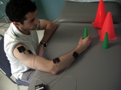
The use of an arm sling during walking training. The sling makes it impossible to use stronger arm. The man must use his weaker arm to hold

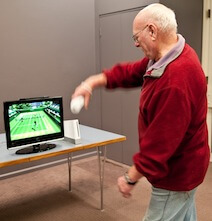
Use of a mitten while writing. The mitten makes it impossible to use the good arm. The woman is forced to use her weaker hand to write

Are there different kinds of constraint-induced therapies?

The term “CIMT” is used to describe a newer technique based on older existing techniques that force the patients to use their weaker arm. These older techniques are called “forced-use” therapy. The goal is to intensively train the weaker arm by using it to perform daily tasks such as preparing meals, engage in fun activities such as throwing a ball or fine motor activities such as writing.

More recently, research has looked at the benefit of modified CIMT (mCIMT), which is based on the same principles as CIMT but with less time wearing the restraint and fewer hours of exercise each day.

What is it used for in people with stroke?

The goal of CIMT is to help regain strength and function on the weaker side of the body, typically the side affected by the stroke. CIMT is used for a variety of purposes. Here we describe its use for arm and hand therapy.

Does it work for stroke?

As already mentioned, this therapy has been examined using high quality research studies and has been found beneficial for arm function in some patients after a stroke – especially those who already have some use of their arm and hand.

Although researchers are not exactly sure of how it works, some experts suggest that CIMT affects the brain by enlarging the brain area controlling the weaker arm. Research studies have reported that patients who receive CIMT have better control of their weaker arm and better ability to perform daily activities such as cooking and dressing when compared to people with stroke who received other forms of arm and hand therapy.

What can I expect?

Your therapist will decide with you what regime is most suitable for you. However, CIMT typically requires you to wear either a large mitten or an arm sling on your stronger arm, many hours a day, seven days a week, for about two weeks or more. The mitten or the sling is worn to encourage you to use your weaker arm and hand to do everyday tasks. In addition, the occupational therapist or physical therapist providing the treatment will do exercises with you and may also give you exercises to do on your own or with a family member or friend. While results can vary from person to person, there is scientific evidence that many people who receive this therapy can have improved use of their weaker arm.

Are there any side effects or risks?

CIMT is usually done by a physical therapist or an occupational therapist at a rehabilitation centre or out-patient clinic. However, many of the exercises must be done outside of treatment time. Family members and friends can be very important in helping you do these exercises. Ask your therapist to give you and your friends/family specific information on exercises.

How long is the treatment period?

Intense, repetitive training of the weaker arm is usually given for 90 percent of waking hours (about 13 hours/day) for a 2-week period. This can be done in the clinic, at home, and wherever else it is safe to do so.

An alternative form of CIMT – modified CIMT – is done for fewer hours and possibly, for more weeks. Consult with your therapist or physician who will help you decide which is right for you – CIMT or mCIMT.

This treatment program requires a good deal of self-discipline and commitment. Individuals with stroke tell us it is hard work! Improvement has been shown to be best for those who spend lots of time using the mitten or the sling.

Who provides the treatment?

CIMT is usually provided by a physical therapist or an occupational therapist at a rehabilitation centre or out-patient clinic. However, many of the exercises must be done outside of treatment time. Family members and friends can be very important in helping you do these exercises. Ask your therapist to give you and your friends/family specific information on exercises.

Is constraint-induced movement therapy for me?

CIMT can be of benefit to those who have lost some of the use of their upper limb following stroke. Studies have looked at the benefit for individuals who have had a stroke very recently, over the past couple of months, and those who have experienced a stroke six or more months ago. There is some positive research that suggests that CIMT may be beneficial for certain patients at all of these times.

Clinician Information

Note: When reviewing the findings, it is important to note that they are always made according to randomized clinical trial (RCT) criteria – specifically as compared to a control group. To clarify, if a treatment is “effective” it implies that it is more effective than the control treatment to which it was compared. Non-randomized studies are no longer included when there is sufficient research to indicate strong evidence (level 1a) for an outcome.

The effectiveness of Constraint-Induced Movement Therapy (CIMT) has been explored as an approach to reducing motor impairment and improving motor activity during functional tasks post-stroke. Fifty-one RCTs, 35 of high quality, 12 of fair quality and four of poor quality have examined the effectiveness of CIMT (restraint for 90% of waking hours and 6 hours of upper limb therapy per day for 2 weeks) or modified CIMT (mCIMT – whereby restraint and/or therapy is provided at a lesser intensity than CIMT) in comparison to traditional upper extremity (UE) therapy OR other forms of UE treatment. Studies have explored the use of CIMT or mCIMT at varying phases of stroke recovery: seven RCTs in the acute phase, 16 RCTs in the subacute phase, and 16 RCTs in the chronic phase of stroke recovery. An additional 12 RCTs examined the use of CIMT or mCIMT with populations of patients where the time since stroke was not specific to one period of recovery.

There is evidence to support CIMT and mCIMT as an effective therapy for patients with upper extremity deficits following a stroke. A systematic review and meta-analysis by Corbetta et al. (2010) updated an earlier Cochrane review by Sirtori et al. (2009) of the efficacy of CIMT, mCIMT and forced use techniques for upper extremity rehabilitation in patients with hemiparesis following stroke. A more recent systematic review and meta-analysis by Shi et al. (2011) compared only mCIMT with conventional rehabilitation (e.g. physiotherapy, occupational therapy, neurodevelopmental therapy, neuromuscular facilitation and daily living retraining).

The results from all reviews indicate a significant effect of CIMT, mCIMT or forced use therapy on arm motor function and impairment. The effect on disability is less conclusive, with Sirtori et al. (2009) reporting a moderate effect of CIMT, mCIMT or forced use therapy immediately post-intervention (but not 3 to 6 months post-intervention), Corbetta et al. (2010) reporting no significant effect of CIMT, mCIMT or forced use therapy on disability, and Shi et al. (2011) reporting a significant effect of mCIMT when disability is measured using the Functional Independence Measure, but not the Barthel Index. While all but two studies included in these three reviews are also reviewed on this website, differentiation of the studies according to the particular type of intervention (CIMT or mCIMT) and stage of stroke, as well as the inclusion of other non-randomized studies, has contributed to variation on StrokEngine in reported outcomes at different stroke stages.

The Cochrane review by Sirtori et al. (2009) concluded that restriction of the hand only, and a treatment protocol of no more than 30 hours indicate a significant effect size. Other studies suggest that functional benefits appear to be confined to a subset of stroke patients with existing active wrist and arm movements.

We have reviewed all of the CIMT and mCIMT studies to identify outcomes according to stage of post-stroke recovery (acute, subacute, chronic). Patients less than one month post-stroke were identified as acute, those between 1 and 6 months post-stroke as subacute, and those greater than 6 months post-stroke as chronic. Intensity and duration of treatment were also important factors in influencing outcome, and these details are included in the review of each study.

Results Table

View results table

Outcomes

Acute phase: mCIMT vs. control or alternative interventions

Depression
Not effective
1b

One high quality RCT (Dromerick et al., 2009) examined the effect of mCIMT on depression among patients with acute stroke. This high quality RCT randomized patients with acute stroke to receive ‘standard’ mCIMT (shaping therapy for 2 hours/day and restraint 6 hours/day), ‘intensive’ mCIMT (shaping therapy for 3 hours/day and restraint 90% of waking hours) or conventional UE therapy. There were no significant between-group differences in depression (Geriatric Depression-15 Scale) at post-treatment (14 days) or follow-up (90 days).

Note: The terms ‘standard’ and ‘intensive’ mCIMT were defined by the authors of this study.

Conclusion: There is moderate evidence (level 1b) from 1 high quality RCT that mCIMT is not more effective than conventional rehabilitation for improving depression among patients with acute stroke.

Note: The high quality RCT also found no difference in depression between different intensities of mCIMT.

Dexterity
Conflicting
4

Two high quality RCTs (Boake et al., 2007; Thrane et al., 2015) have investigated the effect of mCIMT on dexterity in patients with acute stroke.

The first high quality RCT (Boake et al., 2007) randomized patients with acute stroke to receive mCIMT or to receive intensive traditional upper extremity therapy (control group). The mCIMT group received 3 hours of therapy a day and wore a constraint 90% of waking hours. Dexterity was measured with the Grooved Pegboard Test at baseline, at 14-15 days (post-treatment) and at 3 to 4 months post-stroke (follow-up). No significant between-group difference in dexterity was found at either time point.

The second high quality RCT (Thrane et al., 2015) randomized patients with acute stroke to receive mCIMT or usual care. The mCIMT group received rehabilitation for 3 hours/day over 10 consecutive weekdays and wore a mitt on the nonaffected hand for 90% of waking hours. Dexterity was measured with the Nine Hole Peg Test at baseline, 2 weeks (post-treatment) and six months (follow-up). A significant between-group difference in dexterity was found at post-treatment in favour of the mCIMT group compared to the control group. This difference did not remain significant at 6-month follow-up.

Conclusion: There is conflicting evidence (level 4) from 2 high quality RCTs regarding the effect of mCIMT on dexterity. While one high quality RCT found that mCIMT is not more effective than intensive traditional therapy, a second high quality RCT found that mCIMT was more effective than usual care in improving dexterity among patients with acute stroke.

TNote: The significant difference found post-treatment did not remain significant at 6-month follow up.

Functional independence and activities of daily living
Not effective
1a

Three high quality RCTs (Dromerick et al., 2000; Dromerick et al., 2009; Liu et al., 2016) examined the effects of mCIMT on functional independence in patients with acute stroke.

The first high quality RCT (Dromerick et al., 2000) randomized patients with acute stroke to receive mCIMT plus conventional UE therapy or conventional UE therapy alone. The mCIMT group wore a padded mitt 6 hours a day for 14 days and received traditional therapy 2 hours a day. Functional independence in ADL was measured with the Barthel Index and 5 subscales of the Functional Independence Measure (FIM) at discharge from inpatient rehabilitation. A significant between-group difference in upper extremity dressing (FIM), was found in favour of mCIMT compared to conventional UE. There were no other significant between-group differences found in functional independence in ADL (Barthel Index; FIM eating, bathing, grooming and lower body dressing subscales) on discharge.

The second high quality RCT (Dromerick et al., 2009) randomized patients with acute stroke to receive ‘standard’ mCIMT (shaping therapy for 2 hours/day and restraint 6 hours/day), ‘intensive’ mCIMT (shaping therapy for 3 hours/day and restraint 90% of waking hours) or conventional UE therapy. Functional independence was measured with the FIM upper extremity score (sum of the 5 FIM items requiring significant hand and arm use) at baseline, at 14-15 days (post-treatment) and at 3 to 4 months post-stroke (follow-up). No significant between-group differences were found in functional independence at either time point.

Note: The terms ‘standard’ and ‘intensive’ CIMT were defined by the authors of this study.

The third high quality RCT (Liu et al., 2016) randomized patients with acute stroke to receive self-regulated mCIMT (SR-mCIMT), mCIMT or conventional rehabilitation. Participants in the mCIMT groups wore a restraint for 4 hours/day and all participants received individual training for 1 hour/day for 10 days. Functional ability was measured at baseline, 2 weeks (post-treatment) and 4 weeks post-treatment (follow-up) using the Lawton Instrumental Activities of Daily Living Scale (Lawton IADL). There was no signficiant difference in functional ability beween mCIMT and conventional rehabilitation at post-treatment or at follow-up.

Note: There were significant between-group differences in functional ability at post-treatment in favour of SR-mCIMT compared to conventional rehabilitation and in favour of SR-mCIMT compared to mCIMT, but differences did not remain significant at follow-up.

Conclusion: There is strong evidence (level 1a) from 3 high quality RCTs that mCIMT is not more effective than conventional rehabilitation for improving functional independence in patients with acute stroke.

Note: One of the high quality RCTs found a significant difference in the FIM upper extremity dressing score only, in favour of mCIMT compared to conventional rehabilitation.

Note: One high quality RCT found no difference in functional independence between different intensities of mCIMT.

Note: One high quality RCT found that self-regulated mCIMT was more effective than both mCIMT and conventional rehabilitation, although differences did not remain significant at 4 weeks post-treatment.

Motor activity (Upper extremity)
Conflicting
4

Three high quality RCTs (Boake et al., 2007; Thrane et al., 2015; Liu et al., 2016) and 1 fair quality RCT (Page et al., 2005) examined the effects of mCIMT on UE motor activity in patients with acute stroke.

The first high quality RCT (Boake et al., 2007) randomized patients with acute stroke to receive mCIMT or conventional UE therapy. The mCIMT group received therapy 3 hours/day and wore a constraint 90% of waking hours. Upper extremity motor activity was measured with the Motor Activity Log – Amount of Use (MAL-AOU) and – Quality of Movement (MAL-QOM) subscales at baseline, at 14-15 days (post-treatment) and at 3 to 4 months post-stroke (follow-up). There were no significant between-group differences in motor activity at post-treatment or follow up.

The second high quality RCT (Thrane et al., 2015) randomized patients with acute stroke to receive mCIMT or usual care. Participants in the mCIMT group received rehabilitation for 3 hours/day over 10 consecutive weekdays and wore a mitt on the non-affected hand for 90% of waking hours. Upper extremity motor activity was measured according to arm use during functional tasks (arm use ratio) at baseline, 2 weeks (post-treatment) and six months (follow-up). No significant between-group difference were found in functional arm use at either time post.

The third high quality RCT (Liu et al., 2016) randomized patients with acute stroke to receive self-regulated mCIMT (SR-mCIMT), mCIMT or conventional rehabilitation. Participants in the mCIMT groups wore a restraint for 4 hours/day and all participants received individual training for 1 hour/day for 10 days. Upper extremity motor activity was measured at baseline, 2 weeks (post-treatment) and 4 weeks post-treatment (follow-up), using the MAL-AOU and MAL-QOM. At post-treatment there were significant differences in MAL-AOU and MAL-QOM scores in favour of mCIMT compared to conventional rehabilitation; differences did not remain significant at follow-up.

Note: Comparison of SR-mCIMT with conventional rehabilitation showed significant differences in MAL-AOU and MAL-QOM scores at post-treatment and follow-up. Comparison of SR-mCIMT with mCIMT showed significant differences in MAL-AOU scores in favour of SR-mCIMT at post-treatment, but differences did not remain significant at follow-up.

The fair quality RCT (Page et al., 2005) randomized patients with acute stroke to receive mCIMT or conventional rehabilitation. The mCIMT group wore a restraint for 5 hours/day and received 30 minutes of individual therapy 3 days/week for 10 weeks. Motor activity was measured using the Motor Activity Log (MAL) at baseline and 10 weeks (post-treatmeant). At post-treatment the mCIMT group demonstrated a significant improvement in motor activity (MAL).

Note: As this study did not report significant between-group differences these results are not used to determine level of evidence below.

Conclusion: There is conflicting evidence (level 4) among 3 high quality RCTs. While 2 high quality RCTs reported that mCIMT (training 3 hours/day and restraint 90% of waking hours for 2 weeks) is not more effective than conventional rehabilitation, 1 high quality RCT found that mCIMT of less intensity (training 1 hour/day and restraint for 4-5 hours/day for 2 weeks) to be more effective than conventional rehabilitation.

Note: One fair quality RCT also reported significant improvement in motor activity following mCIMT, but did not report between-group differences.

Note: One high quality RCT also found that self-regulated mCIMT was more effective than both mCIMT and conventional rehabilitation, although differences did not remain significant at 1-month follow-up.

Motor function (Upper extremity)
Conflicting
4

Six high quality RCTs (Dromerick et al., 2000; Boake et al., 2007; Dromerick et al., 2009; Thrane et al., 2015; El-Helow et al., 2015; Liu et al., 2016) and 1 fair quality RCT (Page et al., 2005) examined the effectiveness of mCIMT on upper extremity motor function in patients with acute stroke.

The first high quality RCT (Dromerick et al., 2000) randomized patients with acute stroke to receive mCIMT and conventional UE therapy or conventional upper extremity therapy alone. The mCIMT group wore a padded mitt 6 hours/day and received traditional therapy 2 hours/day. Motor function was measured with the Action Research Arm Test (ARAT) at baseline and at post-treatment (14 days). A significant between-group differences in motor function ARAT total score and pinch subscale was found at post-treatment in favour of mCIMT compared to conventional UE therapy alone. No significant between-group differences were found in other ARAT subscales (grasp, grip, gross movement).

The second high quality RCT (Boake et al., 2007) randomized patients with acute stroke to receive mCIMT or conventional UE therapy. The mCIMT group received 3 hours of therapy/day and wore a restraint 90% of waking hours. Motor function was measured with the Fugl Meyer Assessment of Motor Recovery (FMA) and with movements of the affected hand evoked by transcranial magnetic stimulation (TMS) at baseline, at 14-15 days (post-treatment) and at 3 to 4 months post-stroke (follow-up). There was no significant between-group difference in upper extremity motor function at post-treatment or at follow-up.

The third high quality RCT (Dromerick et al., 2009) randomized patients with acute stroke to receive ‘standard’ mCIMT (shaping therapy for 2 hours/day and restraint 6 hours/day), ‘intensive’ mCIMT (shaping therapy for 3 hours/day and restraint 90% of waking hours) or conventional UE therapy. Motor function was measured with the ARAT at baseline, 14 days (post-treatment) and 90 days (follow-up). There were no significant between-group differences in motor function between standard mCIMT and conventional UE therapy at post-treatment or at follow-up. There were significant between-group differences in motor function at post-treatment and follow-up, in favour of standard mCIMT and conventional UE therapy compared to intensive mCIMT.

Note: The terms ‘standard’ and ‘intensive’ CIMT were defined by the authors of this study.

The fourth high quality RCT (Thrane et al., 2015) randomly assigned patients with acute stroke to receive mCIMT or usual care. Participants in the mCIMT group received rehabilitation for 3 hours/day over 10 consecutive weekdays and wore a mitt on the non-affected hand for 90% of waking hours. Motor function was measured with the Wolf Motor Function Test (WMFT) and the FMA-Upper Extremity (FMA-UE) at baseline, 2 weeks (post-treatment) and six months (follow-up). A significant between-group difference was found post-treatment in one subscale of WMFT (performance time) in favour of the mCIMT group compared to the control group. This difference did not remain significant at 6-month follow-up. No significant differences were found in other subscales of the WMFT (functional ability, arm strength and grip strength) or for the FMA-UE at either time point.

The fifth high quality RCT (El-Helow et al., 2015) randomized patients with acute stroke to receive mCIMT or conventional rehabilitation. The mCIMT group wore a restraint for up to 6 hours/day and received shaping intervention for 2 hours/day. Upper extremity motor function was measured with the FMA and ARAT at baseline and 2 weeks (post-treatment). There were significant between-group differences in FMA and ARAT scores at post-treatment, in favour of mCIMT compared to the control group.

The sixth high quality RCT (Liu et al., 2016) randomized patients with acute stroke to receive self-regulated mCIMT (SR-mCIMT), mCIMT or conventional rehabilitation. Participants in the mCIMT groups wore a restraint for 4 hours/day and all participants received individual training for 1 hour/day for 10 days. Upper extremity motor function was measured at baseline, 2 weeks (post-treatment) and 4 weeks post-treatment (follow-up) using the ARAT (total score, grasp, grip, pinch and gross movement subscales) and the FMA-UE (total score, upper arm, wrist, hand and coordination subscales). At post-treatment there were significant between-group differences in upper extremity motor function (ARAT total, grip, pinch; FMA-UE total score only) in favour of mCIMT compared with conventional rehabilitation; at follow-up there was a significant between-group difference on only one measure of upper extremity motor function (FMA-UE hand), in favour of mCIMT.

Note: Comparison of SR-mCIMT with conventional rehabilitation showed significant between-group differences at post-treatment (ARAT total, pinch; FMA-UE total, upper arm, wrist, hand, coordination) and follow-up (ARAT grip, gross movement; FMA-UE wrist, coordination), in favour of SR-mCIMT. Comparison of SR-mCIMT with mCIMT showed significant differences in only 2 measures of upper extremity motor function at post-treatment (FMA-UE total, coordination), and 3 measures at follow-up (ARAT pinch; FMA-UE hand, coordination), in favour of SR-mCIMT.

The fair quality RCT (Page et al., 2005) randomized patients with acute stroke to receive mCIMT or conventional rehabilitation. The mCIMT group wore a restraint for 5 hours, 5 days/week and received 30 minutes of individual therapy 3 days/week. Motor function was measured with the FMA and the ARAT at baseline and 10 weeks (post-treatment). At post-treatment the mCIMT group demonstrated greater improvement in motor function compared to conventional rehabilitation.

Note: As this study did not report significant between-group differences, these results are not used to determine level of evidence below.

Conclusion: There is conflicting evidence (level 4) among 6 high quality RCTs regarding the effectiveness of mCIMT compared to other interventions. Four of the six high quality RCTs reported significant differences on some measures of upper extremity motor function at post-treatment, in favour of mCIMT compared to conventional rehabilitation. Meanwhile, five of the six high quality RCTs also reported no significant differences on other measures of upper extremity motor function between mCIMT and comparison interventions at post-treatment or follow-up time points.

Note: One high quality RCT found that a mCIMT program of higher intensity (restraint for 90% of waking hours and therapy for 3 hours/day) is less effective than a mCIMT program at a lower intensity (restraint for 6 hours/day and therapy for 2 hours/day) or conventional rehabilitation.

Note: One high quality RCT found that self-regulated mCIMT was more effective than both mCIMT and conventional rehabilitation.

Note: One fair quality RCT also reported significant improvement in motor function following mCIMT, but did not report between-group differences.

Pain
Not effective
1b

One high quality RCT (Dromerick et al., 2009) examined the effects of mCIMT on perception of pain among patients with acute stroke. This high quality RCT randomized patients with acute stroke to receive ‘standard’ mCIMT (shaping therapy for 2 hours/day and restraint 6 hours/day), ‘intensive’ mCIMT (shaping therapy for 3 hours/day and restraint 90% of waking hours) or conventional UE therapy. Pain was measured with the Wong-Baker Faces Scale at baseline, 14 days (post-treatment) and 90 days (follow-up). No significant between-group differences were found in pain for any group at post-treatment or follow-up.

Note: The terms ‘standard’ and ‘intensive’ CIMT were defined by the authors of this study.

Conclusion: There is moderate evidence (level 1b) from 1 high quality RCT that mCIMT is not more effective than conventional rehabilitation for alleviating pain among patients with acute stroke.

Note: The high quality RCT also found no difference in pain between different intensities of mCIMT.

Stroke outcomes
Not effective
1A

Two high quality RCTs (Dromerick et al., 2009; Thrane et al., 2015) examined the effects of mCIMT on stroke outcomes in patients with acute stroke.

The high quality RCT (Dromerick et al., 2009) randomized patients with acute stroke to receive ‘standard’ mCIMT (shaping therapy for 2 hours/day and restraint 6 hours/day), ‘intensive’ mCIMT (shaping therapy for 3 hours/day and restraint 90% of waking hours) or conventional UE therapy. Stroke outcomes were measured at baseline, post-treatment (14 days) and follow-up (90 days), using the Stroke Impact Scale (SIS) hand function subtest. There were no significant differences in self-perception of hand function (SIS hand function subscale) at post-treatment (14 days) or follow-up (90 days) between standard mCIMT and conventional UE therapy. However, there were significant between-group differences in SIS hand function scores at follow-up (90 days), in favour of both standard CIMT and conventional UE therapy when compared with high-intensity CIMT.

Note: The terms ‘standard’ and ‘intensive’ CIMT were defined by the authors of this study.

The second high quality RCT (Thrane et al., 2015) randomly assigned patients with acute stroke to receive modified CIMT or usual care. Participants in the mCIMT group received rehabilitation for 3 hours/day over 10 consecutive weekdays and wore a mitt on the nonaffected hand for 90% of waking hours. Stroke outcomes were measured with the SIS hand function, ADL/IADL, participation/role function and global perception of recovery subtests at baseline, 2 weeks (post-treatment) and six months (follow-up). There were no significant between-group differences in stroke outcomes at post-treatment (2 weeks) or at 6-month follow-up.

Conclusion: There is strong evidence (level 1a) from 2 high quality RCTs that mCIMT is not more effective than conventional rehabilitation for improving stroke outcomes in patients with acute stroke.

Note: One quality RCT found that high-intensity mCIMT (restraint for 90% of waking hours and therapy for 3 hours/day) is less effective than low-intensity mCIMT (restraint for 6 hours/day and therapy for 2 hours/day) or conventional rehabilitation for improving self-perception of hand function.

Subacute phase: CIMT vs. control or alternative interventions

Finger dexterity
Not effective
2A

A fair quality RCT (Yoon et al., 2014) has examined the effect of CIMT on dexterity among patients with subacute stroke. This fair quality RCT randomized patients with subacute stroke to receive CIMT, CIMT + mirror therapy, or a control group that received occupational therapy and a self-exercise program. Finger dexterity was measured at baseline and 2 weeks (post-treatment) using the Nine Hole Peg Test (NHPT). There was no significant difference in finger dexterity at post-treatment between CIMT and the control group.

Note: At post-treatment the CIMT + mirror therapy group showed significantly better finger dexterity than the CIMT group and the control group.

Conclusion: There is limited evidence (level 2a) from 1 fair quality RCT that CIMT is not more effective than a comparison intervention (occupational therapy) for improving finger dexterity among patients with subacute stroke.

Note: This fair quality RCT found that CIMT with mirror therapy was more effective than CIMT alone for improving finger dexterity.

Functional independence
Effective
2A

One fair quality RCT (Yoon et al., 2014) has examined the effect of CIMT on functional independence among patients with subacute stroke. This fair quality RCT randomized patients with subacute stroke to receive CIMT, CIMT + mirror therapy, or a control group that received occupational therapy and a self-exercise program. Functional independence was measured at baseline and 2 weeks (post-treatment) using the Korean version of the modified Barthel Index (K-mBI). At post-treatment there was a significant between-group difference in functional independence, in favour of CIMT compared to the control group.

Note: At post-treatment the CIMT + mirror therapy group also showed significantly better K-mBI scores than the control group. There were no significant differences between CIMT and CIMT + mirror therapy at post-treatment.

Conclusion: There is limited evidence (level 2a) from 1 fair quality RCT that CIMT is more effective than comparison interventions (occupational therapy) for improving functional independence among patients with subacute stroke.

Grip strength
Effective
2a

A fair quality RCT (Yoon et al., 2014) has examined the effect of CIMT on grip strength among patients with subacute stroke. This fair quality RCT randomized patients with subacute stroke to receive CIMT, CIMT + mirror therapy, or a control group that received occupational therapy and a self-exercise program. Grip strength was measured at baseline and 2 weeks (post-treatment). There was a significant between-group difference in grip strength at post-treatment, in favour of CIMT compared to the control group.

Note: At post-treatment the CIMT + mirror therapy group showed significantly better grip strength than the CIMT group and the control group.

Conclusion: There is limited evidence (level 2a) from 1 fair quality RCT that CIMT is more effective than a comparison intervention (occupational therapy) for improving grip strength among patients with subacute stroke.

Note: The fair quality RCT found that CIMT with mirror therapy was more effective than CIMT alone for improving grip strength.

Manual dexterity
Effective
2A

A fair quality RCT (Yoon et al., 2014) examined the effect of CIMT on dexterity among patients with subacute stroke. This fair quality RCT randomized patients with subacute stroke to receive CIMT, CIMT + mirror therapy, or a control group that received occupational therapy and a self-exercise program. Manual dexterity was measured at baseline and 2 weeks (post-treatment) using the Box and Block Test (BBT). There was a significant between-group difference in manual dexterity at post-treatment, in favour of CIMT compared to the control group.

Note: At post-treatment the CIMT + mirror therapy group showed significantly better manual dexterity than the CIMT group and the control group.

Conclusion: There is limited evidence (level 2a) from 1 fair quality RCT that CIMT is more effective than a comparison intervention (occupational therapy) for improving manual dexterity among patients with subacute stroke.

Note: The fair quality RCT found that CIMT with mirror therapy was more effective than CIMT alone for improving manual dexterity.

Motor function (Upper extremity)
Not effective
1b

One high quality RCT (Sawaki et al., 2008) and 1 fair quality RCT (Yoon et al., 2014) have examined the effect of CIMT on upper extremity (UE) motor function in patients with subacute stroke.

The high quality RCT (Sawaki et al., 2008) randomized patients with subacute stroke to receive CIMT or usual care. The CIMT group wore a padded mitt on the less-affected limb for at least 90% of waking hours and performed intensive therapy for 6 hours/day, 5 days/week for 2 weeks. Upper extremity motor function was measured using the Wolf Motor Function Test (WMFT) at baseline, 2 weeks (post-treatment) and at 4 months (follow-up). There was a significant between-group difference in only one measure of upper extremity motor function (WMFT grip strength), in favour of the CIMT group compared to the control group. There were no significant between-group differences in other measures of UE motor function (WMFT weight and time-based measures).

The fair quality RCT (Yoon et al., 2014) randomized patients with subacute stroke to receive CIMT, CIMT + mirror therapy, or a control group that received occupational therapy and a self-exercise program. Upper extremity motor function was measured at baseline and 2 weeks (post-treatment) using the Wolf Motor Function Test (WMFT) and the Fugl-Meyer Assessment (FMA) – total score and Upper Extremity score (FMA-UE). At post-treatment there was a significant between-group difference in one measure of upper extremity motor function (WMFT), in favour of CIMT compared to the control group.

Note: At post-treatment the CIMT + mirror therapy group also showed significantly better WMFT scores than the control group.

Conclusion: There is moderate evidence (level 1b) from 1 high quality RCT and 1 fair quality RCT that CIMT is not more effective than comparison interventions (usual care, CIMT with mirror therapy, occupational therapy) in improving UE motor function in patients with subacute stroke.

Note: However the high quality RCT found a difference in WMFT grip strength in favour of CIMT vs. usual care; the fair quality RCT found differences in WMFT scores in favour of CIMT vs. OT and in favour of CIMT + mirror therapy vs. OT.

Stroke recovery
Not effective
2A

One fair quality RCT (Yoon et al., 2014) has examined the effect of CIMT on stroke recovery among patients with subacute stroke. This fair quality RCT randomized patients with subacute stroke to receive CIMT, CIMT + mirror therapy, or a control group that received occupational therapy and a self-exercise program. Stroke recovery was measured at baseline and 2 weeks (post-treatment) using the Brunnstrom stages of stroke recovery. There were no significant between-group differences in stroke recovery at post-treatment.

Conclusion: There is limited evidence (level 2a) from 1 fair quality RCT that CIMT is not more effective than comparison interventions (CIMT and mirror therapy, occupational therapy) for improving stroke recovery among patients with subacute stroke.

Subacute phase: mCIMT vs. control or alternative interventions

Dexterity
Conflicting
4

Four high quality RCTs (Myint et al., 2008; Hammer & Lindmark, 2009b; Brunner, Skouen & Strand, 2012; Treger et al., 2012) have examined the effect of mCIMT on finger dexterity in patients with subacute stroke.

The first high quality RCT (Myint et al., 2008) randomized patients with subacute stroke to receive either mCIMT or conventional occupational and physical therapy. The mCIMT group wore a shoulder sling on the less-affected extremity for 90% of waking hours and received 4 hours daily therapy. Finger dexterity was measured using the Nine Hole Peg Test (NHPT) at baseline, 10 days (post-treatment) and follow-up (12 weeks). There were significant between-group differences in dexterity at post-treatment and follow-up, in favour of mCIMT compared to conventional rehabilitation.

The second high quality RCT (Hammer & Lindmark, 2009b) randomized patients with subacute stroke to a ‘forced use’ group or a control group that received conventional rehabilitation. The forced use group wore a sling to promote forced use of the paretic upper limb for up to 6 hours/day, 5 days/week. Finger dexterity was measured using the 16 Hole Peg Test at baseline, 2 weeks (post-treatment), 1 month (follow-up A) and 3 months (follow-up B). There were no significant between-group differences in dexterity at any time point.

The third high quality RCT (Brunner, Skouen & Strand, 2012) randomized patients with subacute stroke to receive mCIMT or bimanual task-related training. The mCIMT group wore a restraint for 4 hours/day in addition to therapist-directed training for 4 hours/week and self-directed training for 2-3 hours/day for 4 weeks. Finger dexterity was measured at baseline, 4 weeks (post-treatment) and 3-month follow-up using the Nine Hole Peg Test (NHPT). There were no significant between-group differences in finger dexterity at any time point.

The fourth high quality RCT (Treger et al., 2012) randomly assigned patients with subacute stroke to receive mCIMT or conventional rehabilitation. The mCIMT group restrained the non-affected hand during 1-hour rehabilitation sessions and wore a mitten for up to 4 hours each weekday for 2 weeks. Dexterity was measured by a peg task and a ball grasp, carry and release task modified from the Manual Function Test at baseline and 4 weeks. There were significant between-group differences in dexterity at 4 weeks, in favour of mCIMT compared to conventional rehabilitation.

Conclusion: There is conflicting evidence (level 4) between 2 high quality RCTs that found mCIMT to be more effective than comparison interventions (conventional rehabilitation), and a third high quality RCT that found mCIMT was not more effective than the comparison interventions (bimanual task training) for improving finger dexterity in patients with subacute stroke. A fourth high quality RCT found that forced use therapy with conventional rehabilitation was not more effective than conventional rehabilitation alone for improving finger/manual dexterity among patients with subacute stroke.

Functional independence and activities of daily living
Conflicting
4

Three high quality RCTs (Myint et al., 2008, Azab et al., 2009; Treger et al., 2012) examined the effects of mCIMT on functional independence and activities of daily living (ADLs) in patients with subacute stroke.

The first high quality RCT (Myint et al., 2008) randomized patients with subacute stroke to receive mCIMT or conventional occupational and physical therapy. The mCIMT group received 4 hours daily of therapy and wore a shoulder sling on the less-affected extremity for 90% of waking hours. Functional independence was measured using the modified Barthel Index (mBI) at baseline, 2 weeks (post-treatment) and 12 weeks (follow-up). There were no significant between-group differences in ADLs at either time point.

The second high quality RCT (Azab et al., 2009) randomized patients with subacute stroke to receive mCIMT or standard rehabilitation. The mCIMT group wore a mitt on the unaffected hand for 6-7 hours per day and both groups received physical therapy and occupational therapy for 40 mins/session, 3 times/week for the treatment period. Functional independence was measured using the Barthel Index (BI) at baseline, 4 weeks (post-treatment) and 6 months (follow-up). There was a significant between-group difference in functional independence at both time points, in favour of mCIMT compared to standard rehabilitation.

The third high quality RCT (Treger et al., 2012) randomly assigned patients with subacute stroke to receive mCIMT or conventional rehabilitation. The mCIMT group restrained the nonaffected hand during 1-hour rehabilitation sessions and wore a mitten for up to 4 hours each weekday for 2 weeks. Functional independence was measured according to spoon use over 30 seconds, at baseline and at 4 weeks. There was a significant between-group difference in spoon use at 4 weeks, in favour of mCIMT compared to conventional rehabilitation.

Conclusion: There is conflicting evidence (level 4) regarding the effect of mCIMT on functional independence in patients with subacute stroke. One high quality RCT reported that a short-term (2-week) high-intensity mCIMT program was not more effective than conventional rehabiliation, whereas a second high quality RCT reported that a longer (4-week), low-intensity mCIMT program was more effective than conventional rehabilitation. A third high quality RCT also reported that low-intensity mCIMT was more effective than conventional rehabilitation, using a non-standardised measure of functional rehabilitation (spoon use).

Grip strength
Not effective
1b

One high quality RCT (Hammer & Lindmark, 2009b) investigated the effect of mCIMT on hand strength in patients with subacute stroke. This high quality RCT randomized patients with subacute stroke to a ‘forced use’ group or a control group that received conventional rehabilitation. The forced use group wore a sling to promote forced use of the paretic upper limb for up to 6 hours/day, 5 days/week for 2 weeks. Isometric grip strength was measured at baseline, 2 weeks (post-treatment), 1 month (follow-up A) and 3 months (follow-up B). There was no significant between-group difference in grip strength at any time point.

Conclusion: There is moderate evidence (level 1b) from one high quality RCT that mCIMT is not more effective than comparison interventions (conventional rehabilitation) for improving grip strength among patients with subacute stroke.

Motor activity (Upper extremity)
Not effective
1a

Five high quality RCTs (Page et al., 2002; Myint et al., 2008; Hammer & Lindmark, 2009a; Brogardh et al., 2009b; Brogardh & Lexell, 2010 – follow-up study; Brunner, Skouen & Strand, 2012) and one fair quality RCT (Page et al., 2001) examined the effects of mCIMT on UE motor activity in patients with subacute stroke.

The first high quality RCT (Page et al., 2002) randomized patients with subacute stroke to receive mCIMT, traditional therapy, or no treatment. The mCIMT group wore a restraint for 5 hours/day and received 30 minutes each of physical therapy and occupational therapy 3 times/week. Upper extremity motor activity was measured using the Motor Activity Log (MAL) at baseline and at 10 weeks (post-treatment). There were no significant between-group differences in MAL scores at post-treatment.

The second high quality RCT (Myint et al., 2008) randomized patients with subacute stroke to receive either mCIMT or conventional occupational and physical therapy. The mCIMT group received therapy for 4 hours/day and wore a shoulder sling on the less-affected extremity for 90% of waking hours during the 10-day treatment period. Upper extremity motor activity was measured using the MAL Amount of Use (MAL-AOU) and Quality of Movement (MAL-QOM) at baseline, 2 weeks (post-treatment) and 12 weeks (follow-up). There were significant between-group differences in motor activity at both time points, in favour of mCIMT compared to conventional therapy.

The third high quality RCT (Hammer & Lindmark, 2009a) randomized patients with subacute stroke to a ‘forced use’ group or a control group that received conventional rehabilitation alone. The forced use group wore a sling to promote forced use of the paretic upper limb for up to 6 hours/day, 5 days/week for 2 weeks. Upper extremity motor activity was measured using the MAL-AOU and MAL-QOM at baseline, 2 weeks (post-treatment), 1 month (follow-up A) and 3 months (follow-up B). There were no significant between-group differences in motor activity at any time point.

The fourth high quality RCT (Brogardh et al., 2009b) randomized patients with subacute stroke to a mCIMT group that wore a mitt on the less affected arm for 90% of waking hours, or a group that did not wear a mitt. Both groups received 3 hours of therapy for the affected arm for 12 days. Upper extremity motor activity was measured using the MAL at baseline, 2 weeks (post-treatment) and 3 months (follow-up). There were no significant between-group differences in motor activity at either time point.

Further to the study by Brogardh et al, 2009b (Brogardh & Lexell, 2010), no significant between-group differences in upper extremity motor activity (MAL-AOU, MAL-QOM) were seen at 12 months follow-up.

The fifth high quality RCT (Brunner, Skouen & Strand, 2012) randomized patients with subacute stroke to receive mCIMT or bimanual task-related training. The mCIMT group wore a restraint for 4 hours/day in addition to therapist-directed training for 4 hours/week and self-directed training for 2-3 hours/day for 4 weeks. Upper extremity motor activity was measured at baseline, 4 weeks (post-treatment) and 3-month follow-up using the MAL-AOU and MAL-QOM. There were no significant between-group differences in upper extremity motor activity at any time point.

The fair quality RCT (Page et al., 2001) randomized patients with subacute stroke to receive mCIMT, conventional rehabilitation or no therapy. mCIMT comprised 30 minutes each of physiotherapy and occupational therapy 5 days/week for 10 weeks and restraint of the less affected arm 5 hours/day, 5 days/week for 10 weeks. Upper extremity motor activity was measured using the MAL-AOU and MAL-QOM. The mCIMT group demonstrated improved motor activity at post-treatment, whereas the other two groups did not demonstrate substantial improvement.

Note: Statistical data and between-group differences were not reported; accordingly this study is not included in determining level of evidence in the conclusion below.

Conclusion: There is strong evidence (level 1a) from 4 high quality RCTs that mCIMT or forced-use therapy is not more effective than control therapies (e.g. no treatment, conventional rehabilitation, mCIMT training with no mitt use, bimanual task training) for improving UE motor activity in patients with subacute stroke.

Note: However, one high quality RCT did find significant between-group differences in UE motor activity in favour of mCIMT compared to conventional therapy. Also, one fair quality RCT demonstrated improved motor activity at post-treatment for the mCIMT group but as statistical data and between-group differences were not reported, this study is not included in determining level of evidence.

Motor function (Upper extremity)
Not effective
1a

Five high quality RCTs (Page et al., 2002; Myint et al., 2008; Hammer & Lindmark, 2009b; Brogardh et al., 2009b; Brogardh & Lexell, 2010 – follow-up study; Brunner, Skouen & Strand, 2012) and one fair quality RCT (Page et al., 2001) examined the effects of mCIMT on upper extremity (UE) motor function in patients with subacute stroke.

The first high quality RCT (Page et al., 2002) randomized patients with subacute stroke to receive mCIMT, traditional therapy, or no treatment. The mCIMT group wore a restraint for 5 hours/day and received 30 minutes each of physical therapy and occupational therapy 3 times/week for 10 weeks. Motor function was assessed using the Fugl-Meyer Assessment (FMA) and the Action Research Arm Test (ARAT) at baseline, 10 weeks (post-treatment). At post-treatment (10 weeks) there was a significant between-group difference in FMA scores in favour of mCIMT compared to traditional therapy and no therapy at post-treatment. There were no significant differences in ARAT scores between any groups at post-treatment.

The second high quality RCT (Myint et al., 2008) randomized patients with subacute stroke to receive either mCIMT or conventional occupational and physical therapy. The mCIMT group wore a shoulder sling on the less-affected extremity for 90% of waking hours and received 4 hours daily therapy for 10 days. Upper extremity motor function was measured using the ARAT and Functional Test of the Hemiparetic Upper Extremity (FTHUE) at baseline, 10 days (post-treatment) and 12 weeks (follow-up). There were significant between-group differences in ARAT (grasp, grip, pinch, gross movement) and FTHUE scores at post-treatment, and in ARAT (total, grip) and FTHUE scores at follow-up, in favour of mCIMT compared to conventional therapy.

The third high quality RCT (Hammer & Lindmark, 2009b) randomized patients with subacute stroke to a ‘forced use’ group or a control group that received conventional rehabilitation. The forced use group wore a sling to promote forced use of the paretic upper limb for up to 6 hours/day, 5 days/week for 2 weeks. Upper extremity motor function was measured using the FMA-UE, ARAT and Motor Assessment Scale at baseline, 2 weeks (post-treatment), 1 month (follow-up A) and 3 months (follow-up B). There were no significant between-group differences in upper extremity motor function at any time point.

The fourth high quality RCT (Brogardh et al., 2009b) randomized patients with subacute stroke to a mCIMT group that wore a mitt on the less affected arm for 90% of waking hours, or a group that did not wear a mitt. Both groups received 3 hours of therapy for the affected arm for 12 days. Upper extremity motor function was measured using the modified Motor Assessment Scale and the Sollerman Hand Function Test (SHFT) at baseline, 2 weeks (post-treatment), 3 months and 12 months (follows-up). There were no significant between-group differences in upper extremity motor function at post-treatment or at follow-up (see Brogardh & Lexell, 2010).

The fifth high quality RCT (Brunner, Skouen & Strand, 2012) randomized patients with subacute stroke to receive mCIMT or bimanual task-related training. The mCIMT group wore a restraint for 4 hours/day in addition to therapist-directed training for 4 hours/week and self-directed training for 2-3 hours/day for 4 weeks. Upper extremity motor function was measured at baseline, 4 weeks (post-treatment) and 3-month follow-up using the ARAT. There were no significant between-group differences in upper extremity motor function at any time point

The fair quality RCT (Page et al., 2001) randomized patients with subacute stroke to receive mCIMT, conventional rehabilitation or no therapy. mCIMT comprised 30 minutes each of physiotherapy and occupational therapy 5 days/week for 10 weeks and restraint of the less affected arm 5 hours/day, 5 days/week for 10 weeks. Upper extremity motor function was measured using the ARAT, FMA and Wolf Motor Function Test (WMFT) at baseline and 10 weeks (post-treatment). The mCIMT group demonstrated substantial improvements in motor function whereas the other groups did not demonstrate substantial improvements on any measure of upper extremity motor function. Statistical data and between-group differences were not reported; accordingly this study is not included in determining level of evidence in the conclusion below.

Conclusion: There is strong evidence (level 1a) from 4 high quality RCTs that mCIMT or forced use therapy is not more effective than comparison interventions (conventional rehabilitation, no treatment, mCIMT training with no mitt use or bimanual task training) for improving upper extremity motor function among patients with subacute stroke. However, 1 of these high quality RCTs found results in favour of mCIMT on one measure of upper extremity motor function (FMA) but not another (ARAT); and another highquality RCTfound that mCIMT is more effective than control therapy (e.g. conventional rehabilitation).

Note: Studies varied in constraint intensity (from 4 hours/day to 90% of waking hours), frequency of therapy (from 3 to 20 hours/week) and intervention duration (from 10 days to 10 weeks), which is likely to account for discrepancies in results among studies.

Note: One fair quality RCT demonstrated improved motor function at post-treatment for the mCIMT group but as statistical data and between-group differences were not reported, this study is not included in determining level of evidence.

Spasticity
Not effective
1B

One high quality RCT (Hammer & Lindmark, 2009b) examined the effects of mCIMT on upper extremity spasticity in patients with subacute stroke. This high quality study randomized patients with subacute stroke to a ‘forced use’ group that wore a sling on the less affected arm for up to 6 hours/day, 5 days a week for 2 weeks, or to a control group that received conventional rehabilitation alone. Upper extremity spasticity was measured using the Modified Ashworth Scale at baseline, 2 weeks (post-treatment), 1 month (follow-up A) and 3 months (follow-up B). There were no significant between-group differences in upper extremity spasticity at any time point.

Conclusion: There is moderate evidence (level 1b) from one high quality RCT that mCIMT is not more effective than conventional rehabilitation for improving upper extremity spasticityin patients with subacute stroke.

Chronic phase: CIMT vs. control or alternative interventions

Functional independence
Not effective
1B

One high quality RCT (Huseyinsinoglu, Ozdincler, & Krespi, 2012) investigated the effect of CIMT on functional independence in patients with chronic stroke. This high quality RCT randomized patients with chronic stroke to receive CIMT or Bobath Concept therapy. Functional independence was measured using the Functional Independence Measure (FIM self cares and total score) at baseline and 2 weeks (post-treatment). There were no significant between-group differences in functional independence at post-treatment.

Conclusion: There is moderate (level 1b) evidence from 1 high quality RCT that CIMT is not more effective than a comparison intervention (Bobath Concept therapy) for improving functional independence among patients with chronic stroke.

Hand strength
Effective
1b

One high quality RCT (Suputtitada et al., 2004) investigated the effect of CIMT on hand strength in patients with chronic stroke. This high quality RCT randomized patients with chronic stroke to receive CIMT and affected-UE training or bimanual-UE training without restraint. Pinch strength and grip strength were measured by dynamometer at baseline and 2 weeks (post-treatment). At post-treatment there were significant between-group differences in pinch strength only, in favour of the CIMT group compared to the control group.

Conclusion: There is moderate evidence (level 1b) from one high quality RCT that CIMT is more effective than comparison interventions (bimanual upper extremity training) for improving pinch strength – but not grip strength – among patients with chronic stroke.

Kinematics (Upper extremity)
Insufficient evidence
5

One pre-post study (Richards et al., 2008) investigated the effect of CIMT on upper extremity kinematics with 3 patients with chronic stroke and ataxia. This pre-post study assigned patients to wear a mitten restraint during 90% of waking hours. Patients 1 and 2 received therapy 6 hours a day (CIMT) while patient 3 received therapy 3 hours a day (mCIMT). All participants demonstrated improved kinematic reaching values post-intervention, with no significant between-patient differences.

Note: Patients 1 and 2 improved on all kinematic measures: maximum velocity and time to maximum velocity increased, while index of curvature, number of peaks in the velocity profile, and trunk movement decreased. Participant 3 improved on some kinematic measures (smoother velocity profile, increased time to maximum velocity and decreased number of peaks in the velocity profile) but not all (decreased maximum velocity, increased index of curvature).

Conclusion: There is an insufficient evidence (level 5) comparing CIMT and control therapies on upper extremity kinematics in patients with chronic stroke. However, 1 non-experimental study found that CIMT is effective for improving reach kinematics in patients with chronic stroke.

Motor activity (Upper extremity)
Effective
1b

One high quality RCT (Huseyinsinoglu, Ozdincler, & Krespi, 2012), two fair quality RCTs (Wittenberg et al., 2003; Brogårdh & Sjülund, 2006; Brogårdh et al., 2009a – follow-up study) and 2 non-experimental studies (Taub et al., 2006; Kunkel et al., 1999) examined the effectiveness of CIMT on motor activity in patients with chronic stroke.

The high quality RCT (Huseyinsinoglu, Ozdincler, & Krespi, 2012) randomized patients with chronic stroke to receive CIMT or Bobath Concept therapy. Upper extremity motor activity was measured using the Motor Activity Log – Amout of Use (MAL-AOU) and – Quality of Movement (MAL-QOM) at baseline and 2 weeks (post-treatment). There was a significant between-group difference in MAL-AOU and MAL-QOM scores at post-treatment, in favour of CIMT compared to Bobath Concept therapy.

The first fair quality RCT (Wittenberg et al., 2003) randomized patients with chronic stroke to receive either CIMT and task-oriented training or task-oriented training alone. Upper extremity motor activity was measured using the MAL at baseline and 10 days (post-treatment). There were significant gains in MAL scores at post-treatment, in favour of CIMT + task-oriented training compared to task-oriented training alone.

The second fair quality RCT (Brogårdh & Sjülund, 2006) provided patients with chronic stroke with a 2-week CIMT program, then randomized patients to prolonged mitt use for a further 3 months, or no further treatment. All participants demonstrated improved MAL-AOU and MAL-QOM scores after 2 weeks of CIMT, however no significant between-group differences were reported at 3 months.

Note: The authors reported that the small sample size was a limitation of this study.

A 4-year follow-up to this study (Brogårdh et al., 2009a) found a significant improvement in upper extremity motor activity from baseline to 4 years. However, comparison from post-treatment time points (2 weeks and 3 months) to 4-year follow-up revealed a significant decrease in motor activity.

A controlled clinical trial (Taub et al., 2006) assigned patients with chronic stroke to a CIMT group or a control group that received time-matched and interaction-matched physical, cognitive and relaxation exercises. Upper extremity motor activity was measured using the MAL-QOM subtest and the Upper Extremity Actual Amout of Use test (AAUT) at baseline, 2 weeks (post-treatment), 4 weeks (follow-up A) and 2 years (follow-up B). There was a significantly greater improvement in upper extremity motor activity at post-treatment, in favour of CIMT compared to the control group. No significant between-group differences were reported at follow-up time points.

A pre-post study without multiple baselines (Kunkel et al., 1999) assigned patients with chronic stroke to receive CIMT. Upper extremity motor activity was measured using the MAL and AAUT at baseline, 2 weeks (post-treatment) and 3 months (follow-up). Significant improvements in upper extremity motor activity were noted at both time points.

Conclusion: There is moderate evidence (level 1b) from 1 high quality RCT, 1 fair quality RCT and 1 non-experimental study that CIMT is more effective than comparison interventions (e.g. Bobath Concept therapy, task-oriented training, time-matched physical/cognitive/relaxation exercises) for improving upper extremity motor activity in patients with chronic stroke. Further, 1 fairquality RCTand 1 non-experimental study found significant improvements in motor activity following CIMT.

Note: One fair quality RCT found no benefit to prolonged mitt use following CIMT.

Motor function (Upper extremity)
Conflicting
4

Three high quality RCTs (Suputtitada et al., 2004; Huseyinsinoglu, Ozdincler, & Krespi, 2012; Abo et al., 2014), 2 fair quality RCTs (Wittenberg et al., 2003; Brogårdh & Sjülund, 2006; Brogårdh et al., 2009a – follow-up study) and 2 non-experimental studies (Taub et al., 2006; Kunkel et al., 1999) examined the effects of CIMT on upper extremity motor function in patients with chronic stroke.

The first high quality RCT (Suputtitada et al., 2004) randomized patients with chronic stroke to receive CIMT and affected-UE training or bimanual-UE training without restraint. Upper extremity motor function was measured using the Action Research Arm Test (ARAT). At post-treatment, there were significant between-group differences in upper extremity motor function, in favour of CIMT compared to the control group.

The second high quality RCT (Huseyinsinoglu, Ozdincler, & Krespi, 2012) randomized patients with chronic stroke to receive CIMT or Bobath Concept therapy. Upper extremity motor function was measured at baseline and 2 weeks (post-treatment) using the Wolf Motor Function Test – Functional Ability and – Performance Time (WMFT-FA, WMFT-PT) subtests. There was no significant between-group difference in upper extremity motor function at post-treatment.

The third high quality RCT (Abo et al., 2014) randomized patients with chronic stroke to receive CIMT or low-frequency rTMS with intensive OT (NEURO). Upper extremity motor function was measured at baseline and 15 days (post-treatment) using the FMA and WMFT-FA and WMFT-PT. There were significant between-group differences in FMA and WMFT-FA scores at post-treatment, in favour of NEURO compared to CIMT.

The first fair quality RCT (Wittenberg et al., 2003) randomized patients with chronic stroke to receive CIMT and task-oriented training or task-oriented training alone. Upper extremity motor function was measured at baseline and at 2 weeks (post-treatment) using the WMFT and the Assessment of Motor and Process Skills (AMPS), and using Transcranial Magnetic Stimulation and Positron Emission Tomography (PET) scans. There were no significant between-group differences on any measure of upper extremity motor function at post-treatment.

The second fair quality RCT (Brogårdh & Sjülund, 2006) provided patients with chronic stroke with a 2-week CIMT program, then randomized patients to prolonged mitt use for a further 3 months, or no further treatment. Upper extremity motor function was measured at baseline and 2 weeks (post-treatment) using the modified Motor Assessment Scale (MAS) and the Sollerman Hand Function Test (SHFT). All participants demonstrated improved upper extremity motor function after 2 weeks of CIMT. After 3 months there were no significant differences in motor function between prolonged mitt use and no mitt use.

A 4-year follow-up to this study (Brogårdh et al., 2009a) found no significant change in SHFT scores when compared to baseline or post-treatment data. The MAS was not used on retesting.

A controlled clinical trial (Taub et al., 2006) assigned patients with chronic stroke to a CIMT group or a control group that received time-matched and interaction-matched physical, cognitive and relaxation exercises. Upper extremity motor function was measured at baseline and at 2 weeks (post-treatment) using the WMFT-PT and WMFT-FA. There was a significant between-group difference in WMFT–PT scores at post-treatment, in favour of CIMT compared to the control group. There were no significant between-group differences in WMFT-FA scores. Although measures were also taken at 3 follow-up intervals (4 weeks, 3 months, 2 years), statistical data for between-group differences were not reported at these time points.

A pre-post study without multiple baselines (Kunkel et al., 1999) examined the effects of CIMT in patients with chronic stroke. Upper extremity motor function was measured at baseline, 2 weeks (post-treatment) and at 3 months (follow-up) using the ARAT and WMFT. There was a significant improvement in ARAT and WMFT scores at both time points.

Conclusion: There is conflicting evidence (level 4) regarding the effectiveness of CIMT compared to other interventions. Two high quality RCTs and one fair quality RCT reported that CIMT was not more effective than comparison interventions (Bobath Concept therapy, task-oriented training, rTMS+OT); in fact, 1 high quality RCT found that CIMT was less effective than rTMS+OT for improving upper extremity motor function. However, another high quality RCT and a controlled clinical trial reported that CIMT was more effective than comparison interventions (bimanual training, time-matched rehabilitation) on some measures of upper extremity motor function (ARAT and WMFT performance time). Further, 1 pre-post design study found significant improvement in upper extremity motor function following CIMT.

Note: A fair quality RCT found an improved upper extremity motor function after 2 weeks of CIMT, and note that prolonged mitt use following a CIMT program was not more effective than CIMT alone for improving upper extremity motor function in patients with chronic stroke.

Quality of movement (Upper extremity)
Not effective
1B

One high quality RCT (Huseyinsinoglu, Ozdincler, & Krespi, 2012) has investigated the effect of CIMT on quality of movement of the upper extremity in patients with chronic stroke. This high quality RCT randomized patients with chronic stroke to receive CIMT or Bobath Concept therapy. At post-treatment (2 weeks) there was no significant between-group difference in upper extremity quality of movement (Motor Evaluation Scale for Arm in Stroke Patients).

Conclusion: There is moderate (level 1b) evidence from 1 high quality RCT that CIMT is not more effective than a comparison intervention (Bobath Concept therapy) for improving upper extremity quality of movement among patients with chronic stroke.

Sensory discrimination
Not effective
2a

One fair quality RCT (Brogårdh & Sjülund, 2006) examined the effect of CIMT on sensory discrimination in patients with chronic stroke. This fair quality RCT provided patients with a 2-week CIMT program, then randomized patients to prolonged mitt use for a further 3 months, or no further treatment. There were no significant within-group differences in sensory discrimination (Two-Point Discrimination Test) following 2 weeks of CIMT. Further, there were no significant between-group differences in sensory discrimination at 3 months.

Conclusion: There is limited evidence (level 2a) from 1 fairquality RCTthat CIMT does not improve sensory discrimination in chronic stroke. Further, the fairquality RCTfound that prolonged mitt wear is not more effective than control therapy (i.e. no treatment) for improving sensory discrimination in patients with chronic stroke.

Chronic phase: mCIMT vs. control or alternative interventions

Dexterity
Not effective
1b

One high quality RCT (Barzel et al., 2015), 1 poor quality RCT (Kim et al., 2008) and 1 quasi-experimental study (Siebers & Skargren, 2010) investigated the effect of mCIMT on dexterity among patients with chronic stroke.

The high quality RCT (Barzel et al., 2015) randomized patients with chronic stroke to receive home-based mCIMT or conventional rehabilitation. Finger dexterity was measured at baseline, 4 weeks (post-treatment) and 6-month follow-up using the Nine Hole Peg Test. There was no significant between-group difference in finger dexterity any time point.

The poor quality RCT (Kim et al., 2008) randomized patients with chronic stroke to receive mCIMT or a control group (intervention not specified). Patients in the mCIMT group wore a modified opposition restriction orthosis (MORO) on the unaffected hand for at least 5 hours/day, 7 days a week. Dexterity was measured using the Purdue Pegboard Test at baseline and 8 weeks (post-treatment). There were no significant between-group differences in dexterity at post-treatment.

The quasi-experimental study (Siebers & Skargren, 2010) provided patients with chronic stroke with mCIMT that comprised restraint for 90% of waking hours and an individualized training program for 6 hours/weekday. Manual dexterity was measured at baseline, 2 weeks (post-treatment) and 6-month follow-up using the Box and Block Test (BBT). There was a significant improvement in manual dexterity from baseline to post-treatment, but this did not remain significant at 6-month follow-up.

Note: As this study did not report between-group differences these results are not used to determine level of evidence below.

Conclusion: There is moderate evidence (level 1b) from 1 high quality RCT and 1 poorquality RCT that mCIMT is not more effective than comparison interventions (conventional rehabilitation) for improving dexterity among patients with chronic stroke.

Note: One quasi-experimental study reported a significant improvement, in short term, in manual dexterity from baseline to post-treatment but as this study did not report between-group differences these results are not used to determine level of evidence.

Grip strength
Insufficient evidence
5

One quasi-experimental study (Siebers & Skargren, 2010) investigated the effect of mCIMT on grip strength among patients with chronic stroke. This quasi-experimental study provided patients with chronic stroke with mCIMT that comprised restraint for 90% of waking hours and an individualized training program for 6 hours/weekday. Grip strength was measured at baseline, 2 weeks (post-treatment) and 6-month follow-up using the Grippit instrument. There was a significant improvement in grip strength from baseline to post-treatment and this persisted at 6-month follow-up.

Conclusion: There is insufficient evidence (level 5) regarding the effectiveness of mCIMT on grip strength compared to other interventions. However, 1 quasi-experimental study reported improved grip strength among patients with chronic stroke following mCIMT.

Functional independence and activities of daily living
Effective
1A

Four high quality RCTs (Lin et al., 2007, Wu et al., 2007c,, Lin et al., 2009b; Barzel et al., 2015) and 2 fair quality RCTs (Lin et al., 2008, Wu et al., 2012b) examined the effectiveness of mCIMT on functional independence in patients with chronic stroke.

The first high quality RCT (Lin et al., 2007) randomized patients with chronic stroke to either mCIMT or traditional rehabilitation. The mCIMT group had their unaffected hand restricted by a mitt for 6 hours per day and received intensive training on the affected arm for 2 hours per weekday. Functional independence was measured at baseline and 3 weeks (post-treatment) using the Functional Independence Measure (FIM). There were significant between-group differences in functional independence at post-treatment, in favour of mCIMT compared to traditional rehabilitation.

The second high quality RCT (Wu et al., 2007c) randomized patients with chronic stroke to either mCIMT or neurodevelopmental therapy. The mCIMT group wore a constraint on the less affected UE 6 hours a day while receiving 2 hours daily of intensive training. Functional independence was measured at baseline and 3 weeks (post-treatment) using the FIM. The mCIMT group had significantly greater gains in functional independence than the control group at post-treatment.

The third high quality RCT (Lin et al., 2009b) randomized patients with chronic stroke to receive mCIMT, bilateral arm training (BAT), or standard UE therapy (control). The mCIMT group received 2 hours per weekday of UE therapy for 12 days for 3 weeks while wearing a restraint on the unaffected UE for 6 hours per day. Functional independence was measured at baseline and 3 weeks (post-treatment) using the FIM. The mCIMT group showed significantly greater functional independence than the BAT group and the control group at post-treatment.

The fourth high quality RCT (Barzel et al., 2015) randomized patients with chronic stroke to receive home-based mCIMT or conventional rehabilitation. Functional independence was measured at baseline, 4 weeks (post-treatment) and 6-month follow-up using the Barthel Index (BI) and the Instrumental Activities of Daily Living (IADL). There were no significant between-group differences in functional independence on either measure at any time point.

The first fair quality RCT (Lin et al., 2008) randomized patients with chronic stroke to either mCIMT or traditional intervention. The mCIMT group wore restraints on the hand and wrist for 3 hours/weekday and received 2-hour training sessions each weekday. Functional independence was measured at baseline and 3 weeks (post-treatment) using the FIM and the Nottingham Extended Activities of Daily Living (NEADL). The mCIMT group showed significantly greater improvements in functional independence (FIM total, self-care and locomotion domains; NEADL mobility domain only) than the control group at post-treatment.

The second fair quality RCT (Wu et al., 2012b) randomized patients with chronic stroke to receive mCIMT, mCIMT with trunk restraint (mCIMT-TR) or conventional rehabilitation based on neurodevelopmental principles. Both mCIMT groups wore a mitt on the non-affected hand and wrist for 5 hours/day, the mCIMT-TR group wore a harness to restrain the trunk during rehabilitation sessions; all groups received their respective intervention for 2 hours/day, 5 days/week. Functional independence was measured at baseline and 3 weeks (post-treatment) using the Functional Activities Index (FAI). There were significant between-group differences in functional independence in favour of mCIMT compared to conventional rehabilitation (FAI total score only) at post-treatment.

Note: There were also significant between-group differences in favour of mCIMT-TR compared to conventional rehabilitation (FAI outdoor and total scores); there were no significant between-group differences on other measures of functional independence (FAI domestic chores, leisure/work subtests).

Conclusion: There is strong evidence (level 1a) from 3 high quality RCTs and 2 fair qualityRCTs that mCIMT is more effective than control therapies (e.g. conventional rehabilitation, neurodevelopmental therapy, bilateral arm training) for improving functional independence in patients with chronic stroke.

Note: However, one high quality RCT found that mCIMT was not more effective than conventional rehabilitation for improving functional independence among patients with chronic stroke. This high quality RCT used different measures of functional independence to the other RCTs reviewed above.

Kinematics (Upper extremity)
Effective
1a

Four high quality RCTs (Wu et al., 2011; Wu et al., 2007c; Lin et al., 2007; Wu et al., 2012a), 1 fair quality RCT (Wu et al., 2012b) and 2 pre-post studies (Caimmi et al., 2008; Richards et al., 2008) have investigated the effect of mCIMT on upper extremity kinematics in patients with chronic stroke.

The first high quality RCT (Wu et al., 2011) randomized patients with chronic stroke to receive mCIMT, bilateral arm therapy (BAT) or conventional therapy. mCIMT comprised use of a restrictive mitt for 6 hours/weekday. All groups received 2 hours of occupational therapy daily over the intervention period. Unilateral and bilateral reach kinematics were measured at baseline and 3 weeks (post-treatment) according to movement efficiency (normalized movement time – NMT), movement smoothness (normalized movement unit – NMU), peak velocity (PV) and movement strategies (percentage of movement time when peak velocity occurred – PPV). At post-treatment there was a significant between-group difference in unilateral and bilateral NMU in favour of the mCIMT group compared to the control group.

Note: There was also a significant between-group difference in unilateral and bilateral NMU and PV, in favour of the BAT group compared to the control group. There were no differences between groups for other kinematic measures.

The second high quality RCT (Wu et al., 2007c) randomized patients with chronic stroke to either mCIMT or neurodevelopmental therapy (NDT). The mCIMT group wore a constraint on the less affected UE 6 hours/ day while receiving 2 hours daily of intensive training. Kinematic measures were taken at baseline and 3 weeks (post-treatment). The mCIMT group showed more temporally and spatially efficient movement and more preplanned movement during bimanual tasks than the NDT group at post-treatment. During unilateral task performance, the mCIMT group produced more ballistic/preplanned reaching movement than the NDT group but did not produce significant between-group differences in temporal or spatial efficiency.

The third high quality RCT (Lin et al., 2007) randomized patients with chronic stroke to receive mCIMT or traditional rehabilitation. The mCIMT group wore a mitt for 6 hours/day and received intensive training on the affected arm for 2 hours/weekday. Kinematic measures were taken at baseline and 3 weeks (post-treatment) and included reaction time, NMT, PPV, NMU, maximum grip aperture, and percentage of movement time where maximum grip aperture occurs. At post-treatment, significant between-group differences were reported for reaction time and PPV, in favour of mCIMT compared to traditional rehabilitation.

The fourth high quality RCT (Wu et al., 2012a) randomized patients with chronic stroke to receive modified CIMT (mCIMT), modified CIMT with trunk restraint (mCIMT+TR) or conventional rehabilitation. The mCIMT groups received therapy for 2 hours/weekday and wore a mitt for 6 hours/day. Upper extremity kinematics were measured at baseline and 3 weeks (post-treatment) and measures included grasp, shoulder and elbow joint range and trunk movement. There were no significant differences in grasping, joint range and trunk movement during reach-to-grasp between mCIMT and conventional rehabilitation.

Note: There were significant differences in grasp kinematics in favour of mCIMT+TR compared to conventional rehabilitation (active shoulder movement, trunk movement), and in favour of mCIMT+TR compared to mCIMT (active shoulder movement, active elbow movement, trunk movement).

The fair quality RCT (Wu et al., 2012b) randomized patients with chronic stroke to receive modified constraint-induced movement therapy (mCIMT), mCIMT with trunk restraint (mCIMT-TR) or conventional rehabilitation based on neurodevelopmental principles. Both mCIMT groups wore a mitt on the non-affected hand and wrist for 5 hours/day and the mCIMT-TR group wore a harness to restrain the trunk during rehabilitation sessions; all groups received their respective intervention for 2 hours/day, 5 days/week. Kinematic measures including trunk slope (start, middle, end reach) and normalized shoulder and elbow flexion were taken at baseline and 3 weeks (post-treatment). There were no significant differences in kinematic measures at post-treatment between mCIMT and conventional rehabilitation.

Note: there were significant between-group differences in kinematics in favour of mCIMT-TR compared to conventional rehabilitation (trunk slope – start), and in favour of mCIMT-TR compared to mCIMT (normalized shoulder flexion).

The first pre-post study without multiple baselines (Caimmi et al., 2008) tested the suitability of a new method of kinematic analysis for evaluating the effects of mCIMT in patients with chronic stroke. Patients wore a splint on the less affected limb for approximately 80% of waking hours for 14 consecutive days and received physiotherapy and occupational therapy for 1 hour/weekday. Movement of the affected arm was measured using kinematic testing at baseline and 2 weeks (post-treatment). The mCIMT group demonstrated an improvement in the movement efficiency of the affected arm at post-treatment.

The second pre-post study without multiple baselines (Richards et al., 2008) examined the effect of CIMT protocols on UE kinematics in patients with chronic stroke. Subjects wore a mitten restraint during 90% of waking hours. Patients 1 and 2 received therapy 6 hours/day (CIMT) while patient 3 received therapy 3 hours/day (mCIMT). All participants demonstrated improved kinematic reaching values post-intervention, with no significant between-patient differences.

Conclusion: There is strong evidence (level 1a) from 3 high quality RCTs that mCIMT is more effective than control therapies (e.g. bilateral arm training, neurodevelopmental therapy and conventional rehabilitation) for improving upper extremity kinematic measures such as movement smoothness or efficiency, reaction time and reaching values in patients with chronic stroke. Further, 2 pre-post studies reported improvement in upper extremity kinematics following mCIMT.

Note: However, one high quality RCT and one fair quality RCT found that mCIMT was not more effective than conventional rehabilitation for improving upper extremity kinematics in patients with chronic stroke. Further, these RCTs reported that mCIMT with trunk restraint is more effective than mCIMT alone and conventional rehabilitation for improving some measures of upper extremity kinematics.

Motor activity (Upper extremity)
Effective
1A

Seven high quality RCTs (Page et al., 2004; Lin et al., 2007; Wu et al., 2007c; Lin et al., 2009b; Wu et al., 2011; Wu et al., 2012a; Barzel et al., 2015), 4 fair quality RCTs (Page et al., 2008; Lin et al., 2008; Lin et al., 2010; Wu et al., 2012b), 3 poor quality RCTs (Wu et al., 2010; Kim et al., 20080; Atteya, 2004) and 4 non-experimental studies (Barzel et al., 2009; Dettmers et al., 2005; Caimmi et al., 2008; Siebers & Skargren, 2010) have examined the effect of mCIMT on upper extremity (UE) motor activity in patients with chronic stroke.

The first high quality RCT (Page et al., 2004) randomized patients with chronic stroke to receive mCIMT + UE therapy, UE therapy alone, or no therapy. The mCIMT group wore a constraint on the affected extremity for 5 hours/day and received UE therapy for 30 minutes, 3 times/week. Upper extremity motor activity was measured at baseline and 10 weeks (post-treatment) using the Motor Activity Log – Amount of Use and – Quality of Movement (MAL-AOU, MAL-QOM). There were no significant differences between any group at post-treatment (10 weeks).

The second high quality RCT (Lin et al., 2007) randomized patients with chronic stroke to receive mCIMT or conventional rehabilitation. The mCIMT group wore a mitt for 6 hours/day and received intensive training on the affected arm for 2 hours/weekday. Upper extremity motor activity was measured at baseline and 3 weeks (post-treatment) using the MAL. There were significant between-group differences in motor activity at post-treatment, in favour of mCIMT compared to conventional rehabilitation.

The third high quality RCT (Wu et al., 2007c) randomized patients with chronic stroke to receive mCIMT or neurodevelopmental therapy (NDT). The mCIMT group wore a constraint on the less affected UE 6 hours/day and received 2 hours daily of intensive training. Upper extremity motor activity was measured at baseline and 3 weeks (post-treatment) using the MAL-AOU and MAL-QOM. There was a significant between-group difference in motor activity improvement scores from baseline to post-treatment, in favour of mCIMT compared to NDT.

The fourth high quality RCT (Lin et al., 2009b) randomized patients with chronic stroke to receive mCIMT, bilateral arm training (BAT), or standard UE therapy. The mCIMT group received UE therapy 2 hours/weekday and wore a restraint on the unaffected UE for 6 hours/day. Upper extremity motor activity was measured at baseline and 3 weeks (post-treatment) using the MAL-AOU and MAL-QOM. The mCIMT group showed significantly better MAL-AOU and MAL-QOM scores than the BAT group or the control group at post-treatment.

The fifth high quality RCT (Wu et al., 2011) randomized patients with chronic stroke to receive mCIMT, bilateral arm therapy (BAT) or conventional rehabilitation. The mCIMT group wore a restrictive mitt for 6 hours/day, 5 days/week and all groups received occupational therapy for 2 hours/day. Upper extremity motor activity was measured at baseline and 3 weeks (post-treatment) using the MAL-AOU and MAL-QOM. There were significant between-group differences in MAL-AOU and MAL-QOM scores at post-treatment, in favour of the mCIMT group compared to BAT and conventional rehabilitation. There were no significant differences in motor activity between BAT and conventional rehabilitation.

The sixth high quality RCT (Wu et al., 2012a) randomized patients with chronic stroke to receive modified CIMT (mCIMT), modified CIMT with trunk restraint (mCIMT+TR) or conventional rehabilitation. The mCIMT groups wore a mitt on the unaffected hand for 6 hours/day and received intervention for 2 hours/weekday. Upper extremity motor activity was measured at baseline and 3 weeks (post-treatment) using the MAL-AOU and MAL-QOM. There were no significant differences in MAL-AOU or MAL-QOM scores between any group pairing at post-treatment.

The seventh high quality RCT (Barzel et al., 2015) randomized patients with chronic stroke to receive home-based mCIMT or conventional rehabilitation. The mCIMT group wore a glove on the unaffected hand for 2-4 hours/day and performed exercises with the affected arm for 2 hours/weekday. Upper extremity motor activity was measured at baseline, 4 weeks (post-treatment) and 6-month follow-up using the MAL-AOU and MAL-QOM. There was a significant between-group difference in MAL-AOU and MAL-QOM scores at post-treatment and follow-up, in favour of home-based mCIMT compared with conventional rehabilitation.

The first fair quality RCT (Page et al., 2008) randomized patients to receive mCIMT, time-matched rehabilitation, or no treatment. Patients in the mCIMT group received functional practice sessions for 30 minutes/day and restricted us of the less-affected arm for 5 hours/weekday. Upper extremity motor activity was measured at baseline and 10 weeks (post-treatment) MAL-AOU and MAL-QOM. The mCIMT group demonstrated significantly improved MAL-AOU and MAL-QOM scores compared to the other treatment groups at post-treatment.

The second fair quality RCT (Lin et al., 2008) randomized patients with chronic stroke to receive mCIMT or conventional intervention. The mCIMT group wore restraints on the hand and wrist for 3 hours/weekday and received training sessions for 2 hours/weekday. Upper extremity motor activity was measured at baseline and 3 weeks (post-treatment) MAL-AOU and MAL-QOM. There were no significant between-group differences in motor activity at post-treatment.

The third fair quality RCT (Lin et al., 2010) randomized patients with chronic stroke to receive mCIMT or conventional rehabilitation for the same intensity and duration. The mCIMT group wore a restrictive mitt 6 hours/weekday and received upper limb training 2 hours/weekday. Upper extremity motor activity was measured at baseline and 3 weeks (post-treatment) using the MAL-AOU and MAL-QOM. There was a significant between-group difference in MAL-AOU and MAL-QOM scores at post-treatment, in favour of mCIMT compared to conventional rehabilitation.

The fourth fair quality RCT (Wu et al., 2012b) randomized patients with chronic stroke to receive modified constraint-induced movement therapy (mCIMT), mCIMT with trunk restraint (mCIMT-TR) or conventional rehabilitation based on neurodevelopmental principles. Both mCIMT groups wore a mitt on the non-affected hand and wrist for 5 hours/day and the mCIMT-TR group wore a harness to restrain the trunk during rehabilitation sessions; all groups received their respective intervention for 2 hours/weekday. Upper extremity motor activity was measured at baseline and 3 weeks (post-treatment) using the MAL-AOU and MAL-QOM. There were significant between-group differences in MAL-AOU and MAL-QOM scores at post-treatment, in favour of mCIMT compared to conventional rehabilitation.

Note: There were also significant between-group differences in favour of mCIMT-TR compared to conventional rehabilitation (MAL-QOM only).

The first poor quality RCT (Wu et al., 2010) randomized patients with chronic stroke to receive mCIMT with intensive training of the upper extremity for 2 hours/weekday, or bilateral arm therapy training for the same frequency and duration. Upper extremity motor activity was measured at baseline and 3 weeks (post-treatment) using the MAL-AOU and MAL-QOM. The mCIMT group demonstrated improved MAL-QOU and MAL-QOM scores at post-treatment.

Note: Statistical data and between-group differences were not reported; accordingly, this study is not included in determining level of evidence for the effectiveness of mCIMT compared to other interventions.

The second poor quality RCT (Kim et al., 2008) randomized patients to either mCIMT or a control group (intervention not specified). Patients in the CIMT group wore a modified opposition restriction orthosis (MORO) on the unaffected hand at least 5 hours/day. Upper extremity motor activity was measured at baseline and 8 weeks (post-treatment) using the MAL. Improved motor activity was seen post-treatment, although between-group statistical comparisons were not provided.

The third poor quality RCT (Atteya, 2004) randomized patients with chronic stroke to receive mCIMT, conventional rehabilitation or no therapy. Upper extremity motor activity was measured at baseline and 10 weeks (post-treatment) using the MAL-AOU and MAL-QOM. The mCIMT group demonstrated improved MAL-AOU and MAL-QOM scores at post-treatment compared to the control groups.

Note: Statistical data and between-group differences were not reported; accordingly, this study is not included in determining level of evidence for the effectiveness of mCIMT compared to other interventions.

A quasi-experimental study (Barzel et al., 2009) assigned patients with chronic stroke to receive either CIMT (physiotherapy 6 hours/weekday for 2 weeks and splint worn on unaffected hand for a target of 90% of waking hours) or a mCIMT home program (home-based training with a family member for 2 hours/weekday for 4 weeks and splint worn on the unaffected hand for a target of 60% of waking hours). Upper extremity motor activity was measured using the MAL-AOU and MAL-QOM at baseline, post-treatment and 6-month follow-up. There were no significant between-group differences in motor activity at any time point.

A pre-post study with multiple baselines (Dettmers et al., 2005) examined the effects of mCIMT on patients with chronic stroke. Patients underwent intensive motor training of the more-affected arm for 3 hours/day for 20 days. The unaffected arm was restrained for 9.3 hours/day to limit its use. Upper extremity motor activity was measured at baseline, 3 weeks (post-treatment) and at 6-month follow-up using the MAL. Significant improvements in motor activity MAL were found from pre- to post-treatment and were retained at follow-up (6 months).

A pre-post study without multiple baselines (Caimmi et al., 2008) tested the suitability of a new method of kinematic analysis for evaluating the effects of mCIMT in patients with chronic stroke. Patients wore a splint on the less affected limb for approximately 80% of waking hours for 14 consecutive days and received 1 hour of physiotherapy and occupational therapy every weekday. Upper extremity motor activity was measured at baseline and 2 weeks (post-treatment) using the MAL. The mCIMT group demonstrated significantly improved motor activity at post-treatment.

A quasi-experimental study (Siebers & Skargren, 2010) provided patients with chronic stroke with mCIMT that comprised restraint for 90% of waking hours and an individualized training program for 6 hours/weekday. Upper extremity motor activity was measured at baseline, 2 weeks (post-treatment) and 6-month follow-up using the Motor Activity Log – Quality of Movement (MAL-QOM). There was a significant improvement in upper extremity motor activity from baseline to post-treatment but this did not remain significant at 6-month follow-up.

Conclusion: There is strong evidence (level 1a) from 5 high quality RCTs and 3 fair quality RCTs that mCIMT is more effective than other therapies (conventional rehabilitation, neurodevelopmental therapy, bilateral arm therapy or no treatment) for improving upper extremity motor activity in patients with chronic stroke. Further, 3 poor quality RCTs and 3 non-experimental studies found improved motor activity following mCIMT.

Note: However, 2 high quality RCTs and 1 fair quality RCT found that mCIMT was not more effective than control therapies (e.g. conventional rehabilitation or no treatment) for improving upper extremity motor activity in patients with chronic stroke. Also, 1 non experimental study found no improvement in motor activity of the upper extremity after mCIMT.

Note: One of the high quality RCT found that mCIMT alone is not more effective than mCIMT with trunk restraint for improving upper extremity motor activity in patients with chronic stroke and 1 fair quality RCT found that mCIMT alone is as effective as mCIMT with trunk restraint for improving motor activity of the upper extremity, both compared to conventional rehabilitation.

Motor function (Upper extremity)
Effective
1a

Five high quality RCTs (Page et al., 2004; Lin et al., 2009b; Wu et al., 2011; Barzel et al., 2015; Wu et al., 2012a), 5 quality RCTs (Page et al., 2008; Lin et al., 2008; Lin et al., 2010; Hayner, Gibson & Giles, 2010; Wu et al., 2012b), 3 poor quality RCTs (Wu et al., 2010; Kim et al., 2008; Atteya, 2004) and 3 non-experimental studies (Caimmi et al., 2008; Dettmers et al., 2005; Siebers & Skargren, 2010) have examined the effects of mCIMT on upper extremity (UE) motor function in patients with chronic stroke.

The first high quality RCT (Page et al., 2004) randomized patients with chronic stroke to receive mCIMT and UE therapy, conventional UE therapy alone, or no therapy. The mCIMT group wore a constraint on the affected extremity for 5 hours/day and received 30 minutes of UE therapy 3 times/week. Upper extremity motor function was measured at baseline and 10 weeks (post-treatment) using the Fugl-Meyer Assessment (FMA) and the Action Research Arm Test (ARAT). There were significant between-group differences at post-treatment in favour of mCIMT compared to no therapy (FMA, ARAT), and in favour of mCIMT compared to conventional rehabilitation (FMA only).

The second high quality RCT (Lin et al., 2009b) randomized patients with chronic stroke to receive mCIMT, bilateral arm training (BAT), or standard UE therapy. The mCIMT group wore a restraint on the unaffected UE for 6 hours/day and received UE therapy for 2 hours/weekday. Upper extremity motor function was measured at baseline and 3 weeks (post-treatment) using the FMA. At post-treatment there was a significant between-group difference on measures of upper extremity motor function (FMA overall score and distal score) in favour of mCIMT compared to standard UE therapy.

Note: At post-treatment there was also a significant difference in motor function (FMA proximal score only) in favour of the BAT group compared to the mCIMT group.

The third high quality RCT (Wu et al., 2011) randomized patients with chronic stroke to receive mCIMT, bilateral arm therapy (BAT) or conventional therapy. mCIMT comprised use of a restrictive mitt for 6 hours/weekday and all groups received occupational therapy for 2 hours/weekday. Upper extremity motor function was measured at baseline and 3 weeks (post-treatment) using the Wolf Motor Function Test Performance Time (WMFT-PT), Functional Ability (WMFT-FA) and Strength scores. There was a significant between-group difference in WMFT–PT and WMFT-FA scores at post-treatment, in favour of the mCIMT group compared to conventional therapy. There were no significant differences in motor function between the mCIMT and BAT groups, or between the BAT and control groups.

The fourth high quality RCT (Barzel et al., 2015) randomly assigned patients with chronic stroke to receive home-based mCIMT or conventional rehabilitation. The mCIMT group wore a glove on the unaffected hand for 2-4 hours/day and performced exercises with the affected arm for 2 hours/weekday. Upper extremity motor function was measured at baseline, 4 weeks (post-treatment) and 6-month follow-up using the WMFT-PT and WMFT-FA. There was no significant between-group difference in motor function at any time point.

The fifth high quality RCT (Wu et al., 2012a) randomly assigned patients with chronic stroke to receive modified CIMT (mCIMT), modified CIMT with trunk restraint (mCIMT+TR) or conventional rehabilitation. The mCIMT groups wore a mitt on the unaffected hand for 6 hours/day and received intervention for 2 hours/weekday. Upper extremity motor function was measured at baseline and 3 weeks (post-treatment) using the FMA-UE proximal, distal and total scores. There were no significant differences in upper extremity motor function between mCIMT and conventional rehabilitation.

Note: There were no significant differences in upper extremity motor function between mCIMT and mCIMT+TR; there were significant between-group differences in FMA-UE distal and total scores in favour of mCIMT+TR compared to conventional rehabilitation.

The first fair quality RCT (Page et al., 2008) randomized patients with chronic stroke to receive mCIMT, time-matched rehabilitation, or no treatment. The mCIMT group received functional practice sessions for 30 minutes/weekday and restrained the less-affected arm for 5 hours/weekday. Motor function was measured at baseline and 10 weeks (post-treatment) using the FMA and ARAT. The mCIMT group showed significant improvements in motor function (ARAT only) compared to the other 2 groups at post-treatment.

The second fair quality RCT (Lin et al., 2008) randomized patients with chronic stroke to receive mCIMT or traditional intervention. The mCIMT group wore a restraint for 3 hours/weekday and training sessions for 2 hours/weekday. Upper extremity motor function was measured at baseline and 3 weeks (post-treatment) using the FMA. There was a significant between-group difference in motor function (FMA improvement scores) at post-treatment, in favour of mCIMT compared to traditional intervention.

The third fair quality RCT (Lin et al., 2010) randomized patients with chronic stroke to receive either mCIMT or conventional rehabilitation for the same intensity and duration. mCIMT comprised use of a restrictive mitt 6 hours/weekday and upper limb training 2 hours/weekday, 5 days/week. Upper extremity motor function was measured at baseline and 3 weeks (post-treatment) using the FMA upper limb subscale. The mCIMT group demonstrated a significantly greater improvement in motor function than the control group at post-treatment.

The fourth fair quality RCT (Hayner, Gibson & Giles, 2010) randomized patients with chronic stroke to receive mCIMT or bilateral upper extremity training. The mCIMT group wore a restraining mitt 6 hours/day for 10 days. Upper extremity motor function was measured using the WMFT at baseline, 10 days (post-treatment) and at 6-month follow-up. There were no significant between-group differences in motor function at any time point.

Note: A comparison of ‘less impaired’ vs. ‘more impaired’ patients revealed a significant between-group difference in motor function (WMFT), in favour of ‘less impaired’ patients, regardless of intervention group.

The fifth fair quality RCT (Wu et al., 2012b) randomized patients with chronic stroke to receive modified constraint-induced movement therapy (mCIMT), mCIMT with trunk restraint (mCIMT-TR) or conventional rehabilitation based on neurodevelopmental principles. Both mCIMT groups wore a mitt on the non-affected hand and wrist for 5 hours/day and the mCIMT-TR group wore a harness to restrain the trunk during rehabilitation sessions; all groups received their respective intervention for 2 hours/weekday. Upper extremity motor function was measured at baseline and 3 weeks (post-treatment) using the ARAT. At post-treatment there were significant between-group differences in favour of mCIMT compared to conventional rehabilitation (ARAT gross and total scores only).

Note: At post-treatment there was also a significant between-group difference in in favour of mCIMT-TR compared to mCIMT (ARAT grip scores), and in favour of mCIMT-TR compared to conventional rehabilitation (ARAT grip, pinch, gross and total scores).

The first poor quality RCT (Wu et al., 2010) randomized patients with chronic stroke to receive mCIMT for 2 hours/weekday, or bilateral arm therapy training for the same frequency and duration. Upper extremity motor function was measured at baseline and post-treatment (3 weeks) using the ARAT and FMA upper extremity subscale. Patients from both groups demonstrated improved scores on measures of motor function at post-treatment.

Note: Statistical data and between-group differences were not reported; accordingly, this study is not included in determining level of evidence for the effectiveness of mCIMT compared to other interventions.

The second poor quality RCT (Kim et al., 2008) randomized patients with chronic stroke to a mCIMT group or a control group (intervention not specified). Patients in the mCIMT group wore a modified opposition restriction orthosis (MORO) on the unaffected hand at least 5 hours/day. Upper extremity motor function was measured at baseline and 8 weeks (post-treatment) using the Manual Function Test. There were no significant between-group differences in motor function at post-treatment.

The third poor quality RCT (Atteya, 2004) randomized patients with subacute stroke to receive mCIMT, conventional rehabilitation or no therapy. Upper extremity motor function was measured at baseline and 10 weeks (post-treatment) using the FMA, WMFT and ARAT. The mCIMT group demonstrated improved scores on all measures of motor function at post-treatment.

Note: Statistical data and between-group differences were not reported; accordingly, this study is not included in determining level of evidence for the effectiveness of mCIMT compared to other interventions.

A pre-post study without multiple baselines (Caimmi et al., 2008) tested the suitability of a new method of kinematic analysis for evaluating the effects of mCIMT in patients with chronic stroke. The patients wore a splint on the less affected limb for approximately 80% of waking hours for 14 consecutive days and received 1 hour of physiotherapy and occupational therapy every weekday. Motor function was assessed at baseline and 2 weeks (post-treatment) using the WMFT and the Motricity Index upper extremity subtests. At post treatment the patients demonstrated significantly improved within-subject pre-post scores on the WMFT but not on the Motricity Index.

A pre-post study with multiple baselines (Dettmers et al., 2005) examined the effects of mCIMT in patients with chronic stroke. Patients received intensive motor training of the more-affected arm for 3 hours/day for 20 days. The unaffected arm was restrained for 9.3 hours daily to limit its use. Upper extremity motor function was measured at baseline, 3 weeks (post-treatment) and at 6-month follow-up using the WMFT, Frenchay Arm Test, and grip strength. There were significant improvements on all measures of motor function from pre- to post-treatment, and results were retained at 6-month follow-up.

A quasi-experimental study (Siebers & Skargren, 2010) provided patients with chronic stroke with mCIMT that comprised restraint for 90% of waking hours and an individualized training program for 6 hours/weekday. Upper extremity motor function was measured at baseline, 2 weeks (post-treatment) and 6-month follow-up using the Sollerman hand function test. There was a significant improvement in motor function from baseline to post-treatment and this persisted at 6-month follow-up.

Conclusion: There is strong evidence (level 1a) from 3 high quality RCTs and 4 fair quality RCTs that mCIMT is more effective than control interventions (conventional rehabilitation, bilateral arm training or no treatment) for improving upper extremity motor function in patients with chronic stroke. Further, one poor quality RCTs and 3 non-randomized studies also found significant improvement in upper extremity motor function following mCIMT.

Note: However, 2 high quality RCTs and 1 fair quality RCT reported that mCIMT is not more effective than comparison interventions (conventional rehabilitation, mCIMT with trunk restraint or bilateral arm training) for improving upper extremity motor function among patients with chronic stroke. Further, two poor quality RCTs did not find any significant between group difference when comparing mCIMT to a control intervention.

Note: Interestingly, one high quality RCT found a significant between group difference in favour of bilateral arm training when compared to mCIMT for one subscale of upper extremity motor function.

Range of motion
Insufficient evidence
5

One non-randomized study (Siebers & Skargren, 2010) has investigated the effect of mCIMT on range of motion among patients with chronic stroke. This quasi-experimental study provided patients with chronic stroke with mCIMT that comprised restraint for 90% of waking hours and an individualized training program for 6 hours/weekday. Active range of motion on elbow extension and wrist dorsiflexion was measured at baseline, 2 weeks (post-treatment) and 6-month follow-up using a goniometer. There was a significant improvement in elbow extension from baseline to post-treatment and this persisted at 6-month follow-up; there was a significant improvement in wrist dorsiflexion from baseline to post-treatment, but this did not remain significant at 6-month follow-up.

Conclusion: There is insufficient evidence (level 5) regarding the effect of mCIMT on upper extremity range of motion when compared with other interventions. However, 1 non-randomized study reported improved active range of motion among patients with chronic stroke following mCIMT.

Spasticity
Insufficient evidence
5

One non-randomized study (Siebers & Skargren, 2010) has investigated the effect of mCIMT on spasticity among patients with chronic stroke. This quasi-experimental study provided patients with chronic stroke with mCIMT that comprised restraint for 90% of waking hours and an individualized training program for 6 hours/weekday. Spasticity at the elbow and wrist was measured at baseline, 2 weeks (post-treatment) and 6-month follow-up using the Modified Ashworth Scale. There was a significant improvement in wrist spasticity from baseline to post-treatment and this persisted at 6-month follow-up; there was a significant improvement in elbow spasticity from baseline to follow-up.

Conclusion: There is insufficient evidence (level 5) regarding the effect of mCIMT on upper extremity spasticity when compared with other interventions. However, 1 non-randomized study reported improved upper extremity spasticity among patients with chronic stroke following mCIMT.

Stroke outcomes
Effective
1B

Two high quality RCTs (Lin et al., 2009b; Barzel et al., 2015), 1 fair quality RCT (Wu et al., 2012b), and 1 pre-post study with multiple baselines (Dettmers et al., 2005) examined the effects of mCIMT on stroke outcomes in patients with chronic stroke.

The first high quality RCT (Lin et al., 2009b) randomized patients with chronic stroke to receive mCIMT, bilateral arm training (BAT), or standard UE therapy (control). The mCIMT group received UE therapy for 2 hours/weekday and wore a restraint on the unaffected UE for 6 hours/day. Stroke outcomes were measured at baseline and 3 weeks (post-treatment) using the Stroke Impact Scale (SIS). There were significant between-group differences in stroke outcomes at post-treatment, in favour of mCIMT compared to standard UE therapy (SIS overall score, ADL/IADL and hand function domains), and in favour of mCIMT compared to BAT (SIS overall score, ADL/IADL and social participation domains).

The second high quality RCT (Barzel et al., 2015) randomized patients with chronic stroke to receive home-based mCIMT or conventional rehabilitation. Stroke outcomes were measured at baseline, 4 weeks (post-treatment) and 6-month follow-up using the SIS. There was no significant between-group difference in hand function (SIS – Hand function) at post-treatment (4 weeks) or follow-up (6 months post-treatment).

The fair quality RCT (Wu et al., 2012b) randomized patients with chronic stroke to receive modified constraint-induced movement therapy (mCIMT), mCIMT with trunk restraint (mCIMT-TR) or conventional rehabilitation based on neurodevelopmental principles. Both mCIMT groups wore a mitt on the non-affected hand and wrist for 5 hours/day and the mCIMT-TR group wore a harness to restrain the trunk during rehabilitation sessions; all groups received their respective intervention for 2 hours/weekday. Stroke outcomes were measured at baseline and 3 weeks (post-treatment) using the SIS. At post-treatment there were significant between-group differences in stroke outcomes in favour of mCIMT compared to conventional rehabilitation (SIS strength and hand function scores); and in favour of mCIMT compared to mCIMT-TR (SIS strength scores).

Note: There were also significant between-group differences in favour of mCIMT-TR compared to conventional rehabilitation (SIS hand function scores only).

The pre-post study with multiple baselines (Dettmers et al., 2005) examined the effects of CIMT on patients with chronic stroke. Patients underwent intensive motor training of the more-affected arm for 3 hours a day for 20 days. The unaffected arm was restrained for 9 hours daily to limit its use. Stroke outcomes were assessed at baseline and 3 weeks (post-treatment) by the SIS. Participants displayed improved stroke outcomes (SIS physical, social participation and communication domains).

Conclusion: There is moderate evidence (level 1b) from 1 high quality RCT and 1 fair qualityRCT that mCIMT is more effective than control therapies (e.g. bilateral arm training, conventional rehabilitation or mCIMT with trunk restraint) for improving stroke outcomes in patients with chronic stroke. One non-experimental study also found improvements following mCIMT.

Note: One high quality RCT found that home-based mCIMT was not more effective than conventional rehabilitation for improving stroke outcomes, where the only measure used was the SIS hand function domain.

Chronic phase: CIMT vs. mCIMT

Motor activity (Upper extremity)
Not effective
1B

One high quality RCT(Sterr et al., 2002) and 2 non-experimental studies (Barzel et al., 2009; Richards et al., 2008) have compared the effect of CIMT and mCIMT on upper extremity motor activity in patients with chronic stroke.

The high quality RCT (Sterr et al., 2002) randomized patients with chronic stroke to receive CIMT (6 hours of UE therapy) or mCIMT (3 hours of UE therapy). Both groups wore a restraining splint or mitt for 90% of waking hours. Upper extremity motor function was measured at baseline, 2 weeks (post-treatment) and 4-week follow-up using the Motor Activity Log – Amout of Use and – Quality of Movement (MAL-AOU, MAL-QOM). Both groups demonstrated significantly improve MAL-AOU and MAL-QOM scores at post-treatment. Comparison of scores from baseline to 4-week follow-up revealed a greater improvement in motor activity in favour of CIMT compared to mCIMT.

A quasi-experimental study (Barzel et al., 2009) assigned patients with chronic stroke to receive either CIMT (physiotherapy 6 hours/weekday for 2 weeks and splint worn on unaffected hand for a target of 90% of waking hours) or a mCIMT home program (home-based training with a family member for 2 hours/weekday for 4 weeks and splint worn on the unaffected hand for a target of 60% of waking hours). Upper extremity motor activity was measured using the MAL-AOU and MAL-QOM at baseline, post-treatment and 6-month follow-up. There were no significant between-group differences in motor activity at any time point.

A pre-post study without multiple baselines (Richards et al., 2008) examined the effect of CIMT protocols on motor activity in patients with chronic stroke. Subjects wore a mitten restraint during 90% of waking hours. Patients 1 and 2 received therapy 6 hours/day (CIMT) while patient 3 received therapy 3 hours/day (mCIMT). At post-treatment all patients demonstrated improved motor activity (MAL-AOU, MAL-QOM).

Conclusion: There is moderate evidence (level 1b) from 1 high quality RCT that CIMT is not more effective than mCIMT for improving upper extremity motor activity among patients with chronic stroke in the short-term. Further, 2 non-experimental studies found no significant difference on motor activity when comparing CIMT with mCIMT.

Note: However, the high quality RCT found that CIMT was more effective than mCIMT four weeks post-treatment.

Motor function (Upper extremity)
Not effective
1B

One high quality RCT (Sterr et al., 2002) and 2 non-experimental studies (Barzel et al., 2009; Richards et al., 2008) have compared the effects of CIMT and mCIMT on motor function in patients with chronic stroke.

The high quality RCT (Sterr et al., 2002) randomized patients with chronic stroke to receive CIMT (6 hours of UE therapy), or mCIMT (3 hours of UE therapy). Both groups were required to wear a restraining splint or mitt for 90% of waking hours. Upper extremity motor function was measured at baseline and 2 weeks (post-treatment) using the Wolf Motor Function Test (WMFT). There were no significant between-group differences in motor function at post-treatment.

A quasi-experimental study (Barzel et al., 2009) assigned patients with chronic stroke to receive traditional CIMT (restraint for a target of 90% of waking hours and physiotherapy for 6 hours/weekday for 2 weeks) or a mCIMT home program (restraint for a target of 60% of waking hours and home-based training with a family member for 2 hours/weekday for 4 weeks). Upper extremity motor function was measured at baseline, 2 weeks and 6-month follow-up using the WMFT – Performance Time and – Functional Ability scales. There were no significant between-group differences in motor function at post-treatment or follow-up

A pre-post study without multiple baselines (Richards et al., 2008) examined the effect of CIMT protocols on UE function in patients with chronic stroke. Subjects wore a mitten restraint during 90% of waking hours. Patients 1 and 2 received therapy 6 hours/day (CIMT) while patient 3 received therapy 3 hours/day (mCIMT). At post-treatment all patients demonstrated improved motor function (FMA, WMFT).

Conclusion: There is moderate evidence (level 1b) from 1 high quality RCT that CIMT is not more effective than mCIMT for improving upper extremity motor function in patients with chronic stroke. Further, 2 non-experimental studies found no significant difference on motor function when comparing CIMT with mCIMT.

Phase of stroke recovery not specific to one period: CIMT vs. control or alternative interventions

Functional independence
Not effective
1b

One high quality RCT (Dahl et al., 2008) examined the effects of CIMT on functional independence in patients with stroke. This high quality RCT randomized patients between 1 and 92 months post stroke to CIMT plus conventional rehabilitation or conventional rehabilitation alone. Functional independence was measured at baseline, 2 weeks (post-treatment) and 6-month follow-up using the Functional Independence Measure (FIM). There were no significant between-group differences in functional independence at any time point.

Conclusion: There is moderate evidence (level 1b) from 1 high quality RCT that CIMT is not more effective than conventional rehabilitation in improving functional independence in patients with stroke.

Motor activity (Upper extremity)
Conflicting
4

Two high quality RCTs (Wolf et al., 2006, – Wolf et al., 2008, 2 year follow-up of 2006 study participants; Wolf et al., 2010 , crossover follow-up of 2006 study participants –; Dahl et al., 2008) examined the effects of CIMT on motor activity in patients with stroke.

Motor function (Upper extremity)
Conflicting
4

Three high quality RCTs (Wolf et al., 2006, – Wolf et al., 2008, 2 year follow-up of 2006 study participants; Wolf et al., 2010, crossover follow-up of 2006 study participants –; Dahl et al., 2008; Underwood et al., 2006), 1 fair quality RCT (Alberts et al., 2004) and 1 poor quality RCT (Lang, Thompson & Wolf, 2013) examined the effects of CIMT on upper extremity motor function in patients with stroke.

The first high quality RCT (Wolf et al., 2006) randomized patients between 3 and 9 months post stroke to CIMT with intensive UE therapy or usual care. Upper extremity motor function was measured at baseline, 2 weeks (pst-treatment) and 4-month, 8-month and 12-month follow-up using the Wolf Motor Function Test Performance Time (WMFT-PT), Functional Ability (WMFT-FA), grip strength and weight subtests. The CIMT group had significant improvements in WMFT-PT and WMFT-FA scores at post-treatment compared to the usual care group; differences in WMFT-PT scores remained significant at 4-month, 8-month and 12-month follow-up. There was no significant between-group difference in WMFT grip strength or weight scores at post-treatment, but the CIMT group showed significantly larger improvement in scores on these items than the usual care group at 12-month follow-up.

In the follow-up study of the study group described above (Wolf et al., 2008), differences were found to remain significant at 24 months, with further gains in the WMFT grip strength and weight items.

Further to the 2006 study, a crossover RCT (Wolf et al., 2010) was conducted to compare the effects of early CIMT (CIMT provided 3-9 months after stroke) and delayed CIMT (CIMT provided 15-24 months after stroke) among patients with subacute and chronic stroke. Comparison of scores at 12 months revealed a significant between-group difference in motor function (WMFT-PT, WMFT-FA) in favour of the group who had received CIMT compared to those who had not yet commenced CIMT. These results did not remain significant at 24 months, by which stage both groups had received CIMT.

The second high quality RCT (Dahl et al., 2008) randomized patients between 1 and 92 months post stroke to CIMT plus traditional rehabilitation or traditional rehabilitation alone. Upper extremity motor function was measured at baseline, 2 weeks (post-treatment) and 6-month follow-up using the WMFT-PT and WMFT-FA subtests. There was a significant between-group difference post treatment in WMFT-PT and WMFT-FA scores at post-treatment, in favour of the CIMT group. However, differences were no longer significant at follow-up.

The third high quality RCT (Underwood et al., 2006) randomized participants from the EXCITE trial with subacute or chronic stroke to receive CIMT or no CIMT (CIMT delayed to 1 year after enrollment in the study). Upper extremity motor function was measured at baseline and 2 weeks (post-treatment) using the WMFT. There were no significant between-group differences in upper extremity motor function at post-treatment between the group who had received CIMT and those who had not yet commenced CIMT.

The fair quality RCT (Alberts et al., 2004) randomized patients from the EXCITE trial with subacute or chronic stroke to receive CIMT or no CIMT (CIMT delayed to 1 year after baseline assessment). Upper extremity function was measured at baseline and 2 weeks (post-treatment) using the WMFT, Fugl-Meyer Assessment (FMA) and a key turning activity. There was a significant difference in maximum precision grip force in favour of the group that had received CIMT compared to the group that had not yet commenced CIMT. There were no other significant differences between groups.

The poor quality RCT (Lang, Thompson & Wolf, 2013) randomized participants from the EXCITE Trial with subacute to chronic stroke to receive immediate CIMT or delayed CIMT. Upper extremity motor function was measured at baseline, 2 weeks (post-treatment) and 12-month follow-up using the WMFT. There was a significant between-group difference in only one measure of upper extremity motor function (WMFT lift pencil task) at post-treatment, in favour of immediate compared to delayed CIMT. There were no significant between-group differences at follow-up.

Conclusion: There is conflicting evidence (level 4) regarding the effectiveness of CIMT for improving motor function in stroke. While 2 high quality RCTs found that CIMT was more effective than control therapies (e.g. conventional rehabilitation, no-CIMT*), 1 high quality RCT, 1 fair quality RCT and 1 poor quality RCT found that CIMT was not more effective than no-CIMT*.

* Several studies used ‘delayed CIMT’ control groups, which received CIMT approximately 12 months later than the intervention groups. Given that the ‘delayed CIMT’ groups had not yet received CIMT at the point of comparison, these control groups are referred to as ‘no CIMT’ for the purpose of comparison of effectiveness.

NOTE: Results from 1 high quality RCT show that providing CIMT 12 months post-stroke can still benefit upper extremity function.

Pain
Not effective
1B

One high quality RCT (Underwood et al., 2006) examined the effects of CIMT on pain in patients with stroke. This high quality RCT randomized participants from the EXCITE trial with subacute or chronic stroke to receive immediate CIMT or delayed CIMT (1 year after enrollment in the study). Pain was measured at baseline and 2 weeks (post-treatment) using the Fugl-Meyer Assessment joint pain subtest and a non-standardized scale of pain. There were no significant between-group differences in pain at post-treatment.

Conclusion: There is moderate evidence (level 1b) from 1 high quality RCT that CIMT does not cause more pain than a control therapy (no-CIMT*) in subacute or chronic stroke.

* The study used a ‘delayed CIMT’ control group, which received CIMT approximately 12 months later than the intervention group. Given that the control group had not yet received CIMT at 2 weeks, the control group is considered to have received ‘no CIMT’.

Stroke outcomes
Conflicting
4

Two high quality RCTs (Wolf et al., 2006, – Wolf et al., 2008, follow-up of 2006 study group, Wolf et al., 2010, crossover follow-up of 2006 study group –; Dahl et al., 2008) examined the effects of CIMT on stroke outcomes in patients with stroke.

The first high quality RCT (Wolf et al., 2006) randomized patients between 3 and 9 months post stroke to CIMT plus intensive UE therapy or usual care. Stroke outcomes were measured at baseline and at 4-month and 12-month follow-up using the Stroke Impact Scale (SIS – hand function, physical function subtests). There were no significant between-group differences in SIS physical function scores at either follow-up time point. The CIMT recipients displayed larger gains than the control group on the SIS hand function domain at both follow-up time points.

A follow-up study (Wolf et al., 2008), found that differences remained significant at 24 months.

Further to the 2006 study, a crossover RCT (Wolf et al., 2010) was conducted to compare the effects of early CIMT (CIMT provided 3-9 months after stroke) and delayed CIMT (CIMT provided 15-24 months after stroke) among patients with subacute and chronic stroke. At 12 months there were significant between-group differences in stroke outcomes (SIS hand function, ADL/IADL domains), in favour of the group who received CIMT compared to those who had not yet received CIMT. At 24 months, by which stage both groups had received CIMT, there were significant between-group differences in stroke outcomes (SIS hand function, ADL/IADL and communication domains), in favour of the early CIMT compared to the delayed CIMT group.

The second high quality RCT (Dahl et al., 2008) randomized patients between 1 and 92 months post stroke to CIMT plus traditional rehabilitation or traditional rehabilitation alone. Stroke outcomes were measured at baseline, 2 weeks (post-treatment) and 6-month follow-up. There were no significant between-group differences in SIS scores at any time point.

Conclusion: There is conflicting evidence (level 4) between 1 high quality RCT that found CIMT is not more effective than conventional rehabilitation for improving stroke outcomes, and 1 highquality RCTthat found CIMT is more effective than control therapies (usual care, no-CIMT*) for improving stroke outcomes.

* One study used ‘delayed CIMT’ control groups, which received CIMT approximately 12 months later than the intervention groups. Given that the ‘delayed CIMT’ group had not yet received CIMT at 12 months, at this time point the control group is considered to have received ‘no CIMT’. Comparison at 24 months, by which time both groups had received CIMT, is considered ‘early CIMT’ vs. ‘delayed CIMT’.

NOTE: There is evidence from 1 high quality RCT that providing CIMT in the subacute phase of stroke is more effective than delaying CIMT for 12 months after stroke for improving stroke outcomes.

Phase of stroke recovery not specific to one period: mCIMT vs. control or alternative interventions

Functional independence
Effective
1a

Two high quality RCTs (Wu et al., 2007b; Batool et al., 2015) examined the effects of mCIMT on functional independence in patients with stroke.

The first high quality RCT (Wu et al., 2007b) randomized patients between 3 weeks and 35 months since stroke to receive mCIMT or neurodevelopmental therapy (NDT). Functional independence was measured at baseline and 3 weeks (post-treatment) using the Functional Independence Measure (FIM). There were significant between-group differences in functional independence at post-treatment, in favour of mCIMT compared to NDT.

The second high quality RCT (Batool et al., 2015) randomized patients who were 2 weeks to 3 months post-stroke to receive mCIMT or a motor relearning program. The mCIMT group wore a mitt during 2-hour training sessions, 6 times/week. Functional independence was measured at baseline and 3 weeks (post-treatment) using the Functional Independence Measure (FIM) – eating, grooming, bathing, dressing upper body, dressing lower body and total scores. There were significant between-group differences on all FIM domains except FIM – dressing upper body at post-treatment, in favour of mCIMT compared to the motor relearning program.

Conclusion: There is strong evidence (level 1a) from 2 high quality RCTs that mCIMT is more effective than control therapy (e.g. neurodevelopmental therapy, motor relearning program) for improving functional independence following stroke.

Kinematics (Upper extremity)
Effective
1B

One high quality RCT (Wu et al., 2007a) examined the effects of mCIMT on upper extremity kinematics in patients with stroke. This high quality RCT randomized patients between 3 weeks and 35 months since stroke to either mCIMT or neurodevelopmental therapy. Patients in the mCIMT group received intensive functional training of the affected UE for 2 hours/day and wore a restraining mitt on the less-affected UE for 6 hours /day. Kinematic measures were taken at baseline and 3 weeks (post-treatment). At post-treatment the mCIMT group had significantly better strategies for reaching control than the control group on the kinematic movement analysis, specifically on reaction time, movement time, total upper extremity displacement, and smoothness of movement units. There was no significant between-group difference for the kinematic variable peak velocity at post-treatment.

Conclusion: There is moderate evidence (level 1b) from 1 high quality RCT that mCIMT is more effective than control therapy (e.g. neurodevelopmental therapy) for improving upper extremity kinematics in patients with stroke.

Motor activity (Upper extremity)
Conflicting
4

Five high quality RCTs (Wu et al., 2007a, Wu et al., 2007b; Abu Tariah et al., 2010; Khan et al., 2011; Smania et al., 2012) examined the effect of mCIMT on motor activity in patients with stroke.

The first high quality RCT (Wu et al., 2007a) randomized patients between 3 weeks and 35 months since stroke to receive mCIMT or neurodevelopmental therapy. The mCIMT group received intensive functional training of the affected UE for 2 hours/day and wore a restraining mitt on the less-affected UE 6 hours/day. Upper extremity motor activity was measured at baseline and 3 weeks (post-treatment) using the Motor Activity Log – Amount of Use and – Quality of Movement (MAL-AOU, MAL-QOM). There were significant between-group differences in MAL-AOU and MAL-QOM scores at post-treatment, in favour of mCIMT compared to NDT.

The second high quality RCT (Wu et al., 2007b) randomized patients between 3 weeks and 35 months since stroke to receive mCIMT or NDT. The mCIMT group received intensive functional training of the affected UE for 2 hours/day and wore a restraining mitt on the less-affected UE 6 hours/day. Upper extremity motor activity was measured at baseline and 3 weeks (post-treatment) using the MAL-AOU and MAL-QOM. There were significant between-group differences in MAL-AOU and MAL-QOM scores at post-treatment, in favour of mCIMT compared to NDT.

The third high quality RCT (Abu Tariah et al., 2010) randomized patients with subacute or chronic stroke to receive mCIMT or NDT. Upper extremity motor activity was measured at baseline, 2 months (post-treatment) and 6-month follow-up using the MAL-AOU and MAL-QOM. There were no significant differences in motor activity at any time point.

The fourth high quality RCT (Khan et al., 2011) randomized patients with subacute to chronic stroke to receive mCIMT, conventional neurological therapy or therapeutic climbing. The mCIMT group received physiotherapy and occupational therapy for 5 hours/week while wearing a mitt, and an additional 5 hours/week of self-training of repetitive task-oriented activities. Upper extremity motor activity was measured at baseline, 5 weeks (post-treatment) and 6-month follow-up using the MAL-AOU and MAL-QOM. There were no significant between-group differences in upper extremity motor activity at any time point.

The fifth high quality RCT (Smania et al., 2012) randomized patients with subacute to chronic stroke to receive mCIMT or conventional rehabilitation. The mCIMT group wore a splint on the unaffected arm for 12 or more hours/day and received rehabilitation two hours/weekday. Upper extremity motor activity was measured at baseline, 2 weeks (post-treatment) and 3-month follow-up using the MAL-AOU and MAL-QOM. There were significant between-group differences in MAL-AOU and MAL-QOM scores at post-treatment and follow-up, in favour of mCIMT compared to conventional rehabilitation.

Conclusion: There is conflicting evidence (level 4) from 5 high quality RCTs regarding the effectiveness of mCIMT in comparison to other interventions (neurodevelopmental therapy, conventional rehabilitation) for improving upper extremity motor activity following stroke. While 3 high quality RCTs reported significant difference in favour of mCIMT, 2 high quality RCTs found no significant difference in upper extremity motor activity following mCIMT compared to other therapies (neurodevelopmental therapy, conventional neurological therapy, therapeutic climbing). Interestingly, these studies provided mCIMT over a longer time period (5 weeks, 2 months) than the contrasting studies (2-3 weeks).

Motor function (Upper extremity)
Effective
1A

Six high quality RCTs (Wu et al., 2007a, Wu et al., 2007b; Abu Tariah et al., 2010; Khan et al., 2011;Smania et al., 2012; Batool et al., 2015) and 1 fair quality RCT (Wang et al., 2011) examined the effects of mCIMT on motor function in patients with stroke.

The first high quality RCT (Wu et al., 2007a) randomized patients between 3 weeks and 35 months since stroke to receive mCIMT or neurodevelopmental therapy (NDT). Patients in the mCIMT group received intensive functional training of the affected UE for 2 hours/day and wore a restraining mitt on the less-affected UE 6 hours/day. Upper extremity motor function was measured at baseline and 3 weeks (post-treatment) using the Fugl-Meyer Assessment (FMA). There was a significant between-group difference in motor function at post-treatment, in favour of mCIMT compared to NDT.

The second high quality RCT (Wu et al., 2007b) randomized patients between 3 weeks and 35 months since stroke to receive mCIMT or NDT. Patients in the mCIMT group received intensive functional training of the affected UE for 2 hours/day and wore a restraining mitt on the less-affected UE 6 hours/day. Upper extremity motor function was measured at baseline and 3 weeks (post-treatment) using the FMA. There was a significant between-group difference in motor function at post-treatment, in favour of mCIMT compared to NDT.

The third high quality RCT (Abu Tariah et al., 2010) randomized patients with subacute or chronic stroke to receive mCIMT or NDT for 2 hours/day, 7 days/week. Upper extremity motor function was measured at baseline, 2 months (post-treatment) and 6-month follow-up using the Wolf Motor Function Test – Performance Time, Functional Ability (WMFT-PT, WMFT-FA) and the Fugl-Meyer Assessment (FMA). There was a significant between-group difference in WMFT–FA scores at post-treatment in favour of mCIMT compared to NDT. No significant difference in WMFT–PT or FMA was seen between groups at either time point.

The fourth high quality RCT (Khan et al., 2011) randomized patients with subacute to chronic stroke to receive mCIMT, conventional neurological therapy or therapeutic climbing. The mCIMT group received physiotherapy and occupational therapy for 5 hours/week while wearing a mitt, and an additional 5 hours/week of self-training of repetitive task-oriented activities. Upper extremity motor function was measured at baseline, 5 weeks (post-treatment) and 6-month follow-up using the WMFT-PT and WMFT-FA. There was a significant between-group difference in one measure of upper extremity motor function (WMFT – Time) at post-treatment (approximately 32 days) and at 6-month follow-up, in favour of mCIMT compared to therapeutic climbing. There were no significant differences in WMFT–FA scores at either time point.

The fifth high quality RCT (Smania et al., 2012) randomized patients with subacute to chronic stroke to receive modified constraint-induced movement therapy or conventional rehabilitation. The mCIMT group wore a splint on the unaffected arm for 12 or more hours/day and received rehabilitation two hours/weekday. Upper extremity motor function was measured at baseline, 2 weeks (post-treatment) and 3-month follow-up using the WMFT-PT and WMFT-FA. There was a significant between-group difference in WMFT-FA scores at post-treatment and follow-up, in favour of mCIMT compared to conventional rehabilitation. There was no significant between-group difference in WMFT-PT scores at either time point.

The sixth high quality RCT (Batool et al., 2015) randomized patients who were 2 weeks to 3 months post-stroke to receive mCIMT or a motor relearning program. The mCIMT group wore a mitt during 2-hour training sessions, 6 times/week. Upper extremity motor function was measured at baseline and 3 weeks (post-treatment) using the Motor Assessment Scale (MAS) – upper arm function, hand movements, advanced hand activities and total scores. There were significant between-group differences on all MAS domains at post-treatment, in favour of mCIMT compared to the motor relearning program.

The fair quality RCT (Wang et al., 2011) randomized patients with acute or subacute stroke to receive mCIMT, intensive conventional rehabilitation, or conventional rehabilitation. The mCIMT group received occupational therapy 3 hours/weekday and wore a resting hand splint 90% of the time. Upper extremity motor function was measured at baseline and at 2 weeks and 4 weeks (post-treatment) using the WMFT-PT and WMFT-FA. There were significant between-group differences in WMFT-FA scores at 2 weeks, and in WMFT-PT scores at 4 weeks, in favour of mCIMT compared to conventional rehabilitation. There were no significant differences between mCIMT and intensive conventional rehabilitation, or between intensive conventional rehabilitation and conventional rehabilitation.

Conclusion: There is strong evidence (level 1a) from 6 high quality RCTs and 1 fair quality RCT that mCIMT is more effective than control therapies (neurodevelopmental therapy, conventional rehabilitation, therapeutic climbing, motor relearning program) for improving upper extremity motor function in patients with stroke.

Note: One high quality RCT found no significant difference between mCIMT and conventional neurological therapy; one fair quality RCT found no significant difference between mCIMT and intensive conventional rehabilitation.

Range of motion
Not effective
1b

One high quality RCT (Khan et al., 2011) investigated the effect of mCIMT on upper extremity range of motion in patients with stroke. This high quality RCT randomized patients with subacute to chronic stroke to receive mCIMT, conventional neurological therapy, or therapeutic climbing. The mCIMT group received physiotherapy and occupational therapy for 5 hours/week while wearing a mitt, and an additional 5 hours/week of self-training of repetitive task-oriented activities. Upper extremity active range of motion (shoulder flexion) was measured using a goniometer at baseline, 5 weeks (post-treatment) and 6-month follow-up. There were no significant between-group differences in active range of motion on shoulder flexion at any time point.

Conclusion: There is moderate evidence (level 1b) from 1 high quality RCT that mCIMT is not more effective than comparison interventions (conventional neurological therapy, therapeutic climbing) for improving upper extremity range of motion in patients with stroke.

Shoulder pain
Effective
1B

One high quality RCT (Khan et al., 2011) investigated the effect of mCIMT on shoulder pain among patients with stroke. This high quality RCT randomized patients with subacute to chronic stroke to receive conventional neurological therapy, modified CIMT or therapeutic climbing. The mCIMT group received physiotherapy and occupational therapy for 5 hours/week while wearing a mitt, and an additional 5 hours/week of self-training of repetitive task-oriented activities. Pain was measured at baseline, 5 weeks (post-treatment) and 6-month follow-up using the Chedoke McMaster Impairment Inventory. There were significant between-group differences in pain at post-treatment and follow-up, in favour of mCIMT compared to therapeutic climbing. There were significant between-group differences in pain at follow-up only in favour of mCIMT compared to conventional neurological therapy.

Conclusion: There is moderate evidence (level 1b) from 1 high quality RCT that mCIMT is more effective than comparison intervention (therapeutic climbing) for reducing shoulder pain in patients with stroke.

Note: mCIMT was also found to be more effective than conventional neurological therapy for minimizing shoulder pain long-term.

Spasticity (Upper extremity)
Not effective
1B

One high quality RCT (Smania et al., 2012) has examined the effect of mCIMT on spasticity following stroke. This high quality RCT randomized patients with subacute to chronic stroke to receive mCIMT or conventional rehabilitation. The mCIMT group received rehabilitation two hours/weekday and wore a splint on the unaffected arm for 12 or more hours/day. Spasticity was measured at baseline, 2 weeks (post-treatment) and 3-month follow-up using the Ashworth Scale. There were no significant between-group differences in upper extremity spasticity at post-treatment or follow-up.

Conclusion: There is moderate evidence (level 1b) from 1 high quality RCT that mCIMT is not more effective than conventional rehabilitation for improving spasticity following stroke.

Strength (Upper extremity)
Not effective
1B

One high quality RCT (Khan et al., 2011) investigated the effect of mCIMT on upper extremity strength in patients with stroke. This high quality RCT randomized patients with subacute to chronic stroke to receive conventional neurological therapy, modified CIMT or therapeutic climbing. Modified CIMT comprised group-based physiotherapy and occupational therapy for 5 hours/week while wearing a mitt, and an additional 5 hours/week of self-training of repetitive task-oriented activities. Upper extremity strength was measured at baseline, 5 weeks (post-treatment) and 6-month follow-up using the WMFT-Strength domain and a hand-held dynamometer. There were no significant between-group differences in isometric strength on shoulder or elbow flexion/extension at either time point.

Conclusion: There is moderate evidence (level 1b) from 1 high quality RCT that mCIMT is not more effective than comparison interventions (conventional neurological therapy, therapeutic climbing) for improving upper extremity strength in patients with stroke.

Stroke outcomes
Effective
1B

One high quality RCT (Wu et al., 2007b) has examined the effect of mCIMT on stroke outcomes. This high quality RCT randomized patients between 3 weeks and 35 months since stroke to receive mCIMT or neurodevelopmental therapy (NDT). Stroke outcomes were measured at baseline and 3 weeks (post-treatment) using the Stroke Impact Scale (SIS). There were significant between-group differences in stroke outcomes (SIS strength, ADL/IADL and stroke recovery domains) at post-treatment, in favour of mCIMT compared to NDT. There were no significant between-group differences in other SIS domains (hand function, memory and thinking, emotion, communication, participation, mobility).

Conclusion: There is moderate evidence (level 1b) from 1 high quality RCT that mCIMT is more effective than control therapy (e.g. neurodevelopmental therapy) for improving some stroke outcomes.

References

Abo, M., Kakuda, W., Momosaki, R., Harashima, H., Kojima, M., Watanabe, S., Sato, T., Yokoi, A., Umemori, T., & Sasanuma, J. (2014). Randomized, multicenter, comparative study of NEURO versus CIMT in poststroke patients with upper limb hemiparesis: the NEURO-VERIFY Study. International Journal of Stroke, 9(5), 607-12. http://www.ncbi.nlm.nih.gov/pubmed/24015934

Abu Tariah, H., Almalty, A-M., Sbeih, Z., & Al-Oraibi, S. (2010). Constraint induced movement therapy for stroke Abu Tariah, H., Almalty, A-M., Sbeih, Z., & Al-Oraibi, S. (2010). Constraint induced movement therapy for stroke survivors in Jordan: a home-based model. International Journal of Therapy and Rehabilitation, 17(12), 638-46.

Alberts, J.L., Butler, A.J., & Wolf, S.L. (2004). The effects of constraint-induced therapy on precision grip: A preliminary study. Neurorehabilitation and Neural Repair, 18(4), 250-8. http://www.ncbi.nlm.nih.gov/pubmed/15537995

Atteya, A-A.A. (2004). Effects of modified constraint induced therapy on upper limb function in sub-acute stroke patients. Neurosciences, 9(1), 24-9. http://www.ncbi.nlm.nih.gov/pubmed/23377299

Azab M, Al-Jarrah M, Nazzal M, Maayah M, Sammour MA, Jamous M. (2009). Effectiveness of constraint-induced movement therapy (CIMT) as home-based therapy on Barthel Index in patients with chronic stroke. Top Stroke Rehabil, 16(3), 207-211. http://www.ncbi.nlm.nih.gov/pubmed/19632965

Bang, D.H., Shin, W.S., & Choi, H.S. (2015). Effects of modified constraint-induced movement therapy combined with trunk restraint in chronic stroke: A double-blinded randomized controlled pilot trial. NeuroRehabilitation, 37(1), 131-7. http://www.ncbi.nlm.nih.gov/pubmed/26409698

Barzel A, Liepert J, Haevernick K, Eisele M, Ketels G, Rijntjes M, van den Bussche H. Comparison of two types of Constraint-Induced Movement Therapy in chronic stroke patients: A pilot study. Restor Neurol Neurosci. 2009;27(6):673-80. https://www.ncbi.nlm.nih.gov/pubmed/20042791

Barzel, A., Ketels, G., Stark, A., Tetzlaff, B., Daubmann, A., Wegscheider, K., van den Bussche, H., & Scherer, M. (2015). Home-based constraint-induced movement therapy for patients with upper limb dysfunction after stroke (HOMECIMT): a cluster-randomised, controlled trial. Lancet Neurology, 14(9), 893-902. http://www.ncbi.nlm.nih.gov/pubmed/26231624

Batool., S., Soomro, N., Amjad, F., & Fauz, R. (2015). To compare the effectiveness of constraint induced movement therapy versus motor relearning programme to improve motor function of hemiplegic upper extremity after stroke. Pakistan Journal of Medical Sciences, 31(5), 1167-71. http://www.ncbi.nlm.nih.gov/pubmed/26649007

Boake C., Noser E., Ro,T., Barabiuk S., Gaber M., Johnson R., Salmeron E.T., Tran T.M., Lai J.M., Taub E., Moye L.A., Grotta J.C. & Levin, H.S. (2007). Constraint-induced movement therapy during early stroke rehabilitation. Neurological Neural Repair, 21, 14-24. http://www.ncbi.nlm.nih.gov/pubmed/17172550

Brogårdh, C. & Lexell, J. (2010). A 1-year follow-up after shortened constraint-induced movement therapy with and without mitt poststroke. Archives of Physical Medicine and Rehabilitation, 91, 460-4. http://www.ncbi.nlm.nih.gov/pubmed/20298840

Brogårdh, C., & Sjülund, B.H. (2006). Constraint-induced movement therapy in patients with stroke: a pilot study on effects of small group training and of extended mitt use. Clinical Rehabilitation, 20, 218-27. http://www.ncbi.nlm.nih.gov/pubmed/16634340

Brogårdh, C., Sjülund, B.H., & Lexell, J. (2009a). What is the long-term benefit of constraint-induced movement therapy? A four-year follow-up. Clinical Rehabilitation, 23, 418-23. http://www.ncbi.nlm.nih.gov/pubmed/19349341

Brogårdh, C., Vestling M., & B.H.Sjölund. (2009b). Shortened constraint-induced movement therapy in subacute stroke – No effect of using a restraint: a randomized controlled study with independent observers. J Rehabil Med, 41, 231-236. http://www.ncbi.nlm.nih.gov/pubmed/19247541

Brunner IC, Skouen JS, Strand LI. Is modified constraint-induced movement therapy more effective than bimanual training in improving arm motor function in the subacute phase post stroke? A randomized controlled trial. Clin Rehabil. 2012 Dec;26(12):1078-86. https://www.ncbi.nlm.nih.gov/pubmed/22561098

Brunner, I.C., Skouen, J.S., & Strand, L.I. (2012). Is modified constraint-induced movement therapy more effective than bimanual training in improving upper arm motor function in the subacute phase post stroke? A randomized controlled trial. Clinical Rehabilitation, 26(12), 1078-86. http://www.ncbi.nlm.nih.gov/pubmed/22561098

Caimmi M., Carda S., Giovanzana C., Maini E.S., Sabatini A.M., Smania N. & Molteni F. (2008).Using kinematic analysis to evaluate constraint-induced movement therapy in chronic stroke patients. Neurorehabil Neural Repair, 22, 31-39. http://www.ncbi.nlm.nih.gov/pubmed/17595381

Corbetta, D., Sirtori, V., Moja, L., & Gatti, R. (2010). Constraint-induced movement therapy in stroke patients: systematic review and meta-analysis. European Journal of Physical Rehabilitation and Medicine, 46, 537-44. http://www.ncbi.nlm.nih.gov/pubmed/21224785

Dahl A.E., Askim T., Stock R., Langørgen E., Lydersen S. & Indredavik B., (2008). Short- and long-term outcome of constraint-induced movement therapy after stroke: A randomized controlled feasibility trial. Clinical Rehabilitation, 22, 436-447. http://www.ncbi.nlm.nih.gov/pubmed/18441040

Dettmers C., Teske U., Hamzei F., Uswatte G., Taub E., & Weiller, C. (2005). Distributed form of constraint-induced movement therapy improves functional outcome and quality of life after stroke. Arch Phys Med Rehabil, 86(2), 204-209. http://www.ncbi.nlm.nih.gov/pubmed/15706544

Dromerick A. W., Edwards D. F., & Hahn, M. (2000). Does the application of constraint-induced movement therapy during acute rehabilitation reduce arm impairment after ischemic stroke? Stroke, 31(12), 2984-2988. http://www.ncbi.nlm.nih.gov/pubmed/11108760

Dromerick A.W, Lang C.E, Birkenmeier, R.L., Wagner J.M., Miller J.P., Videen T.O., Powers W.J., Wolf S.L., Edwards D.F. (2009) Very Early Constraint-Induced Movement during troke Rehabilitation (VECTORS): a single-center RCT. Neurology, 73(3), 195. http://www.ncbi.nlm.nih.gov/pubmed/19458319

El-Helow, M.R., Zamzam, M.L., Fathalla, M.M., El-Badawy, M.A., El Nahhas, N., El-Nabil, L.M., Awad, M.R., & Von Wild, K. (2015). Efficacy of modified constraint-induced movement therapy in acute stroke. European Journal of Physical and Rehabilitation Medicine, 51(4), 371-9. http://www.ncbi.nlm.nih.gov/pubmed/25030204

Hammer, A. & Lindmark, B. (2009a). Is forced use of the paretic upper limb beneficial? A randomized pilot study during sub-acute post-stroke recovery. Clinical Rehabilitation, 23, 424-33. http://www.ncbi.nlm.nih.gov/pubmed/19321522

Hammer, A. M. & Lindmark, B. (2009b). Effects of forced use on arm function in the sub-acute phase after stroke: A randomized, clinical pilot study. Physical Therapy, 89(6), 526-39. http://www.ncbi.nlm.nih.gov/pubmed/19372172

Hayner K, Gibson G, Giles GM. Comparison of constraint-induced movement therapy and bilateral treatment of equal intensity in people with chronic upper-extremity dysfunction after cerebrovascular accident. Am J Occup Ther. 2010 Jul-Aug;64(4):528-39. https://www.ncbi.nlm.nih.gov/pubmed/20825123

http://www.ncbi.nlm.nih.gov/pubmed/25229024

Huseyinsinoglu, B.E., Ozdincler, A.R., & Krespi, Y. (2012). Bobath Concept versus constraint-induced movement therapy to improve arm functional recovery in stroke patients: A randomized controlled trial. Clinical Rehabilitation, 26(8), 705-15. http://www.ncbi.nlm.nih.gov/pubmed/22257503

Khan, C.M., Oesch, P.R., Gamper, U.N., Kool, J.P., & Beer, S. (2011). Potential effectiveness of three different treatment approaches to improve minimal to moderate arm and hand function after stroke – a pilot randomized clinical trial. Clinical Rehabilitation, 25(11), 1032-41. http://www.ncbi.nlm.nih.gov/pubmed/21788267

Kim D.G., Cho Y.W., Hong J.H., Song J.C., Chung H.-A., Bai D.-S., Lee C.-H. & Jang S.H. (2008). Effect of constraint-induced movement therapy with modified opposition restriction orthosis in chronic hemiparetic patients with stroke. NeuroRehabilitation, 23(3), 239-244. http://www.ncbi.nlm.nih.gov/pubmed/18560140

Kunkel A, Kopp B, Müller G, Villringer K, Villringer A, Taub E, Flor H. Constraint-induced movement therapy for motor recovery in chronic stroke patients. Arch Phys Med Rehabil. 1999 Jun;80(6):624-8. https://www.ncbi.nlm.nih.gov/pubmed/10378486

Lang, K.C., Thompson, P.A., & Wolf, S.L. (2013). The EXCITE Trial: Reacquiring upper-extremity task performance with early versus late delivery of constraint therapy. Neurorehabilitation and Neural Repair, 27(7), 654-63. http://www.ncbi.nlm.nih.gov/pubmed/23542218

Lin K., Chang Y., Wu C., Chen Y. (2009b). Effects of constraint-induced therapy versus bilateral arm training on motor performance, daily functions, and quality of life in stroke survivors. Neurorehabilitation and Neural Repair, 23(5), 441-448. http://www.ncbi.nlm.nih.gov/pubmed/19118130

Lin K., Wu C., Liu J.S. (2008). A randomized controlled trial of constraint-induced movement therapy after stroke. Acta Neurochir Suppl, 101, 61-64. http://www.ncbi.nlm.nih.gov/pubmed/18642635

Lin K.C., Wu C.Y., Liu J.S., Chen Y.T., Hsu C.J. (2009a). Constraint-Induced Therapy Versus Dose-Matched Control Intervention to Improve Motor Ability, Basic/Extended Daily Functions, and Quality of Life in Stroke. Neurorehabilitation and Neural Repair, 23(2), 160-165. http://www.ncbi.nlm.nih.gov/pubmed/18981188

Lin K.C., Wu C.Y., Wei T.H., Lee C.Y., & Liu J.S. (2007). Effects of modified constraint – induced movement therapy on reach to grasp movements and functional performance after chronic stroke: A randomized controlled study. Clinincal Rehabilitation, 21(12), 1075-1086. http://www.ncbi.nlm.nih.gov/pubmed/18042603

Lin, K-C., Chung, H-Y., Wu, C-Y., Liu, H-L., Hsieh, Y-W., Chen, I-H., Chen, C-L., Chuang, L-L., Liu, J-S. & Wai, Y-Y. (2010). Constraint-induced therapy versus control intervention in patients with stroke. A functional magnetic resonance imaging study. American Journal of Physical Medicine and Rehabilitation, 89, 177-85. http://www.ncbi.nlm.nih.gov/pubmed/20173425

Liu KP, Balderi K, Leung TL, Yue AS, Lam NC, Cheung JT, Fong SS, Sum CM, Bissett M, Rye R, Mok VC. A randomized controlled trial of self-regulated modified constraint-induced movement therapy in sub-acute stroke patients. Eur J Neurol. 2016 Aug;23(8):1351-60. doi: 10.1111/ene.13037. Epub 2016 May 19. https://www.ncbi.nlm.nih.gov/pubmed/27194393

Myint J., Yuen G., Kng C., Wong A., Chow K., & Li H. (2008). A study of constraint-induced movement therapy in subacute stroke patients in Hong Kong. Clinical Rehabilitation, 22, 112-124. http://www.ncbi.nlm.nih.gov/pubmed/18212033

Page S. J., Levine P., & Leonard, A. C. (2005). Modified constraint-induced therapy in acute stroke: a randomized controlled pilot study. Neurorehabil Neural Repair, 19(1), 27-32. http://www.ncbi.nlm.nih.gov/pubmed/15673841

Page S. J., Sisto S., Johnston M. V., & Levine, P. (2002). Modified constraint-induced therapy after subacute stroke: A preliminary study. Neurorehabil Neural Repair, 16(3), 290-295. http://www.ncbi.nlm.nih.gov/pubmed/12234091

Page S. J., Sisto S., Levine P., & McGrath, R. E. (2004). Efficacy of modified constraint-induced movement therapy in chronic stroke: A single-blinded randomized controlled trial. Arch Phys Med Rehabil, 85(1), 14-18. http://www.ncbi.nlm.nih.gov/pubmed/14970962

Page S.J., Levine P., Leonard A., Szaflarski J.P., & Kissela, B.M. (2008). Modified constraint-induced therapy in chronic stroke: Results of a single-blinded randomized controlled trial. Physical Therapy, 88(3), 333-340. http://www.ncbi.nlm.nih.gov/pubmed/18174447

Page, S.J., Sisto, SA., Levine, P., Johnston, M.V., & Hughes, M. (2001). Modified constraint induced therapy: a randomized feasibility and efficacy study. Journal of Rehabilitation Research and Development, 38(5), 583-90. http://www.ncbi.nlm.nih.gov/pubmed/11732835

Ploughman M., & Corbett, D. (2004). Can forced-use therapy be clinically applied after stroke? An exploratory randomized controlled trial. Arch Phys Med Rehabil, 85(9), 1417-1423. http://www.ncbi.nlm.nih.gov/pubmed/15375810

Richards L, Senesac C, McGuirk T, Woodbury M, Howland D, Davis S, Patterson T. Response to intensive upper extremity therapy by individuals with ataxia from stroke. Top Stroke Rehabil. 2008 May-Jun;15(3):262-71. https://www.ncbi.nlm.nih.gov/pubmed/18647730

Sawaki L., Butler A.J., Leng X., Wassenaar P.A., Mohammad Y.M., Blanton S., Sathian K., Nichols-Larsen D.S., Wolf S.L., Good D.C. & Wittenberg G.F. (2008). Constraint-induced movement therapy results in increased motor map area in subjects 3 to 9 months after stroke. Neurorehabilitation and Neural Repair. 22(5), 505-513. http://www.ncbi.nlm.nih.gov/pubmed/18780885

Shi, Y. X., Tian, J. H., Yang, K. H., & Zhao, Y. (2011). Modified constraint-induced movement therapy versus traditional rehabilitation in patients with upper-extremity dysfunction after stroke: A systematic review and meta-analysis. Archives of Physical Medicine and Rehabilitation, 92, 972-982. http://www.ncbi.nlm.nih.gov/pubmed/21621674

Siebers, A. & Skargren, O.E. (2010). The effect of modified constraint-induced movement therapy on spasticity and motor function of the affected arm in patients with chronic stroke. Physiotherapy Canada, 62(4), 388-96. http://www.ncbi.nlm.nih.gov/pmc/articles/PMC2958081/

Sirtori, V., Corbetta, D., Moja, L., & Gatti, R. (2009). Constraint-induced movement therapy for upper extremities in stroke patients. Cochrane Database of Systematic Reviews, 4. Art. No.: DC004433. DOI: 10.1002/14651858.CD004433.pub2. http://www.ncbi.nlm.nih.gov/pubmed/19821326

Smania, N., Gandolfi, M., Paolucci, S., Iosa, M., Ianes, P., Recchia, S., Giovanzana, C., Molteni, F., Avesani, R., di Paolo, P., Zaccala, M., Agostini, M., Tassorelli, C., Fiaschi, A., Primon, D., Ceravolo, M.G., & Farina, S. (2012). Reduced-intensity modified constraint-induced movement therapy versis conventional therapy for upper extremity rehabilitation after stroke: A multicenter trial. Neurorehabilitation and Neural Repair, 26(9), 1035-45. http://www.ncbi.nlm.nih.gov/pubmed/22661278

Sterr A., Elbert T., Berthold I., Kolbel S., Rockstroh B., & Taub, E. (2002). Longer versus shorter daily constraint-induced movement therapy of chronic hemiparesis: An exploratory study. Arch Phys Med Rehabil, 83(10), 1374-1377. http://www.ncbi.nlm.nih.gov/pubmed/12370871

Suputtitada A., Suwanwela N.C. & Tumvitee S. (2004). Effectiveness of constraint-induced movement therapy in chronic stroke patients. J Med Assoc Thai, 87(12), 1482-90. http://www.ncbi.nlm.nih.gov/pubmed/15822545

Taub E., Miller N. E., Novack T. A., Cook E. W., 3rd, Fleming W. C., Nepomuceno C. S., et al. (1993). Technique to improve chronic motor deficit after stroke. Arch Phys Med Rehabil, 74(4), 347-354. http://www.ncbi.nlm.nih.gov/pubmed/8466415

Taub, E., Uswatte, G., King, D.K., Morris, D., Crago, J.E., & Chatterjee, A. (2006). A placebo-controlled trial of constraint-induced movement therapy for upper extremity after stroke. Stroke, 37, 1045-9. http://www.ncbi.nlm.nih.gov/pubmed/16514097

Thrane, G., Askim, T., Stock, R., Indredavik, B., Gjone, R., Erichsen, A., & Anke, A. (2015). Efficacy of constraint-induced movement therapy in early stroke rehabilitation: A randomized controlled multisite trial. Neurorehabilitation and Neural Repair, 29(6), 517-25. http://www.ncbi.nlm.nih.gov/pubmed/25398726

Treger, I., Aidinof, L., Lehrer, H., & Kalichman, L. (2012). Modified constraint-induced movement therapy improved upper limb function in subacute poststroke patients: A small-scale clinical trial. Topics in Stroke Rehabilitation, 19(4), 287-93. http://www.ncbi.nlm.nih.gov/pubmed/22750958

Underwood, J., Clark, P.C., Blanton, S., Aycock, D.M., & Wolf, S.L. (2006). Pain, fatigue and intensity of practice in people with stroke who are receiving constraint-induced movement therapy. Physical Therapy, 86(9), 1241-50. http://www.ncbi.nlm.nih.gov/pubmed/16959672

van der Lee J. H., Wagenaar R. C., Lankhorst G. J., Vogelaar T. W., Deville W. L., & Bouter, L. M. (1999). Forced use of the upper extremity in chronic stroke patients: results from a single-blind randomized clinical trial. Stroke, 30(11), 2369-2375. http://www.ncbi.nlm.nih.gov/pubmed/10548673

Wang, Q., Zhao, J-l., Zhu, Q-x., Li, J., & Meng, P-p. (2011). Comparison of conventional therapy, intensive therapy and modified constraint-induced movement therapy to improve upper extremity function after stroke. Journal of Rehabilitation Medicine, 43, 619-25. http://www.ncbi.nlm.nih.gov/pubmed/21603848

Wittenberg G. F., Chen R., Ishii K., Bushara K. O., Taub E., Gerber L.H., Hallett M., Cohen L.G. (2003). Constraint-induced therapy in stroke: magnetic-stimulation motor maps and cerebral activation. Neurorehabil Neural Repair, 17(1), 48-57. http://www.ncbi.nlm.nih.gov/pubmed/12645445

Wolf S.L., Winstein C.J., Miller J.P., Thompson P.A., Taub E., Uswatte G., Morris D., Blanton S., Nichols-Larsen D., Clark P.C. (2008). Retention of upper limb function in stroke survivors who have received constraint-induced movement therapy: the EXCITE randomised trial. The Lancet Neurology, 7(1), 33-40. http://www.ncbi.nlm.nih.gov/pubmed/18077218

Wolf SL, Winstein CJ, Miller JP, Taub E, Uswatte G, Morris D, Giuliani C, Light KE, Nichols-Larsen D (2006). Effect of constraint-induced movement therapy on upper extremity function 3 to 9 months after stroke: The EXCITE randomized clinical trial. JAMA; 296 (17): 2095-2104. http://www.ncbi.nlm.nih.gov/pubmed/17077374

Wolf, S.L., Thompson, P.A., Winstein, C.J., Miller, J.P., Blanton, S.R., Nichols-Larsen, D.S., Morris, D.M., Uswatte, G., Taub, E., Light, K.E., Sawaki, L. (2010). The EXCITE stroke trial: Comparing early and delayed constraint-induced movement therapy. Stroke, 41, 2309-15. http://www.ncbi.nlm.nih.gov/pubmed/20814005

Wu C.Y, Chen C.L., Tang S.F., Lin K.C., & Huang Y.Y. (2007a). Kinematic and clinical analysis of upper-extremity movement after constraint-induced movement therapy in patients with stroke: A randomized controlled trial. Archives of Physical Medicine and Rehabilitation, 88, 964-970. http://www.ncbi.nlm.nih.gov/pubmed/17678656

Wu C.Y., Chen C.L., Tsai W.C., Lin K.C., Chou, S.H. (2007b). A randomized controlled trial of modified constraint-induced movement therapy for elderly stroke survivors: Changes in motor impairment, daily functioning, and quality of life. Arch Phys Med Rehabil, 88, 273-278. http://www.ncbi.nlm.nih.gov/pubmed/17321816

Wu C.Y., Lin K.C., Chen H.C., Chen I.H., Hong W.H. (2007c). Effects of modified constraint-induced movement therapy on movement kinematics and daily function in patients with stroke: A kinematic study of motor control mechanisms. Neurorehabil Neural Repair, 21, 460. http://www.ncbi.nlm.nih.gov/pubmed/17601803

Wu, C., Chen, Y., Chen, H., Lin, K., & Yeh, I. (2012a). Pilot trial of distributed constraint-induced therapy with trunk restraint to improve poststroke reach to grasp and trunk kinematics. Neurorehabilitation and Neural Repair, 26(3), 247-55. http://www.ncbi.nlm.nih.gov/pubmed/21903975

Wu, C., Chen, Y., Lin, K., Chao, C., & Chen, Y. (2012b). Constraint-induced therapy with trunk restraint for improving functional outcomes and trunk-arm control after stroke: a randomized controlled trial. Physical Therapy, 92(4), 483-92. http://www.ncbi.nlm.nih.gov/pubmed/22228607

Wu, C-y., Chuang, L-l., Lin, K-c., Chen, H-c., & Tsay, P-k. (2011). Randomized trial of distributed constraint-induced therapy versus bilateral arm training for the rehabilitation of upper-limb motor control and function after stroke. Neurorehabilitation and Neural Repair, 25, 130-9. http://www.ncbi.nlm.nih.gov/pubmed/20947493

Wu, C-Y., Hsieh, Y-W., Lin, K-C., Chuang, L-L., Chang, Y-F., Liu, H-L., Chen, C-L., Lin, K-W., & Wai, Y-Y. (2010). Brain reorganization after bilateral arm training and distributed constraint-induced therapy in stroke patients: A preliminary functional magnetic resonance imaging study. Chang Gung Medical Journal, 33, 628-38. http://www.ncbi.nlm.nih.gov/pubmed/21199608

Yoon, J.A., Koo, B.I., Shin, M.J., Shin, Y.B., Ko, H.Y., & Shin, Y.I. (2014). Effect of constraint-induced movement therapy and mirror therapy for patients with subacute stroke. Annals of Rehabilitation Medicine, 38(4), 458-66

Excluded Studies

Bang, D.H., Shin, W.S., & Choi, H.S. (2015) Effects of modified constraint-induced movement therapy combined with trunk restraint in chronic stroke: A double-blinded randomized controlled pilot trial. NeuroRehabilitation, 37(1), 131-7.
Reason for exclusion: Both groups received mCIMT.

Fuzaro, A.C., Guerreiro, C.T., Galetti, F.C., Juca, R.B.V.M., & de Araujo, J.E. (2012). Modified constraint-induced movement therapy and modified forced-use therapy for stroke patients are both effective to promote balance and gait improvements. Revista Brasileira de Fisioterapia [Brazilian Journal of Physical Therapy], 16(2), 157-65.
Reason for exclusion: Outcomes pertained to balance, gait and lower extremity function.

Gauthier, L.V., Taub, E., Perkins, C., Ortmann, M., Mark, V.W., & Uswatte, G. (2008). Remodeling the brain: Plastic structural brain changes produced by different motor therapies after strokeStroke, 39, 1520-5.
Reason for exclusion: Both groups received some form of mCIMT.

Krawczyk, M., Sidaway, M., Radwariska, A., Zaborska, J., Ujma, R., & Czlonkowska, A. (2012). Effects of sling and voluntary constraint during constraint-induced movement therapy for the arm after stroke: a randomized, prospective, single-centre, blinded observer rated study. Clinical Rehabilitation, 26(11), 990-8.
Reason for exclusion: Both groups received some form of constraint therapy.

Lima, R.C.M., Michaelsen, S.M., Nascimento, L.R., Polese, J.C., Pereira, N.D., & Teixeira-Salmela, L.F. (2014). Addition of trunk restraint to home-based modified constraint-induced movement therapy does not bring additional benefits in chronic stroke individuals with mild and moderate upper limb impairments: a pilot randomized controlled trial. NeuroRehabilitation, 35, 391-404.
Reason for exclusion: Both groups received mCIMT.

Functional Electrical Stimulation – Hemiplegic Shoulder

Evidence Reviewed as of before: 29-10-2010
Author(s)*: Marc-André Roy, MSc; Nicol Korner-Bitensky, PhD; Robert Teasell, MD; Norine Foley, BASc; Sanjit Bhogal, MSc; Jamie Bitensky, MScOT; Mark Speechley, MD
Patient/Family Information Table of contents

Introduction

Functional electrical stimulation (FES), also called functional neuromuscular stimulation (FNS), is a technique used to replace or assist a voluntary muscle contraction during a functional task by applying low-level electrical current to the nerves that control muscles or directly over the motor end-plate of the muscle (just like a pacemaker makes a heart beat).

The term “FES” is commonly used to describe electrical stimulation used as a treatment modality for loss of shoulder function, pain, spasticity and subluxation following stroke. The U.S. AHCPR Post Stroke Rehabilitation Guidelines defines FES as “bursts of electrical stimulation applied to the nerves or muscles affected by the stroke, with the goal of strengthening muscle contraction and improving motor control.”

Neuromuscular electrical stimulation, or simply “electrical stimulation” (ES), is a modality used for strengthening muscles. ES may be considered a FES when a muscle contraction is facilitated during a functional task. Despite the use of all three terms in the literature (FES, FNS and ES) the applications to the hemiplegic shoulder all focus on the stimulation of the supraspinatus and deltoid muscles. Therefore, this module includes the modalities that elicit muscular contraction of the rotator cuff muscles. TENS and other therapeutic electrical stimulation that do not elicit muscular contraction are reviewed in other modules.

Theoretically, FES should help to compensate or facilitate flaccid shoulder muscles, which in turn should reduce the risk of shoulder subluxation, by involuntary muscle contractions. The effectiveness of FES in improving function, tone, EMG activity and in reducing pain and subluxation has been reported.

Patient/Family Information

Author: Marc-André Roy, MSc.

What is functional electrical stimulation (FES)?

The term FES is used to describe a technique used to make a muscle move using electrical current. While this may seem bizarre or unnatural to some, we should point out that the body naturally uses electrical current to make muscles move! To do this, the brain sends these currents through our nerves. The nerves relay the message to the muscle and then the muscle shortens.

FES works in a very similar way. Current is applied to either the skin over the nerve or over the muscle belly to cause a muscle contraction (muscle shortening). So people paralyzed by injuries to their necks or backs may be able to move their muscles and maybe even do things such as walking thanks to FES.

However, this module will look at FES used for a different purpose. Strokes often result in loss of function, pain, spasticity (stiffness) and subluxation (joint out of socket) in the shoulder. This module of StrokEngine reviews the use of FES for reducing or reversing these effects of the stroke in the shoulder.

There are also types of electrical stimulation that do not cause muscle contraction. These are described elsewhere (see TENS).

Are there different kinds?

Despite the use of different terms (functional electrical stimulation, functional neuromuscular stimulation and electrical stimulation), these all have the same goal: to make the shoulder muscles contract in order to increase function and decrease pain, spasticity (stiffness) and subluxation (joint out of socket). Different terms are often used to describe the same technique.

Why use FES applied to the shoulder after stroke?

Loss of shoulder function, pain, spasticity, subluxation and shoulder pain after stroke are common and can profoundly affect quality of life. FES is used for increasing shoulder function and for preventing pain and dysfunction in hemiplegic stroke patients.

Does it work for stroke?

Research has studied how FES can help stroke patients with a hemiplegic shoulder:

  • Muscle function: There is limited evidence that FES in combination with regular physical therapy can improve muscle function. There is moderate evidence that FES treatment does not prevent the loss of shoulder function after a stroke.
  • Shoulder Subluxation: There is limited evidence that FES improves and prevents shoulder subluxation after stroke. There is conflicting evidence as to whether there is a lasting effect after the treatment is stopped.
  • Shoulder Pain: There is moderate evidence that FES does reduce shoulder pain post-stroke when there is shoulder subluxation.
  • Shoulder Muscle Tone: There is limited evidence that FES treatment, in combination with conventional physical therapy, can improve tone in the shoulder of post-stroke patients.
  • Shoulder EMG Activity: There is limited evidence that FES treatment, in combination with conventional physical therapy, can improve EMG activity in the shoulder of post-stroke patients.

What can I expect?

Small square stickers (electrodes) are placed over the centre of the muscle belly. Wires connect the electrodes to a stimulator, a small machine that produces the current. The stimulation is usually started at a very low level causing a tingling “pins & needles” feeling on the skin. The current will then slowly be increased after each stimulation until it is strong enough to make the muscle contract. This level (the smallest current necessary to make the muscle contract) will be used for the treatment. Although some people find it uncomfortable, it is usually well tolerated. Treatment times may vary. However, the time is usually divided into 3 to 5 sessions per day ranging in duration from 45 minutes to 2 hours, 5 to 7 days per week.

Side effects/risks?

The electrodes can irritate the skin, but this is not common. Using hypoallergenic electrodes or changing the type of stimulation used can often solve this problem. After the treatment, there may be pink marks on the skin under the electrodes. Usually the pink marks fade out within an hour. Although very rare, this type of therapy can increase spasticity (muscle tightness).

Some people should not use FES. These include people with: epilepsy, poor skin condition, hypersensitivity to the electrical stimulation, cancer, cardiac pacemakers.

Who provides the treatment?

Physical therapists will usually provide the FES treatment. However, due to the long duration of the stimulation it is possible for the treatment to be done at home after discharge from the hospital. This will require having a stimulator at home.

How many treatments?

Some patients continue to use FES for many years. To maximize the benefits after stroke, it should be used for at least 6 weeks.

How much does it cost? Does Insurance Pay For It?

Although the cost of an FES machine varies, some systems are relatively inexpensive. Rental or lease options bring the cost down to the equivalent of 1 or 2 clinical visits per month.

Is FES for me?

While there haven’t been many high quality studies of FES for treating the hemiplegic shoulder, most of the studies have reported good results from its use. More specifically, FES has been reported to improve function, reduce subluxation, reduce pain, reduce tone and increase EMG activity.

Clinician Information

Note: When reviewing the findings, it is important to note that they are always made according to randomized clinical trial (RCT) criteria – specifically as compared to a control group. To clarify, if a treatment is “effective” it implies that it is more effective than the control treatment to which it was compared. Non-randomized studies are no longer included when there is sufficient research to indicate strong evidence (level 1a) for an outcome.

Five studies (two high quality RCTs and three fair quality RCTs) have explored the use of Functional electrical stimulation (FES) applied to the shoulder in patients with stroke. These studies have reported significant improvement in muscle tone, EMG activity and a reduction in shoulder subluxation and pain.

Results Table

View results table

Outcomes

Global health status
Not effective
1b

One high quality RCT (Church et al., 2006) has investigated the use of FES for improving global health status in patients with acute stroke.

The high quality RCT (Church et al., 2006) investigated the use of FES applied to the hemiplegic shoulder to improve global health status (Nottingham E-ADL Index, Nottingham Health Profile) in 176 patients with acute stroke. Those randomized to the FES group received 4 weeks (3 times daily for an hour each) of FES applied to the shoulder, while the control group received sham stimulation. Both groups also received standard stroke unit care. No significant between group difference on global health status was seen at 3 months.

Conclusion: There is moderate evidence (Level 1b) from one high quality RCT that FES in addition to standard stroke unit care is not more effective than sham stimulation in addition to standard stroke unit care for improving global health status in patients with acute stroke.

Preventative measure for shoulder function
Not effective
1B

One high quality RCTs (Linn et al. 1999) investigated the use of electrical stimulation (FES) as a preventative measure for shoulder function loss in 40 subjects with no preexisting shoulder subluxation. No difference was reported in motor function for those receiving FES and conventional therapy versus conventional therapy alone, as assessed using the upper arm section of the Motor Assessment Scale.

Conclusion: There is moderate evidence (Level 1b) based on one high quality RCT, that FES treatment does not prevent the loss of shoulder function after a stroke.

Preventative measure for shoulder subluxation
Effective
1B

One high quality RCT (Linn et al., 1999) has investigated the use of FES for preventing shoulder subluxation in patients with acute stroke.

The high quality RCT (Linn et al., 1999) investigated the use of FES for preventing shoulder subluxation (measured by X-ray) in 40 patients with acute stroke. Those randomized to the FES group received stimulation 4 times per day for 4 weeks, while the control group received no treatment. Both groups also received conventional occupational therapy and physical therapy. Immediately after treatment, the FES group had significantly less subluxation and pain compared to the control group, however, no significant between group differences in subluxation were reported at follow-up (8 weeks and 3 months).

Conclusion: There is moderate evidence (Level 1b) from one high quality RCT that FES in addition to conventional therapy is more effective than conventional therapy alone for preventing shoulder subluxation in patients with acute stroke, however, the gains were not maintained at follow-up.

Shoulder EMG activity
Effective
2A

One fair quality RCT reported in two publications (Faghri et al., 1994, Faghri and Rodgers, 1997) investigated the use of FES for improving electromyographic (EMG) activity of the shoulder in patients with acute stroke.

The fair quality RCT (Faghri et al., 1994, Faghri and Rodgers, 1997), investigated the use of FES for improving EMG activity of the shoulder in 26 patients with acute stroke. Those randomized to the FES group received 1.5 to 6 hours of stimulation daily for 6 weeks while the control group received sham stimulation. Both groups also received standard physical therapy. At 5 weeks during treatment and at post- intervention, there was a significant difference in favor of the experimental group in EMG activity of the shoulder. This difference was not maintained at the 6 week follow-up, however, the significant difference in favour of the experimental group re-emerged at the 12 week follow-up (Faghri and Rodgers, 1997).

Conclusion: There is limited evidence (Level 2a) from one fair quality RCT that FES in combination with conventional physiotherapy is more effective than sham stimulation in combination with conventional physiotherapy for improving shoulder EMG activity in patients with acute stroke.

Shoulder impairment
Not effective
1b

One high quality RCT (Church et al., 2006) investigated the use of FES for improving shoulder impairment in patients with acute stroke.

The high quality RCT (Church et al., 2006) investigated the use of FES applied to the hemiplegic shoulder to improve shoulder impairment (Frenchay Arm Test, arm section of the Motricity Index) in 176 patients with acute stroke. Those randomized to the FES group received 4 weeks (3 times daily for an hour each) of FES applied to the shoulder, while the control group received sham stimulation. Both groups also received standard stroke unit care. Following the 4-week intervention, there were no significant differences between the groups on shoulder impairment on both measures, however, at 3 months, there was a significant difference in favor of the control group, where they experienced a greater reduction in shoulder impairment than the FES group.

Conclusion: There is moderate evidence (Level 1b) from one high quality RCT that FES in combination with conventional therapy is not more effective than sham stimulation in combination with conventional therapy for improving shoulder impairment in patients with acute stroke.

Shoulder muscle tone
Effective
2a

One fair quality RCT reported in two publications (Faghri et al., 1994, Faghri and Rodgers, 1997) has investigated the use of FES for improving shoulder muscle tone in patients with acute stroke.

The fair quality RCT (Faghri et al., 1994, Faghri and Rodgers, 1997) investigated the use of FES for improving shoulder muscle tone (Modified Gross Clinical Scales and the Modified Ashworth Clinical Scale) in 26 patients with acute stroke. Those randomized to the FES group received 1.5 to 6 hours of stimulation daily for 6 weeks while the control group received sham stimulation. Both groups also received 6 weeks of standard physical therapy. At 2-4 weeks during treatment and at post-treatment there was a significant difference in favour of the experimental group in shoulder muscle tone compared to the control group. This difference was not maintained at 6 week follow-up, however, the significant difference in favor of the experimental group re-emerged at the 12 week follow-up (Faghri and Rodgers, 1997).

Conclusion: There is limited evidence (Level 2a) from one fair quality RCT that FES in combination with conventional therapy is more effective than sham stimulation in combination with conventional therapy for improving shoulder muscle tone in patients with acute stroke.

Shoulder pain (with subluxation)
Effective
2A

One fair quality RCT reported in two publications (Faghri et al., 1994, Faghri and Rodgers, 1997) and an additional fair quality RCT (Chantraine et al., 1999) have investigated the use of FES for improving shoulder pain associated with shoulder subluxation in patients with acute stroke.

The first fair quality RCT (Faghri et al., 1994, Faghri and Rodgers, 1997) investigated the use of FES for improving shoulder pain (measured with a goniometer) in 26 patients with acute stroke. Those randomized to the FES group received 1.5 to 6 hours of stimulation daily for 6 weeks while the control group received sham stimulation. Both groups also received standard physical therapy. After the 6 week intervention, there was a significant difference in favor of the experimental group in shoulder pain (a reduction) compared to the control group, which was maintained at the 6 and 12 week follow-up (Faghri and Rodgers, 1997).

The second fair quality RCT (Chantraine et al., 1999) investigated the use of FES for improving shoulder pain (measured by a 10-cm visual analog scale) associated with subluxation (measured by X-ray) in 120 patients with acute stroke. Those assigned to the FES group received stimulation applied to the affected limb for 130 minutes per session in the first week, and increased by five minutes each week for 5 weeks, while the control group received no treatment. Both groups also received conventional Bobath therapy. At 3, 6, 12, and 24 months post-intervention, the FES group had significantly less shoulder pain compared to the control group.

Conclusion: There is limited (Level 2a) evidence from two fair quality RCTs that FES in combination with conventional therapy is more effective than conventional therapy (alone or in combination with sham stimulation) for decreasing pain associated with shoulder subluxation in patients with acute stroke.

Shoulder pain (without subluxation)
Not effective
1a

Two high quality RCTs (Linn et al., 1999, Church et al., 2006) have investigated the use of FES for improving shoulder pain in patients without subluxation in acute stroke.

The first high quality RCT (Linn et al., 1999) investigated the use of FES for improving shoulder pain (five-point pain scale) without subluxation in 40 patients with acute stroke. Those randomized to the FES group received stimulation 4 times per day for 4 weeks, while the control group received no treatment. Both groups received conventional occupational and physical therapy. At 4 weeks (immediately post-intervention) 8 weeks, and 3 month follow-up, there were no significant between group differences on the five-point pain scale.

The second high quality RCT (Church et al., 2006) investigated the use of FES applied to the hemiplegic shoulder to improve shoulder pain (upper limb pain assessed by a visual analogue scale – VAS) in 176 patients with acute stroke without subluxation. Those randomized to the FES group received 4 weeks (3 times daily for an hour each) of FES applied to the shoulder, while the control group received sham stimulation. Both groups also received standard stroke unit care. Following the 4-week intervention and at 3 months, there were no significant differences between the groups in upper limb pain.

Conclusion: There is strong evidence (Level 1a) from two high quality RCTs that FES in combination with conventional therapy is not more effective than conventional therapy (alone or in combination with sham stimulation) for improving shoulder pain in those without subluxation in acute stroke.

Shoulder subluxation in hemiplegic shoulder
Effective
1B

One high quality RCT (Linn et al. 1999) investigated the use of FES for preventing shoulder subluxation in subjects with no preexisting shoulder subluxation. The treatment group received electrical stimulation 4 times per day for 4 weeks in addition to conventional occupational and physical therapy. The control group received conventional occupational and physical therapy only. The treatment group had significantly less subluxation and pain after the treatment period. However, no significant differences between groups were reported at follow-up (8 weeks).

Conclusion: Based on the findings of one high quality RCT, there is moderate evidence (Level 1b) that FES helps prevent shoulder subluxation after stroke. However, there is no evidence of a lasting effect.

Treatment for shoulder subluxation
Effective
2A

Three fair quality RCTs (Faghri et al., 1994, Chantraine et al., 1999, Wang et al., 2000) have investigated the effectiveness of FES for reducing shoulder subluxation in patients with acute or chronic stroke.

The first fair quality RCT (Faghri et al., 1994) investigated the use of FES for reducing shoulder subluxation (measured by X-ray) in 26 patients with acute stroke. Those randomized to the FES group received 1.5 to 6 hours of stimulation daily for 6 weeks while the control group received sham stimulation. Both groups also received 6 weeks of standard physical therapy. At post-treatment, there was a significantly different reduction in subluxation in favor of the experimental group compared to the control group. This difference was not maintained at the 6 week follow-up.

The second fair quality RCT (Chantraine et al., 1999) investigated the use of FES for improving shoulder subluxation (measured by X-ray) in 120 patients with acute stroke. Those assigned to the FES group received stimulation applied to the affected limb for 130 minutes per session in the first week, and increased by five minutes each week for 5 weeks, while the control group received no treatment. Both groups also received conventional Bobath therapy. At 6 weeks, and 12 and 24 months post-intervention a significant reduction in shoulder subluxation was found in the experimental group compared to the control group.

The third fair quality RCT (Wang et al., 2000) investigated the use of FES for reducing shoulder subluxation (measured by X-ray) in 32 patients with acute (called short duration) and chronic (called long duration) post-stroke hemiplegia. Participants in the acute and chronic phase were further randomly assigned to either an experimental group that received FES 5 times a week for 6 weeks, followed by 6 weeks of standard rehabilitation, followed by an additional 6 weeks of FES, or to a control group that received no stimulation. After the first 6 weeks, significantly reduced shoulder subluxation was found in the FES group versus the control group but only for those in the acute phase: not for those in the chronic phase. These results were maintained after the second 6 weeks of FES treatment.

Conclusion: There is limited evidence (Level 2a) from three fair quality RCTs that FES in combination with conventional therapy is more effective than conventional therapy (alone or in combination with sham stimulation) for reducing shoulder subluxation in patients with acute stroke.

Treatment of shoulder function
Not effective
1A

Two high quality RCTs (Church et al., 2006, Linn et al., 1999), one fair quality RCT reported in two publications (Faghri et al., 1994, Faghri and Rodgers, 1997) and an additional fair quality RCT (Chantraine et al., 1999) have investigated the use of FES for improving shoulder function in patients with acute stroke.

The first high quality RCT (Church et al., 2006) investigated the use of FES applied to the hemiplegic shoulder to improve shoulder functioning (Action Research Arm Test- ARAT) in 176 patients with acute stroke. Those randomized to the FES group received 4 weeks (3 times daily for an hour each) of FES applied to the shoulder, while the control group received sham stimulation. Both groups also received standard stroke unit care. Following the 4-week intervention, no significant differences were observed between the two groups on the ARAT. However, at 3 months, the control group performed significantly better than the intervention group on the ARAT – grasp and gross movement sections.

The second high quality RCT (Linn et al., 1999) investigated the use of FES for improving shoulder function (Pain-free range of passive lateral rotation assessment and the Motor Assessment Scale- MAS) in 40 patients with acute stroke. Those randomized to the FES group received stimulation 4 times per day for 4 weeks, while the control group received no treatment. Both groups also received conventional occupational and physical therapy. At post-treatment and at 8 week and 3 month follow-up, there were no significant between group differences on the pain-free range of passive lateral rotation assessment, or on the upper arm section of the MAS.

The first fair quality RCT (Faghri et al., 1994, Faghri and Rodgers, 1997) investigated the use of FES for improving shoulder function (Modified Bobath Assessment Chart) in 26 patients with acute stroke. Those randomized to the FES group received 1.5 to 6 hours of stimulation daily for 6 weeks while the control group received sham stimulation. Both groups also received standard physical therapy. At weeks 4 and 5 during treatment and at post-treatment (6 weeks) there was a significant difference in favor of the experimental group in shoulder function compared to the control group. This was not maintained at the 6 week follow-up, however, the significant difference re-emerged at the 12 week follow-up (Faghri and Rodgers, 1997).

The second fair quality RCT (Chantraine et al., 1999) investigated the use of FES for improving shoulder function (range of motion) in 120 patients with acute stroke. Those assigned to the FES group received stimulation applied to the affected limb for 130 minutes per session in the first week, and increased by five minutes each week for 5 weeks, while the control group received no treatment. Both groups also received conventional Bobath therapy. At 6 weeks, 12, and 24 months post-intervention, the intervention group showed significantly better range of motion in the shoulder compared to the control group.

Conclusion: There is strong evidence (Level 1a) from two high quality RCTs that FES in combination with conventional therapy is not more effective than conventional therapy (alone or in combination with sham stimulation) for improving shoulder function in patients with acute stroke.

Note: However, two fair quality RCTs found a significant difference in favor of the experimental group in shoulder function compared to the control group but this was not maintained at the 6 week follow-up in one of the RCT.

Upper arm girth
Not effective
1B

One high quality RCT (Linn et al., 1999) and one fair quality RCT (Faghri et al., 1994) have investigated the use of FES for maintaining upper arm girth in patients with acute stroke.

The high quality RCT (Linn et al., 1999) investigated the use of FES for maintaining upper arm girth in 40 patients with acute stroke. Those randomized to the FES group received stimulation 4 times per day for 4 weeks, while the control group received no treatment. Both groups also received conventional occupational therapy and physical therapy. At post-treatment and at 8 week and 3 month follow-up, there were no significant between group differences on the measurement of upper arm girth.

The fair quality RCT (Faghri et al., 1994) investigated the use of FES for improving upper arm girth in 26 patients with acute stroke. Those randomized to the FES group received 1.5 to 6 hours of stimulation daily for 6 weeks while the control group received sham stimulation. Both groups also received standard physical therapy. After the 6 week intervention and at the 6 week follow-up, there was no significant difference between the groups on upper arm girth.

Conclusion: There is moderate evidence (Level 1b) from one high quality RCT that FES in combination with conventional therapy is not more effective than conventional therapy (alone or in combination with sham stimulation) for maintaining upper arm girth in patients with acute stroke.

References

Chantraine A., Baribeault A., Uebelhart D., & Gremion, G. (1999). Shoulder pain and dysfunction in hemiplegia: effects of functional electrical stimulation. Arch Phys Med Rehabil, 80(3), 328-331.

Church C., Price C., Pandyan A., Huntley S., Curless R., Rodgers H. (2006). Randomized controlled trial to evaluate the effect of surface neuromuscular electrical stimulation to the shoulder after acute stroke. Stroke, 37(12), 2995-3001.

Faghri P. D., Rodgers M. M., Glaser R. M., Bors J. G., Ho C., & Akuthota, P. (1994). The effects of functional electrical stimulation on shoulder subluxation, arm function recovery, and shoulder pain in hemiplegic stroke patients. Arch Phys Med Rehabil, 75(1), 73-79.

Faghri PD, Rodgers MM. (1997) The effects of functional electrical stimulation – augmented physical therapy program in the functional recovery of hemiplegic arm in stroke patients, J Clin Kinesiol, 51, 9-15.

Linn S. L., Granat M. H., & Lees, K. R. (1999). Prevention of shoulder subluxation after stroke with electrical stimulation. Stroke, 30(5), 963-968.

Wang R. Y., Chan R. C., & Tsai, M. W. (2000). Functional electrical stimulation on chronic and acute hemiplegic shoulder subluxation.Am J Phys Med Rehabil, 79(4), 385-390; quiz 391-384.

Functional Electrical Stimulation – Upper Extremity

Evidence Reviewed as of before: 26-10-2010
Author(s)*: Jamie Bitensky, MSc. OT; Nicol Korner-Bitensky, Ph. D OT
Patient/Family Information Table of contents

Introduction

Functional electrical stimulation (FES), also called functional neuromuscular stimulation (FNS), is a technique used to replace or help a muscle contraction during a functional activity by applying electrical current to the nerves that control muscles. The goal of this treatment modality is to strengthen muscle contraction and improve motor control. The most familiar type of electrical stimulation is probably the use of pacemakers to control heart contractions.

Neuromuscular electrical stimulation, or simply ‘electrical stimulation’ (ES), is a modality used primarily for strengthening muscles, without the purpose of integrating a functional task as done with FES. Despite the use of all three terms in the literature (FES, FNS and ES), these modalities basically focus on eliciting muscular contractions.

This module summarizes the electrical stimulation modalities used to elicit muscular contraction of the upper extremities (FES of the shoulder is reviewed independently). Transcutaneous electrical nerve stimulation (TENS) and other therapeutic electrical stimulation that do not elicit muscular contraction are reviewed in other modules. The effectiveness of FES for improving functional independence/burden of care, strength, spasticity, range of motion, hand function, motor function and reaction time has been reported.

Patient/Family Information

What is functional electrical stimulation (FES)?

Functional electrical stimulation (FES) is a technique that causes a muscle to contract through the use of an electrical current. While this might sound strange, we should point out that the body naturally uses electrical current to make muscles move! Normally, when a part of the body needs to move, the brain sends electrical signals through the nervous system. The nerves, acting like electrical wires, relay these signals to the muscles, directing them to contract. This contraction causes the body part – for example, the elbow, wrist or finger joints – to move. After a stroke, some of these electrical signals do not function as well as they should.

When using FES as an intervention after a stroke, the therapist applies an electrical current to either the skin over the nerve, or over the bulk of the muscle, to cause a muscle contraction

The idea behind FES is that this intervention allows the muscles that are paralyzed or partially paralyzed to move.

This module will look at the use of FES for loss of function, pain or spasticity (stiffness) of the arm, wrist and hand. Two other modules in StrokEngine focus on FES for the leg and on the shoulder. There is also an intervention using electrical stimulation that does not cause muscle contraction. This is called Transcutanious Electrical Neuromuscular Stimulation (TENS) and it is described in another module of StrokEngine (soon to come).

Are there different kinds of FES?

Yes, and you will see different names including: functional electrical stimulation, functional neuromuscular stimulation and electrical stimulation. But, they all have the same goal: to stimulate muscle contraction which in turn may lead to an increase in function, strength, and movement as well as a decrease in pain and spasticity. Also, FES may provide benefits such as increased reaction time and improved hand function (dexterity).

Why use FES for the arm and hand after stroke?

Loss of arm and hand function, movement, and strength are common after a stroke. Pain and spasticity are also common after a stroke. FES may be useful for increasing arm and hand function and for preventing pain and dysfunction after a stroke.

Does it work for stroke?

Researchers have studied how FES can help patients with stroke who experience a weak or painful hand.

  • Hand function and dexterity: Both in acute and chronic individuals with stroke, researchers have found that FES is helpful in re-training hand function and dexterity.
  • Functional independence: Research studies have shown that FES for the arm and hand were not effective in improving overall functional independence. In other words, patients who were treated with FES did not necessarily improve in their ability to take care of themselves.
  • Strength: The research in this area is still inconclusive meaning we don’t know if FES is better than other treatments for strengthening the hand after a stroke.
  • Spasticity (stiffness): There is limited evidence that FES reduces spasticity for patients in the chronic stroke phase.
  • Range of motion (movement of joints): FES has not been shown to be effective for increasing movements of the hand and arm soon after a stroke (acute patients). But for clients who are in rehabilitation (in the sub-acute or chronic phases), FES applied to specific muscles has been shown to be more effective than regular therapy for increasing movement.
  • Motor function (general functioning of muscles and nerves): There is conflicting evidence as to whether FES treatment, in combination with conventional physical therapy, can improve motor function in patients with acute stroke. However, for patients with sub-acute stroke, FES treatment combined with task specific exercises has been shown to improve motor function. For patients with chronic stroke, it has been shown that FES treatment in combination with conventional therapy does not improve motor function.
  • Reaction time (how fast you move your hand in response to instructions): Researchers found that FES does help to improve reaction time in those with an acute stroke. But, for more chronic stroke patients, FES does not seem to be more beneficial than regular therapy.

What can I expect?

Small square stickers (electrodes) are placed over the centre of the bulk of the muscle. Wires connect the electrodes to a stimulator, a small machine that produces the current. The stimulation is usually started at a very low level causing a tingling “pins and needles” feeling on the skin. The current will then slowly be increased after each stimulation until it is strong enough to make the muscle contract. This level (the smallest current necessary to make the muscle contract) will be used for the treatment.

Although some people find the treatment uncomfortable, it is usually well tolerated because FES may give some discomfort, but it is virtually painless. Treatment times may vary. However, the time is usually divided into a number of daily sessions. FES treatments are usually done for 30 – 45 minutes, but once you are setup, you can typically perform the treatments on your own or with a family member.

Side effects/risks?

The electrodes can irritate the skin, but this is not common. Using non-latex hypoallergenic electrodes can often solve this problem. Some people may find that certain types of electrical stimulations are irritating, but this can be easily fixed by changing the level of the current. After the treatment, there may be pink marks left on the skin where the electrodes were placed, but these usually fade within an hour. Although very rare, this type of therapy can increase spasticity (muscle tightness).

Some people should not use FES.

NOTE: People with epilepsy, poor skin condition, hypersensitivity to the electrical stimulation, cancer, and cardiac pacemakers should not receive FES treatment.

Who provides the treatment?

Physical therapists or occupational therapists will usually provide the FES treatment. However, due to the long duration of the stimulation it is possible for the treatment to be done at home after discharge from the hospital. This will require having a stimulator at home. If it so happens that you are provided with a home stimulator, family members or friends will be given instructions on how to assist with treatments. Usually, once the electrodes are placed, the rest of the procedure is very simple.To operate an FES machine, you simply switching it on and increase (slowly and gradually) the intensity of the current on a knob – just like switching on a radio and increasing the volume.

NOTE: Consult with your therapist or medical professional on the exact use of specific models of FES equipment.

How many treatments?

Some patients continue to use FES for many years. To maximize the benefits after stroke, it should be used for at least 6 weeks.

How much does it cost? Does insurance pay for it?

Although the cost of an FES machine varies, some systems are relatively inexpensive. Rental or lease options bring the cost down to the equivalent of 1 or 2 clinical visits per month. Some insurance plans cover the purchase or rental of such equipment. Check with your insurance company.

Is FES for me?

While there have not been many high quality studies on FES for treating the arm and hand, those available generally report good overall results. More specifically, some studies on FES have reported it to be “ineffective” for independence in function, strength, spasticity or range of motion. These studies have compared FES to standard therapy. However, FES, when used on its own, does help recovery after stroke, although the differences between regular therapy and FES therapy might not have been large. There is clear evidence that there are benefits to using FES in comparison to regular therapy, including improved motor hand functioning and dexterity, and increased reaction times.

Clinician Information

Note: When reviewing the findings, it is important to note that they are always made according to randomized clinical trial (RCT) criteria – specifically as compared to a control group. To clarify, if a treatment is “effective” it implies that it is more effective than the control treatment to which it was compared. Non-randomized studies are no longer included when there is sufficient research to indicate strong evidence (level 1a) for an outcome.

Fourteen studies (12 RCTs, one non-RCT and one review article) have examined the efficacy of functional electrical stimulation (FES) as a means to improve hemiparetic upper extremity function post-stroke. Specifically, studies have investigated the effect of ES for improving muscle strength, spasticity, range of motion (ROM), motor function, manual dexterity, activities of daily living (ADL), reaction time and hand function post-stroke.

Results Table

View results table

Outcomes

Acute Phase

Functional independence
Not effective
1a

Three high quality RCTs (Mangold et al., 2009; Powell et al., 1999; Chae et al., 1998) have investigated the effectiveness of FES for improving functional independence in patients with acute stroke.

The first high quality RCT (Mangold et al., 2009) investigated the effectiveness of FES for improving functional independence in 23 patients with acute and sub-acute stroke. The participants were assigned to receive FES and conventional occupational therapy or conventional therapy only. Both groups received 45 minutes of occupational therapy, 3 to 5 times per week for 4 weeks, where the intervention group replaced 3 of their sessions with FES. Outcomes were measured at post-treatment and at six months. At post-treatment there was a significant difference between the groups in favour of the stimulation group on the Extended Barthel Index. Scores were not reported at the six month follow-up.

The second high quality RCT (Powell et al., 1999) investigated the use of FES for improving functional independence in 60 patients with acute stroke. The participants were assigned to receive either FES in addition to conventional (Bobath) therapy or conventional (Bobath) therapy only. Sessions were given for 30 mins/day, 3 times a week for 8 weeks. Outcomes were measured at eight weeks and a 32 week follow-up. At eight weeks and at a 32 week follow-up, there were no significant between group differences on the Barthel Index or the Rankin scale.

The third high quality RCT (Chae et al., 1998) investigated the effectiveness of FES for improving functional independence in 46 patients with acute stroke. The participants were randomized to receive either FES to produce wrist and finger extension exercises or sham stimulation (control group). The sessions were 1 hour a day for 15 sessions (3 weeks). Outcomes were measured at post-treatment (four weeks) and at a 12 week follow-up. At both the four and 12 week assessment, there were no significant between group differences on the Functional Independence Measure (FIM).

Conclusion: There is strong evidence (Level 1a) from two high quality RCTs that FES in combination with conventional therapy is not more effective than conventional therapy (alone or in combination with sham stimulation) for improving functional independence in patients with acute stroke.

Note: However, one high quality RCT found a significant difference between the groups in favour of the stimulation group on the Extended Barthel Index but scores were not reported at the six month follow-up.

Hand function and dexterity
Effective
1B

One high quality RCT (Powell et al., 1999) and one fair quality RCT (Alon et al., 2008) have investigated the effectiveness of FES for improving hand function and dexterity in patients with acute stroke.

The high quality RCT (Powell et al., 1999) investigated the use of FES for improving hand function and dexterity in 60 patients with acute stroke. The participants were assigned to receive either FES in addition to conventional (Bobath) therapy or conventional (Bobath) therapy only. Sessions were given for 30 mins/day, 3 times a week for 8 weeks. Outcomes were measured at eight weeks and at 32 weeks. At post-treatment (eight weeks) there was a significant difference in favour of the stimulation group on hand function and dexterity measured on the grip and grasp items of the Action Research Arm test. This difference was not maintained at the 32 week follow-up.

The fair quality RCT (Alon et al., 2008) investigated the effectiveness of FES for improving hand function and dexterity in 26 patients with acute stroke. The participants were assigned to receive either FES in combination with task-specific exercise or task-specific exercise only. Sessions were given 2 times/day for 30 minutes, 5 days/week for 12 weeks. The FES group practiced the exercises while receiving FES as well as received additional FES without exercises for up to an additional 90 minutes twice a day. Outcomes were measured at baseline and at 12 weeks. At 12 weeks, there were no significant between group differences on hand function and dexterity, measured by the Box and Blocks test, or the Jebsen-Taylor light object lift test.

Conclusion: There is moderate evidence (Level 1b) from one high quality RCT that FES in addition to conventional therapy is more effective than conventional therapy only for improving hand function and dexterity in patients with acute stroke. However these improvements were not maintained in the long-term.

Note: However, one fair quality RCT did not find any significant between group differences on hand function and dexterity.

Motor function
Not effective
1A

Three high quality RCTs (Mangold et al., 2009; Powell et al., 1999.; Chae et al., 1998) and one fair quality RCT (Alon et al., 2008) have investigated the effectiveness of FES for improving motor function in patients with acute stroke.

The first high quality RCT (Mangold et al., 2009) investigated the effectiveness of FES for improving motor function in 23 patients with acute and sub-acute stroke. The participants were assigned to receive FES and conventional occupational therapy or conventional therapy only. Both groups received 45 minutes of occupational therapy, 3 to 5 times per week for 4 weeks, where the intervention group replaced 3 of their sessions with FES. Outcomes were measured at post-treatment (four weeks). At post-treatment there was no significant difference between the groups on the Chedoke McMaster Stroke Assessment. Scores at the six month follow-up were not reported.

The second high quality RCT ( Powell et al., 1999) investigated the effectiveness of FES for improving motor function in 60 patients with acute stroke. The participants were assigned to receive either FES in addition to conventional (Bobath) therapy or conventional (Bobath) therapy only. Sessions were given for 30 mins/day, three times a week for eight weeks. Outcomes were measured at eight weeks and at a 32 week follow-up. At post treatment (8 weeks) and at the 32 week follow-up, there were no significant between group differences on the Nine-hole peg test.

The third high quality RCT ( Chae et al., 1998) investigated the effectiveness of FES for improving motor function in 46 patients with acute stroke. The participants were randomized to receive either FES to produce wrist and finger extension exercises or sham stimulation (control group). The sessions were 1 hour a day for 15 sessions (3 weeks). Outcomes were measured at four weeks and at 12 weeks. At post-treatment, there was a significant between group difference in favour of the experimental group on the Fugl-Meyer Assessment, however, this difference was not maintained at the 12 week follow-up.

The fair quality RCT (Alon et al., 2008) investigated the effectiveness of FES for improving motor function in 26 patients with acute stroke. The participants were assigned to receive either FES in combination with task-specific exercise or task-specific exercise only. Sessions were given 2 times/day for 30 minutes, 5 days/week for 12 weeks. The FES group practiced the exercises while receiving FES as well as received additional FES without exercises for up to an additional 90 minutes twice a day. Outcomes were measured at post-treatment (12 weeks). At post-treatment, there was a significant between group difference on the upper extremity section of the modified Fugl-Meyer Assessment in favour of the experimental group.

Conclusion: There is strong evidence (Level 1a) from two high quality RCTs that FES in addition to conventional therapy or no therapy is not more effective than conventional therapy or no therapy alone for improving motor function in patients with acute stroke.

Note: However, one high quality RCT and one fair quality RCT found a significant difference between the groups in favour of the stimulation group (this difference was not maintained at the 12 week follow-up for the high quality RCT).

Range of motion
Not effective
1B

One high quality RCT (Powell et al., 1999) has investigated the effectiveness of FES for improving range of motion in patients with acute stroke.

The high quality RCT (Powell et al., 1999) investigated the effectiveness of FES for improving range of motion in 60 patients with acute stroke. The participants were assigned to receive either FES in addition to conventional (Bobath) therapy or conventional (Bobath) therapy only. Sessions were given for 30 mins/day, 3 times a week for 8 weeks. Outcomes were measured at eight weeks, and at a 32 weeks follow-up. At post-treatment and at the 32 week follow-up, there were no significant between group differences on passive and active range of motion.

Conclusion: There is moderate evidence (Level 1b) from one high RCT that FES in addition to conventional therapy is not more effective than conventional therapy alone for improving range of motion in patients with acute stroke.

Reaction time
Effective
1b

One high quality RCT (Powell et al., 1999) has investigated the effectiveness of FES for improving reaction time in patients with acute stroke.

The high quality RCT (Powell et al., 1999) investigated the effectiveness of FES for improving reaction time in 60 patients with acute stroke. The participants were assigned to receive either FES in addition to conventional (Bobath) therapy or conventional (Bobath) therapy only. Sessions were given for 30 mins/day, 3 times a week for 8 weeks. Outcomes were measured at post-treatment (eight weeks) and at a 32 weeks. At post-treatment and at the 32 week follow-up, there was a significant difference between the groups in favour of the experimental group on reaction time.

Conclusion: There is moderate evidence (Level 1b) from one high quality RCT that FES in addition to conventional therapy is more effective than conventional therapy only for improving reaction time in patients with acute stroke.

Spasticity
Not effective
1A

Two high quality RCTs (Mangold et al., 2009, Powell et al., 1999) have investigated the effectiveness of FES for improving spasticity in patients with acute stroke.

The first high quality RCT (Mangold et al., 2009) investigated the effectiveness of FES for improving spasticity in 23 patients with acute and sub-acute stroke. The participants were assigned to receive FES and conventional occupational therapy or conventional therapy only. Both groups received 45 minutes of occupational therapy, 3 to 5 times per week for 4 weeks, where the intervention group replaced 3 of their sessions with FES. Outcomes were measured at four weeks. At post-treatment, there was no significant between group difference on the Modified Ashworth Scale. Scores at the six month follow-up were not reported.

The second high quality RCT (Powell et al., 1999) investigated the effectiveness of FES for improving spasticity in 60 patients with acute stroke. The participants were assigned to receive either FES in addition to conventional (Bobath) therapy or conventional (Bobath) therapy only. Sessions were given for 30 mins/day, 3 times a week for 8 weeks. Outcomes were measured at eight weeks and at a 32 week follow-up. At post-treatment and at the 32 week follow-up, there was no significant difference in spasticity between the groups measured by the Modified Ashworth Scale.

Conclusion: There is strong evidence (Level 1a) from two high quality RCTs that FES in addition to conventional therapy is not more effective than conventional therapy alone for improving spasticity in patients with acute stroke.

Strength
Not effective
1B

One RCT (Powell et al., 1999) has investigated the effectiveness of FES for improving strength in patients with acute stroke.

The high quality RCT (Powell et al., 1999) investigated the effectiveness of FES for improving strength in 60 patients with acute stroke. The participants were assigned to receive either FES in addition to conventional (Bobath) therapy or conventional (Bobath) therapy only. Sessions were given for 30 mins/day, 3 times a week for 8 weeks. Outcomes were measured at eight weeks, and at a 32 weeks follow-up. At post-treatment and at a 32 week follow-up, there was no significant difference in grip strength between the groups.

Conclusion: There is moderate evidence (Level 1b) from one high quality RCT that FES in addition to conventional therapy is not more effective than conventional therapy alone for improving strength in patients with acute stroke.

Subacute Phase

Functional independence
Effective
1b

One high quality RCT (Francisco et al., 1998) has investigated the effectiveness of FES for improving functional independence in patients with sub-acute stroke.

The high quality RCT (Francisco et al., 1998) investigated the effectiveness of FES for improving functional independence in 9 patients with sub-acute stroke. The participants were assigned to receive either EMG-triggered FES in combination with conventional therapy or conventional therapy only. The sessions were 30 minutes a day, 5 days a week for the duration of the participants stay in the hospital. Outcomes were measured at the end of each participants hospital stay. At post-treatment there was a significant difference in favour of the experimental group on the self-care items of the Functional Independence Measure (FIM).

Conclusion: There is moderate evidence (Level 1b) from one high quality RCT that EMG-triggered FES in combination with conventional therapy is more effective than conventional therapy alone for improving functional independence in patients with sub-acute stroke.

Hand function and dexterity
Not effective
2A

One fair quality RCT (Hemmen & Seelen, 2007) has investigated the effectiveness of FES for improving hand function and dexterity in patients with sub-acute stroke.

The fair quality RCT (Hemmen & Seelen, 2007) investigated the effectiveness of FES for improving hand function and dexterity in 27 patients with sub-acute stroke. The participants were assigned to receive either movement imagery-assisted electromyography (EMG)-triggered feedback, or electrostimulation. Both groups received their treatments for 30 minutes a day, 5 days a week for 12 weeks in addition to conventional therapy. Outcomes were measured at post-treatment (three months) and at a 12 month follow-up. A significant increase in arm-hand function within each group was found at post-treatment and at the 12 month follow-up within each group, however, there was no significant difference found between the groups on the Action Research Arm Test.

Conclusion: There is limited evidence (Level 2a) from one fair quality RCT that imagery-assisted electromyography (EMG)-triggered feedback in combination with conventional therapy is not more effective than electrostimulation in combination with conventional therapy for improving hand function and dexterity in patients with sub-acute stroke.

Motor function
Effective
1B

One high quality RCT (Francisco et al., 1998) and one fair quality RCT (Hemmen & Seelen, 2007) have investigated the effectiveness of FES for improving motor function in patients with sub-acute stroke.

The high quality RCT (Francisco et al., 1998) investigated the effectiveness of FES for improving motor function in 9 patients with sub-acute stroke. The participants were assigned to receive either EMG-triggered FES in combination with conventional therapy or conventional therapy only. The sessions were 30 minutes a day, 5 days a week for the duration of the participants stay in the hospital. Outcomes were measured at the end of each participants hospital stay. At post-treatment there was a significant difference in favour of the experimental group on the upper limb section of the Fugl-Meyer Assessment.

The fair quality RTC (Hemmen & Seelen, 2007) investigated the effectiveness of FES for improving hand function and dexterity in 27 patients with sub-acute stroke. The participants were assigned to receive either movement imagery-assisted electromyography (EMG)-triggered feedback, or electrostimulation. Both groups received their treatments for 30 minutes a day, 5 days a week for 12 weeks in addition to conventional therapy. Outcomes were measured at post-treatment (three months) and at a 12 week follow-up. At post-treatment and at a 12 month follow-up, there was no significant difference found between the groups on the Brunnstrom Fugl-Meyer.

Conclusion: There is moderate evidence (Level 1b) from one high quality RCT that EMG-triggered FES in addition to conventional therapy is more effective than conventional therapy alone for improving motor function in patients with sub-acute stroke.

Note: However, one fair quality RCT did not find any significant between group differences on motor function.

Range of motion
Effective
2a

One fair quality RCT (Bowman et al., 1979) has investigated the effectiveness of FES for improving range of motion in patients with sub-acute stroke.

The fair quality RCT (Bowman et al., 1979) investigated the effectiveness of FES for improving range of motion in 30 patients with sub-acute stroke. The participants were assigned to receive either positional feedback stimulation in addition to conventional therapy, or conventional therapy only. Sessions were 30 minutes a day, 5 days a week, for 4 weeks. Outcomes were measured at post-treatment (four weeks). At post-treatment, there was a significant difference in favour of the group receiving positional feedback stimulation in addition to conventional therapy on selective range of motion.

Conclusion: There is limited evidence (Level 2a) from one fair quality RCT that positional feedback stimulation in addition to conventional therapy is more effective than conventional therapy alone for improving range of motion in patients with sub-acute stroke.

Chronic Phase

Electromyographic measures
Effective
2a

One fair quality RCT (Hara et al., 2008) has investigated the effectiveness of FES for improving electromyographic measures in patients with chronic stroke.

The fair quality RCT (Hara et al., 2008 ) investigated the effectiveness of daily power-assisted FES for improving electromyographic measures in 20 patients with chronic stroke. The experimental group received, at home, power-assisted FES and standard therapy, and the control group received standard therapy alone. The FES treatment was conducted for 1 hour a day for 5 months. Standard therapy was received once a week for 40 minutes over the 5 months. Outcomes were assessed at post-treatment (5 months). At post-treatment there was a statistically significant between-group difference in favor of the group receiving FES on electromyographic measures.

Conclusion: There is limited evidence (Level 2a) from one fair quality RCT that power-assisted FES in combination with conventional therapy is more effective than conventional therapy alone for improving electromyographic measures in patients with chronic stroke.

Functional independence
Not effective
1b

One high quality RCT (Chan et al., 2009) has investigated the effectiveness of FES for improving functional independence in patients with chronic stroke.

The high quality RCT (Chan et al., 2009) has investigated the effectiveness of FES for improving functional independence in 20 patients with chronic stroke. The participants were assigned to receive either FES in combination with bilateral upper limb motor training and conventional therapy, or bilateral upper limb training and conventional therapy only. 15 sessions were given, each one 20 minutes in length. Each session was preceded by 10 minutes of stretching and followed by 60 minutes of conventional occupational therapy. Outcomes were measured at the end of the 15 sessions. At post-treatment there was no difference between the groups on the Functional Independence Measure (FIM).

Conclusion: There is moderate evidence (Level 1b) from one high quality RCT that FES in combination with bilateral motor training and conventional therapy is not more effective than bilateral motor training and conventional therapy only for improving functional independence in patients with chronic stroke.

Hand function and dexterity
Effective
1A

One meta-analysis (Bolton et al., 2004), two high quality RCTs ( Cauraugh & Kim, 2003a , Cauraugh & Kim, 2003b ) and three fair quality RCTs ( Cauraugh et al., 2000 , Cauraugh & Kim, 2002 , Hara et al., 2008) have investigated the effectiveness of FES for improving hand function and dexterity in patients with chronic stroke.

The meta-analysis (Bolton et al., 2004) examined five studies to assess the effect of EMG-triggered neuromuscular stimulation on arm and hand function post-stroke. 84% of the patient population were in the chronic stage of recovery, and the remainder were acute and sub-acute. The total number of individuals studied was 47 in the treatment groups and 39 in the control groups. Subjects in the control group received usual stroke therapy (i.e. ROM stretching, neuromuscular facilitation, and functional training of the affected upper extremity). Overall, there was a significant difference between the patients treated with FES compared to those that received conventional therapy on the Box and Blocks Test.

The first high quality RCT (Cauraugh & Kim, 2003a) investigated the effectiveness of FES for improving hand function and dexterity in 26 patients with chronic stroke. The participants were assigned to receive 1) 0 seconds of active neuromuscular stimulation (control group), 2) 5 seconds of active neuromuscular stimulation or 3) 10 seconds of neuromuscular stimulation applied to the back of the impaired forearm. In addition, all participants received bilateral movement training. All participants completed 4 days (90 minutes per day) of rehabilitation training over a 2-week period. Each session consisted of 3 sets of 30 active neuromuscular stimulation trials (either 0, 5 or 10 sec depending on group assignment) along with 3 sets of 30 bilateral extension movements of the hand. At post-treatment, there was a significant difference between the 10 second group and the 5 second group on the number of blocks moved on the Box and Blocks Test.

The second high quality RCT (Cauraugh & Kim, 2003b) investigated the effectiveness of FES for improving hand function and dexterity in 34 patients with chronic stroke. The participants were assigned to receive 1) blocked practice

 
(repetitive movements) in combination with active neuromuscular stimulation; (2) random practice (different movements) in combination with active stimulation; or (3) no active neuromuscular stimulation intervention (control). Extension of the wrist/fingers joint, elbow joint, and shoulder joint were performed by all subjects for 2, 90-minute sessions a week for a period of 2 weeks. At post-treatment there was a significant difference between the blocked practice

 
group and the control group, in favour of the blocked practice

 
group, and between the random practice group and the control group (in favour of the random practice group) on the number of blocks moved on the Box and Blocks Test.

The first fair quality RCT (Cauraugh et al., 2000) investigated the effectiveness of FES for improving hand function and dexterity in 11 patients with chronic stroke. The participants were assigned to receive 1) electrical stimulation during voluntary extension of the wrist/fingers, 2) voluntary extension of the wrist/fingers alone. Both groups received passive range of motion (ROM) and stretching exercises prior to the treatment. All participants completed 3 days (60 minutes per day) of rehabilitation training over a 2-week period. Each session consisted of 2 sets of 30 active neuromuscular stimulation trials. The control group also performed voluntary extension of the wrist/fingers for 60 minutes per day (2 sets of 30 movements). At post-treatment, there was a significant difference in favour of the stimulation group on the number of blocks moved on the Box and Blocks Test.

The second fair quality RCT (Cauraugh & Kim, 2002) investigated the effectiveness of FES for improving hand function and dexterity in 25 patients with chronic stroke. The participants were assigned to receive 1) EMG-triggered stimulation and bilateral movement, 2) EMG-triggered stimulation and unilateral movement or 3) a control group that performed wrist and finger exercises only. All participants completed 4 days (90 minutes per day) of rehabilitation training over a 2-week period. Each session consisted of 3 sets of 30 active neuromuscular stimulation trials along with either bilateral or unilateral training (extensions of the wrist/fingers); the control group executed 90 voluntary wrist/finger extensions of the impaired hand per session without active stimulation. At post-treatment, there was a significant difference in favour of the stimulation and bilateral training group compared to the stimulation and unilateral training group and the control group on the number of blocks moved on the Box and Blocks Test. There was also a significant difference between the stimulation and unilateral training group compared to the control group on the number of blocks moved on the Box and Blocks Test.

The third fair quality RCT (Hara et al., 2008) investigated the effectiveness of FES for improving hand function and dexterity in 20 patients with chronic stroke. The experimental group received, at home, power-assisted FES and standard therapy, and the control group received standard therapy alone. The FES treatment was conducted for 1 hour a day for 5 months. Standard therapy was received once a week for 40 minutes over the 5 months. At post-treatment, there was a significant difference in favour of the FES group compared to the control group on both the 10 Cup Moving Test & Nine-Hole-Peg Test.

Conclusion: There is strong evidence (level 1a) from one meta-analysis, two high quality RCTs and three fair quality RCTs that FES in combination with conventional therapy or training is more effective than conventional therapy or training only for improving hand function and dexterity in patients with chronic stroke.

Motor function
Effective
1A

One meta-analysis (Bolton et al., 2004), one high quality RCT ( Chan et al., 2009 ), one fair quality RCT (Cauraugh et al., 2000) and one pre-post study (Gritsenko & Prochazka, 2004) have investigated the effectiveness of FES for improving motor function in patients with chronic stroke.

The meta-analysis (Bolton et al., 2004) examined five studies to assess the effect of EMG-triggered neuromuscular stimulation on arm and hand function post-stroke. 84% of the patient population were in the chronic stage of recovery, and the remainder were acute and sub-acute. The total number of individuals studied was 47 in the treatment groups and 39 in the control groups. Subjects in the control group received usual stroke therapy (i.e. ROM stretching, neuromuscular facilitation, and functional training of the affected upper extremity). Overall, there was a significant difference between the patients treated with FES compared to those that received conventional therapy on the Fugl-Meyer Assessment, Box and Blocks Test and the Rivermead Motor Assessment.

The high quality RCT (Chan et al., 2009) has investigated the effectiveness of FES for improving motor function in 20 patients with chronic stroke. The participants were assigned to receive either FES in combination with bilateral upper limb motor training and conventional therapy, or bilateral upper limb training and conventional therapy only. 15 sessions were given, each one 20 minutes in length. Each session was preceded by 10 minutes of stretching and followed by 60 minutes of conventional occupational therapy. Outcomes were measured after the 15 sessions. At post-treatment there was a significant difference between the groups in favour of the stimulation group on the Functional Test for the Hemiplegic Upper Limb (FTHUE) and on the Fugl-Meyer Assessment.

The fair quality RCT (Cauraugh et al., 2000) investigated the effectiveness of FES for improving motor function in 11 patients with chronic stroke. The participants were assigned to receive 1) electrical stimulation during voluntary extension of the wrist/fingers, 2) voluntary extension of the wrist/fingers alone. Both groups received passive range of motion (ROM) and stretching exercises prior to the treatment. All participants completed 3 days (60 minutes per day) of rehabilitation training over a 2-week period. Each session consisted of 2 sets of 30 active neuromuscular stimulation trials. The control group also performed voluntary extension of the wrist/fingers for 60 minutes per day (2 sets of 30 movements). Outcomes were measured at post-treatment (two weeks). At post-treatment there was no significant difference between the groups on the Fugl-Meyer Assessment and the Motor Assessment Scale.

The pre-post study (Gritsenko & Prochazka, 2004) investigated the effectiveness of FES for improving motor function in 6 patients with chronic stroke. The participants received FES-assisted exercise therapy (reaching, grasping and moving) for 12 sessions of one hour each. Outcomes were measured at post-treatment (after 12 sessions) and at a two month follow-up. At post-treatment and at the two month follow-up, there were no significant improvements on the Fugl-Meyer Assessment and Motor Activity Log, however, these patients did show significant improvements between pre-and post-assessment on the Wolf Motor Function Test following therapy.

Conclusion: There is strong evidence (level 1a) from one meta-analysis and one high quality RCT that FES in combination with voluntary movement, bilateral motor training or conventional therapy is more effective than bilateral motor training or conventional therapy only for improving motor function in patients with chronic stroke.

Note: However, one fair quality RCT did not find any significant between group differences on the Fugl-Meyer Assessment and the Motor Assessment Scale.

Range of motion
Effective
1B

One high quality RCT (Chan et al., 2009) and one fair quality RCT (Hara et al., 2008) has investigated the effectiveness of FES for improving range of motion in patients with chronic stroke.

The high quality RCT (Chan et al., 2009) has investigated the effectiveness of FES for improving range of motion in 20 patients with chronic stroke. The participants were assigned to receive either FES in combination with bilateral upper limb motor training and conventional therapy, or bilateral upper limb training and conventional therapy only. 15 sessions were given, each one 20 minutes in length. Each session was preceded by 10 minutes of stretching and followed by 60 minutes of conventional occupational therapy. Outcomes were measured after 15 sessions. At post-treatment there was a significant difference in favour of the stimulation group on active range of motion of the wrist, however no difference was found between the groups on forward reaching distance.

The fair quality RCT (Hara et al., 2008) investigated the effectiveness of FES for improving range of motion in 20 patients with chronic stroke. The experimental group received, at home, power-assisted FES and standard therapy, and the control group received standard therapy alone. The FES treatment was conducted for one hour a day for five months. Standard therapy was received once a week for 40 minutes over the five months. Outcomes were measured at post-treatment (five months). At post-treatment, there was a significant difference in favour of the stimulation group on measures of range of motion (measured by goniometry).

Conclusion: There is moderate evidence (level 1b) from one high quality and one fair quality RCT that FES in addition to bilateral motor training or conventional therapy is more effective than bilateral motor training or conventional therapy only for improving active range of motion in patients with chronic stroke.

Reaction time
Conflicting
4

Two high quality RCTs (Cauraugh & Kim, 2003a, Cauraugh & Kim, 2003b) and two fair quality RCTs (Cauraugh et al., 2000; Cauraugh & Kim, 2002) have investigated the effectiveness of FES for improving reaction time in patients with chronic stroke.

The first high quality RCT (Cauraugh & Kim, 2003a) investigated the effectiveness of FES for improving reaction time in 26 patients with chronic stroke. The participants were assigned to receive 1) 10 seconds of neuromuscular stimulation applied to the back of the impaired forearm, 2) 5 seconds of active neuromuscular stimulation or 3) 0 seconds of active neuromuscular stimulation (control group). In addition, all participants received bilateral movement training. All participants completed 4 days (90 minutes per day) of rehabilitation training over a 2-week period. Each session consisted of 3 sets of 30 active neuromuscular stimulation trials (either 0, 5 or 10 sec depending on group assignment) along with 3 sets of 30 bilateral extension movements of the hand. Outcomes were measured at post-treatment (two weeks). At post-treatment, there were no significant differences between the groups on reaction time.

The second high quality RCT (Cauraugh & Kim, 2003b) investigated the effectiveness of FES for improving reaction time in 34 patients with chronic stroke. The participants were assigned to receive 1) blocked practice

 
(repetitive movements) in combination with active neuromuscular stimulation; (2) random practice (different movements) in combination with active stimulation; or (3) no active neuromuscular stimulation intervention (control). Extension of the wrist/fingers joint, elbow joint, and shoulder joint were performed by all subjects for 2, 90-minute sessions a week for a period of two weeks. At post-treatment (two weeks) there was a significant difference between in favour of the two stimulation groups compared to the control group on reaction time. No significant difference was found between the blocked and random practice stimulation groups.

The first fair quality RCT (Cauraugh et al., 2000 investigated the effectiveness of FES for improving reaction time in 11 patients with chronic stroke. The participants were assigned to receive 1) electrical stimulation during voluntary extension of the wrist/fingers, 2) voluntary extension of the wrist/fingers alone. Both groups received passive range of motion (ROM) and stretching exercises prior to the treatment. All participants completed 3 days (60 minutes per day) of rehabilitation training over a 2-week period. Each session consisted of 2 sets of 30 active neuromuscular stimulation trials. The control group also performed voluntary extension of the wrist/fingers for 60 minutes per day (2 sets of 30 movements). Outcomes were measured at post-treatment (2 weeks). At post-treatment there were no significant differences between the groups on reaction time.

The second fair quality RCT (Cauraugh & Kim, 2002) investigated the effectiveness of FES for improving reaction time in 25 patients with chronic stroke. The participants were assigned to receive 1) EMG-triggered stimulation and bilateral movement, 2) EMG-triggered stimulation and unilateral movement or 3) a control group that performed wrist and finger exercises only. All participants completed 4 days (90 minutes per day) of rehabilitation training over a 2-week period. Each session consisted of 3 sets of 30 active neuromuscular stimulation trials along with either bilateral or unilateral training (extensions of the wrist/fingers); the control group executed 90 voluntary wrist/finger extensions of the impaired hand per session without active stimulation. Outcomes were measured at post-treatment (two weeks). At post-treatment there was a significant difference in favour of the two stimulation groups compared to the control group on reaction time.

Conclusion: There is conflicting evidence (Level 4) from two high quality RCTs and two fair quality RCTs of whether FES in combination with conventional therapy or training is more effective than conventional therapy or training alone for improving reaction time in patients with chronic stroke.

Note: These studies have small sample sizes which may, in part, be contributing to the divergent findings.

Spasticity
Not effective
1B

One high quality RCT ( Chan et al., 2009 ) and one fair quality RCT (Hara et al., 2008) have investigated the effectiveness of FES for improving spasticity in patients with chronic stroke.

The high quality RCT (Chan et al., 2009) has investigated the effectiveness of FES for improving spasticity in 20 patients with chronic stroke. The participants were assigned to receive either FES in combination with bilateral upper limb motor training and conventional therapy, or bilateral upper limb training and conventional therapy only. 15 sessions were given, each one 20 minutes in length. Each session was preceded by 10 minutes of stretching and followed by 60 minutes of conventional occupational therapy. Outcomes were measured after the 15 sessions. At post-treatment there was no significant difference between the groups on the Modified Ashworth Scale.

The fair quality RCT (Hara et al., 2008) investigated the effectiveness of FES for improving spasticity in 20 patients with chronic stroke. The experimental group received, at home, power-assisted FES and standard therapy, and the control group received standard therapy alone. The FES treatment was conducted for 1 hour a day for five months. Standard therapy was received once a week for 40 minutes over the five months. Outcomes were measured at post-treatment (five months). At post-treatment, there was a significant difference between the groups in favour of the stimulation group on the Modified Ashworth Scale.

Conclusion: There is moderate evidence (level 1b) from one high quality RCT that FES in addition to bilateral motor training or conventional therapy is not more effective than bilateral motor training or conventional therapy only for improving spasticity in patients with chronic stroke.
Note:
However, one fair quality RCT found a significant difference between the groups in favour of the stimulation group on the Modified Ashworth Scale.

Strength
Effective
1a

One meta-analysis (Glanz et al., 1996), three high quality RCTs ( Chan et al., 2009 ; Cauraugh & Kim, 2003a; Cauraugh & Kim, 2003b) and two fair quality RCTs (Cauraugh et al., 2000; Cauraugh & Kim, 2002) have investigated the effectiveness of FES for improving strength in patients with chronic stroke.

The meta-analysis (Glanz et al., 1996) examined four randomized controlled trials to assess the effectiveness of functional electrical stimulation (FES) therapy for improving muscular strength in patients with chronic stroke. The range of mean time since onset of symptoms for the individuals in the four studies included was 1.5 months to 29.2 months. The treatment group received FES for a muscle in their hemiparetic extremity (ankle, knee or wrist), along with standard physical therapy. The control group received physical therapy alone for all studies except one, where these patients received a sham treatment. All four studies generated a positive effect size (0.63), where patients who received FES had significant improvements in muscle strength of the hemiparetic extremity (ankle, knee or wrist) at post-treatment, in comparison to those who received standard physical therapy or even a sham treatment.

The first high quality RCT (Chan et al., 2009) has investigated the effectiveness of FES for improving strength in 20 patients with chronic stroke. The participants were assigned to receive either FES in combination with bilateral upper limb motor training and conventional therapy, or bilateral upper limb training and conventional therapy only. 15 sessions were given, each one 20 minutes in length. Each session was preceded by 10 minutes of stretching and followed by 60 minutes of conventional occupational therapy. Outcomes were measured after the 15 sessions. At post-treatment there was no difference between the groups on grip power.

The second high quality RCT (Cauraugh & Kim, 2003a) investigated the effectiveness of FES for improving strength in 26 patients with chronic stroke. The participants were assigned to receive 1) 0 seconds of active neuromuscular stimulation (control group), 2) 5 seconds of active neuromuscular stimulation or 3) 10 seconds of neuromuscular stimulation applied to the back of the impaired forearm. In addition, all participants received bilateral movement training. All participants completed 4 days (90 minutes per day) of rehabilitation training over a 2-week period. Each session consisted of 3 sets of 30 active neuromuscular stimulation trials (either 0, 5 or 10 sec depending on group assignment) along with 3 sets of 30 bilateral extension movements of the hand. Outcomes were measured at two weeks. At post-treatment there were no significant differences between the groups on sustained contraction of wrist extension.

The third high quality RCT (Cauraugh & Kim, 2003b) investigated the effectiveness of FES for improving strength in 34 patients with chronic stroke. The participants were assigned to receive 1) blocked practice (repetitive movements) in combination with active neuromuscular stimulation; (2) random practice (different movements) in combination with active stimulation; or (3) no active neuromuscular stimulation intervention (control). Extension of the wrist/fingers joint, elbow joint, and shoulder joint were performed by all subjects for 2, 90-minute sessions a week for a period of 2 weeks. Outcomes were measured at two weeks. At post-treatment there was a significant difference in favour of both stimulation groups compared to the control group on sustained contraction of wrist extension.

The first fair quality RCT (Cauraugh et al., 2000) investigated the effectiveness of FES for improving strength in 11 patients with chronic stroke. The participants were assigned to receive 1) electrical stimulation during voluntary extension of the wrist/fingers, 2) voluntary extension of the wrist/fingers alone. Both groups received passive range of motion (ROM) and stretching exercises prior to the treatment. All participants completed 3 days (60 minutes per day) of rehabilitation training over a 2-week period. Each session consisted of 2 sets of 30 active neuromuscular stimulation trials. The control group also performed voluntary extension of the wrist/fingers for 60 minutes per day (2 sets of 30 movements). Outcomes were measured at post-treamtent (two weeks). At post-treatment there was a significant difference between the groups in favour of the stimulation group on sustained contraction of wrist extension.

The second fair quality RCT (Cauraugh & Kim, 2002) investigated the effectiveness of FES for improving strength in 25 patients with chronic stroke. The participants were assigned to receive 1) EMG-triggered stimulation and bilateral movement, 2) EMG-triggered stimulation and unilateral movement or 3) a control group that performed wrist and finger exercises only. All participants completed 4 days (90 minutes per day) of rehabilitation training over a 2-week period. Each session consisted of 3 sets of 30 active neuromuscular stimulation trials along with either bilateral or unilateral training (extensions of the wrist/fingers); the control group executed 90 voluntary wrist/finger extensions of the impaired hand per session without active stimulation. Outcomes were measured at post-treatment (two weeks). At post-treatment, there were no significant differences between the groups on sustained contraction of wrist extension.

Conclusion: There is strong evidence (level 1a) from one meta-analysis, one high quality RCT and one fair quality RCT that FES in addition to bilateral motor training or conventional therapy is more effective than bilateral training or conventional therapy alone for improving strength in patients with chronic stroke.

Note: However, two high quality RCTs and one fair quality RCT did not find any significant between group differences on the grip power or the sustained contraction of wrist extension.

References

Alon G., Levitt A.F., McCarthy P.A., Alan F. (2008). Functional electrical stimulation (FES) may modify the poor prognosis of stroke survivors with severe motor loss of the upper extremity: a preliminary study. American Journal of Physical Medicine and Rehabilitation, 87(8), 627-636.

Bolton D. A., Cauraugh J. H., & Hausenblas, H. A. (2004). Electromyogram-triggered neuromuscular stimulation and stroke motor recovery of arm/hand functions: a meta-analysis. J Neurol Sci, 223(2), 121-127.

Bowman B. R., Baker L. L., & Waters, R. L. (1979). Positional feedback and electrical stimulation: an automated treatment for the hemiplegic wrist. Arch Phys Med Rehabil, 60(11), 497-502.

Cauraugh J. H., & Kim, S. (2002). Two coupled motor recovery protocols are better than one: electromyogram-triggered neuromuscular stimulation and bilateral movements. Stroke, 33(6), 1589-1594.

Cauraugh J. H., & Kim, S. B. (2003a). Chronic stroke motor recovery: duration of active neuromuscular stimulation. J Neurol Sci, 215(1-2), 13-19.

Cauraugh J. H., & Kim, S. B. (2003b). Stroke motor recovery: Active neuromuscular stimulation and repetitive practice schedules. J Neurol Neurosurg Psychiatry, 74(11), 1562-1566.

Cauraugh J. H., Kim S. B., & Duley, A. (2005). Coupled bilateral movements and active neuromuscular stimulation: intralimb transfer evidence during bimanual aiming. Neurosci Lett, 382(1-2), 39-44.

Cauraugh J., Light K., Kim S., Thigpen M., & Behrman, A. (2000). Chronic motor dysfunction after stroke: recovering wrist and finger extension by electromyography-triggered neuromuscular stimulation. Stroke, 31(6), 1360-1364.

Chae J, Bethoux F, Bohine T, Dobos L, Davis T, Friedl A. Neuromuscular stimulation for upper extremity motor and functional recovery in acute hemiplegia. Stroke 1998; 29(5): 975-979.

Chan M.K.L., Tong R.K.Y., Chung K.Y.K. (2009). Bilateral upper limb raining with functional electric stimulation in patients with chronic stroke. Neurorehabilitation and Neural Repair, 23(4), 357-365.

Francisco G., Chae J., Chawla H., Kirshblum S., Zorowitz R., Lewis G., et al. (1998). Electromyogram-triggered neuromuscular stimulation for improving the arm function of acute stroke survivors: a randomized pilot study. Arch Phys Med Rehabil, 79(5), 570-575.

Glanz M., Klawansky S., Stason W., Berkey C., & Chalmers, T. C. (1996). Functional electrostimulation in poststroke rehabilitation: a meta-analysis of the randomized controlled trials. Arch Phys Med Rehabil, 77(6), 549-553.

Gritsenko V., & Prochazka, A. (2004). A functional electric stimulation-assisted exercise therapy system for hemiplegic hand function. Arch Phys Med Rehabil, 85(6), 881-885.

Hara Y., Ogawa S., Tsujiuchi K., & Muraoka Y. (2008). A home-based rehabilitation program for the hemiplegic upper extremity by power-assisted functional electrical stimulation. Disability and Rehabilitation, 30, 296-304.

Hemmen B., Seelen H.A.M. (2007). Effects of movement imagery and electromyography-triggered feedback on arm-hand function in stroke patients in the sub-acute phase. Clinical Rehabilitation, 21, 587-594.

Mangold S, Schuster C, Keller T, Zimmermann-Schlatter A, Ettlin T. (2009). Motor training of upper extremity with functional electrical stimulation in early stroke rehabilitation. Neurorehabil Neural Repair, 23(2): 184-90.

Powell J., Pandyan A. D., Granat M., Cameron M., & Stott, D. J. (1999). Electrical stimulation of wrist extensors in poststroke hemiplegia. Stroke, 30(7), 1384-1389.

Motor Imagery / Mental Practice

Evidence Reviewed as of before: 01-06-2017
Author(s)*: Tatiana Ogourtsova, MSc BSc OT; Annabel McDermott, OT; Angela Kim, B.Sc.; Adam Kagan, B.Sc.; Emilie Belley B.A. Psychology, B.Sc PT; Mathilde Parent-Vachon Bsc PT; Josee-Anne Filion; Alison Nutter; Marie Saulnier; Stephanie Shedleur, Bsc PT; Tsz Ting Wan, BSc PT; Elissa Sitcoff, BA BSc; Nicol Korner-Bitensky, PhD OT
Expert Reviewer: Stephen Page, PhD (C)
Patient/Family Information Table of contents

Introduction

Motor imagery or mental practice/mental imagery/mental rehearsal involves activation of the neural system while a person imagines performing a task or body movement without actually physically performing the movement. Motor imagery has been used after a stroke to attempt to treat loss of arm, hand and lower extremity movement, to help improve performance in activities of daily living, to help improve gait, and to minimize the effects of unilateral spatial neglect. Motor imagery can be used in the acute phase, subacute phase or chronic phase of rehabilitation. It has been shown that while motor imagery is beneficial by itself, it is most effective when used in addition to physical practice. In fact, many of the first studies on motor imagery were designed to investigate whether motor imagery improved motor performance in athletes. Brain scanning techniques have shown that similar areas of the brain are activated during motor imagery and physical movement. In addition, motor imagery has been shown in one study to help the brain reorganize its neural pathways, which may help promote learning of motor tasks after a stroke.

Patient/Family Information

Authors: Tatiana Ogourtsova, MSc BSc OT, Annabel McDermott, OT, Erica Kader; Emilie Belley, BA Psychology, BSc PT; Josee-Anne Filion; Alison Nutter; Mathilde Parent-Vachon; Marie Saulnier; Stephanie Shedleur, Bsc PT; Tsz Ting Wan, BSc PT; Elissa Sitcoff, BA BSc; Nicol Korner-Bitensky, PhD OT

What is motor imagery?

Motor imagery is a form of therapy that can be used to strengthen the arms, hands, feet and legs which may be weakened by stroke. In motor imagery, we mentally rehearse the movement of the affected body parts, without ever actually attempting to perform the movement. In other words, you imagine doing the motion in your mind. For example, you may imagine hitting a golf ball or drinking a cup of tea. Researchers have shown that this “mental rehearsal” actually works, as it stimulates the brain areas responsible for making the weaker arm or leg move.

Courtesy of Dr. Stephen Page and his team at Drake Center and University of Cincinnati

What is motor imagery used for?

It has been used to improve strength, increase hip movements, and improve postural control in the elderly, as well as treat people who have health problems, including injury to the spinal cord, Parkinson’s disease, or fibromyalgia (general muscle pain). It is especially useful for people with problems with the arms, legs, and hands.

Are there different types of motor imagery?

There are two distinct types of motor imagery:

  • Kinaesthetic motor imagery – imagining the feeling associated with performing a movement.
  • Visual motor imagery – imagining the movement itself.

What can I expect from a motor imagery session?

An example of a motor imagery session for a person with a weakened arm might include:

  • 5 minutes of listening to a tape recording of relaxation techniques
  • 20 minutes of exercises related to motor imagery. In week one the mental imagery training involves using computer images and movies to analyze steps and sequences required to successfully complete a task ie. reaching for a cup or turning a page in a book. In week two, patients are trained to identify problems they are having with the tasks and correct them using mental imagery. In the third week, they practice the corrected tasks mentally as well as perform the actual tasks.
  • The session concludes with time given to the individual to refocus on the room around them.

Does it Work for Stroke?

Experts have done experiments to compare mental imagery with other treatments, to see if mental imagery helps people who have had a stroke.

In individuals with ACUTE stroke (up to 1 month after stroke), 1 high quality study and one fair quality study found that mental imagery:

  • Was more helpful than the usual treatment alone for improving self-care skills (e.g. dressing and shopping);
  • Was as helpful as other treatments for improving thinking skills (e.g. attention) and motor function of the arms and legs.

In individuals with SUBACUTE stroke (1 month to 6 months after stroke), 2 high quality studies and 1 fair quality study found that mental imagery:

  • Was more helpful than the usual treatment alone for improving walking speed;
  • Was as helpful as other treatments for improving self-care skills (e.g. dressing) and physical skills of the arms and legs, including mobility, dexterity and grip strength.

In individuals with CHRONIC stroke (more than 6 months after stroke), 10 high quality studies, 6 fair quality studies in 1 poor quality study found that mental imagery:

  • Was more helpful than the usual treatment alone for improving balance, walking speed, and motor function of the arms and legs;
  • Was as helpful as other treatments for improving self-care skills (e.g. dressing and shopping) and spasticity.

When can motor imagery be used after stroke?

Motor imagery techniques can be started at any time following a stroke. However, it is believed that the treatments would be most useful in the first 6 to 18 months after a stroke when the majority of post-stroke recovery occurs.

Are there any risks to me?

There are no specific risks involved in participating in motor imagery. Motor imagery is actually quite easy to do at home, and many people find it a fun and relaxing way of having additional therapy.

How do I begin?

Your rehabilitation therapist should be able to provide you with a program to meet your individual needs. She/He can guide you as to:

  • how many times a week you should do motor imagery exercises,
  • what specific activities and movements you should do,
  • what activities you should not do,
  • how long each motor imagery session should be,
  • how to change activities as you improve.

How much does it cost? Do I need special equipment?

Motor imagery is inexpensive and accessible. Insurance will cover the services that you will receive in the hospital or rehabilitation centre. Once you are home you can continue this treatment on your own. No special equipment is required.

Clinician Information

Note: When reviewing the findings, it is important to note that they are always made according to randomized clinical trial (RCT) criteria – specifically as compared to a control group. To clarify, if a treatment is “effective” it implies that it is more effective than the control treatment to which it was compared. Non-randomized studies are no longer included when there is sufficient research to indicate strong evidence (level 1a) for an outcome.

The present module compiled results from 30 RCTs – 16 high quality RCTs, 12 fair quality RCTs and two low quality RCTs – and one non-randomized quasi experimental study. A Cochrane review by Barclay-Goddard et al. (2011) and three systematic reviews by Harris & Hebert (2015), Nilsen, Gillen & Gordon (2010), and Braun et al. (2006) were also reviewed to ensure completeness of results.

Studies were excluded if: (1) they were not RCTs and outcomes within those studies could be found in RCTs; (2) both groups were receiving a form of mental imagery training; and/or (3) no between-group analyses were performed.

Studies included in this review used mental imagery across all stages of stroke recovery, although most studies included individuals in the chronic phase or mixed phases of recovery (acute/subacute/chronic). Overall, mental imagery was often provided in combination with other interventions (e.g. conventional rehabilitation, physical therapy, occupational therapy, electrical stimulation or modified-Constraint Induced Movement Therapy – mCIMT). While in many instances it was found to achieve similar results to other interventions, mental imagery was shown to be more effective than comparison interventions in improving outcomes such as:

  • Acute stroke – functional independence and instrumental activities of daily living;
  • Subacute stroke – gait speed;
  • Chronic stroke – balance, gait speed, lower extremity motor function, mobility and stroke outcomes.

Note: Mental imagery, motor imagery or mental rehearsal are used interchangeably in this module.

Results Table

View results table

Outcomes

Acute phase

Functional independence
Effective
1b

One high quality RCT (Liu et al., 2004) investigated the effect of mental imagery on functional independence in patients with acute stroke. This high quality RCT randomized patients to receive mental imagery + activity of daily living (ADL) training or ADL training alone. Functional independence of trained and untrained tasks was measured by a 7-point Likert Scale at post-treatment (3 weeks) and at follow-up (1 month). Significant between-group differences in functional independence (trained and untrained tasks) were found at post-treatment, favoring mental imagery + ADL training vs. ADL training alone. Significant between-group differences in functional independence (trained tasks only) were found at follow-up, favoring mental imagery + ADL training vs. ADL training alone.
Note: In this study, mental imagery training was aimed at creating a strategy to correct ADLs in general, rather than to improve a particular movement.

Conclusion: There is moderate evidence (Level 1b) from one high quality RCT that mental imagery + ADL training is more effective than a comparison intervention (ADL training alone) in improving functional independence in patients with acute stroke.

Instrumental activities of daily living (IADLs)
Effective
2a

One fair quality RCT (Liu et al., 2009) investigated the effect of mental imagery on instrumental activities of daily living (IADLs) in patients with acute stroke. This fair quality RCT randomized patients to receive mental imagery training or conventional functional rehabilitation. IADLs (trained: sweeping, tidying, cooking, going outdoors, going to a shop; untrained: cooking, cleaning, visiting a resource center) were measured at post-treatment (3 weeks). There were significant between-group differences in performance of 3/5 trained tasks (tidying, cooking, going outdoors) and 2/3 untrained tasks (cleaning, visiting a resource center) at post-treatment, favoring mental imagery training vs. conventional functional rehabilitation.

Conclusion: There is limited evidence (Level 2a) from one fair quality RCT that mental imagery training is more effective than a comparison intervention (conventional functional rehabilitation) in improving IADLs in patients with acute stroke.

Motor function - lower extremity
Not effective
1b

One high quality RCT (Liu et al., 2004) investigated the effect of mental imagery on lower extremity motor function in patients with acute stroke. This high quality RCT randomized patients to receive mental imagery + activity of daily living (ADL) training or ADL training alone. Lower extremity motor function was measured by the Fugl-Meyer Assessment – Lower Extremity at post-treatment (3 weeks). No significant between-group differences were found.

Conclusion: There is moderate evidence (Level 1b) from one high quality RCT that mental imagery + ADL training is not more effective than a comparison intervention (ADL training alone) in improving lower extremity motor function in patients with acute stroke.

Motor function - upper extremity
Not effective
1b

One high quality RCT (Liu et al., 2004) investigated the effects of mental imagery on upper extremity motor function in patients with acute stroke. This high quality RCT randomized patients to receive mental imagery + activity of daily living (ADL) training or ADL training alone. Upper extremity motor function was measured by the Fugl-Meyer Assessment – Upper Extremity at post-treatment (3 weeks). No significant between-group differences were found.

Conclusion: There is moderate evidence (Level 1b) from one high quality RCT that mental imagery + ADL training is not more effective than a comparison intervention (ADL training alone) in improving upper extremity motor function in patients with acute stroke.

Sensation
Not effective
1b

One high quality RCT (Liu et al., 2004) investigated the effect of mental imagery on sensation in patients with acute stroke. This high quality RCT randomized patients to receive mental imagery + activity of daily living (ADL) training or ADL training alone. Sensation was measured by the Fugl-Meyer Assessment – Sensation subtest at post-treatment (3 weeks). No significant between-group differences were found.

Conclusion: There is moderate evidence (Level 1b) from one high quality RCT that mental imagery + ADL training is not more effective than a comparison intervention (ADL training) in improving sensation in patients with acute stroke.

Sustained visual attention
Not effective
1b

One high quality RCT (Liu et al., 2004) investigated the effects of mental imagery on sustained visual attention in patients with acute stroke. This high quality RCT randomized patients to receive mental imagery + activity of daily living (ADL) training or ADL training alone. Sustained attention was measured by the Color Trails Test at post-treatment (3 weeks). No significant between-group differences were found.

Conclusion: There is moderate evidence (Level 1b) from one high quality RCT that mental imagery + ADL training is not more effective than a comparison intervention (ADL training alone) in improving sustained attention in patients with acute stroke.

Subacute phase

Dexterity
Not effective
1b

One high quality RCT (Ietswaart et al., 2011) investigated the effect of mental imagery on dexterity in patients with subacute stroke. This high quality RCT randomized patients to receive mental rehearsal training, non-motor mental rehearsal training or conventional rehabilitation. Dexterity was measured by a timed manual dexterity task at post-treatment (4 weeks). No significant between-group differences were found.

Conclusion: There is moderate evidence (Level 1b) from one high quality RCT that mental rehearsal training is not more effective than comparison interventions (non-motor mental rehearsal training, conventional rehabilitation) in improving dexterity in patients with subacute stroke.

Functional independence
Not effective
1b

One high quality RCT (Ietswaart et al., 2011) investigated the effect of mental imagery on functional independence in patients with subacute stroke. This high quality RCT randomized patients to receive mental rehearsal training, non-motor mental rehearsal training or conventional rehabilitation. Functional independence was measured by the Barthel Index and the Modified Functional Limitations Profile at post-treatment (4 weeks). No significant between-group differences were found on any of the measures.

Conclusion: There is moderate evidence (Level 1b) from one high quality RCT that mental rehearsal training is not more effective than comparison interventions (non-motor mental rehearsal training, conventional rehabilitation) in improving functional independence in patients with subacute stroke.

Gait speed
Effective
1b

One high quality RCT (Oostra et al., 2015) investigated the effect of mental imagery on gait speed in patients with subacute stroke. This high quality RCT randomized patients to receive lower extremity mental imagery practice or muscle relaxation. Gait speed was measured by the 10 Meter Walking Test at post-treatment (6 weeks). Significant between-group differences were found at post-treatment, favoring lower extremity mental imagery practice vs. muscle relaxation.

Conclusion: There is moderate evidence (Level 1b) from one high quality RCT that lower extremity mental imagery practice is more effective than a comparison intervention (muscle relaxation) in improving gait speed in patients with subacute stroke.

Grip strength
Not effective
1b

One high quality RCT (Ietswaart et al., 2011) investigated the effect of mental imagery on grip strength in patients with subacute stroke. This high quality RCT randomized patients to receive mental rehearsal training, non-motor mental rehearsal training or conventional rehabilitation. Grip strength was measured with a dynamometer at post-treatment (4 weeks). No significant between-group differences were found.

Conclusion: There is moderate evidence (Level 1b) from one high quality RCT that mental rehearsal training is not more effective than comparison interventions (non-motor mental rehearsal training, conventional rehabilitation) in improving grip strength in patients with subacute stroke.

Motor function - lower extremity
Not effective
1b

One high quality RCT (Oostra et al., 2015) investigated the effect of mental imagery on lower extremity motor function in patients with subacute stroke. This high quality RCT randomized patients to receive lower extremity mental imagery practice or muscle relaxation. Lower extremity motor function was measured by the Fugl-Meyer Assessment – Lower Extremity (far transfer) at post-treatment (6 weeks). No significant between-group differences were found.

Conclusion: There is moderate evidence (Level 1b) from one high quality RCT that lower extremity mental imagery practice is not more effective than a comparison intervention (muscle relaxation) in improving lower extremity motor function in patients with subacute stroke.

Motor function - upper extremity
Not effective
1b

One high quality RCT (Ietswaart et al., 2011) and one fair quality RCT (Riccio et al., 2010) investigated the effect of mental imagery on upper extremity motor function in patients with subacute stroke.

The high quality RCT (Ietswaart et al., 2011) randomized patients to receive mental rehearsal training, non-motor mental rehearsal training or conventional rehabilitation. Upper extremity motor function was measured by the Action Research Arm Test at post-treatment (4 weeks). No significant between-group differences were found.

The fair quality RCT (Riccio et al., 2010) randomized patients to receive mental rehearsal training + conventional rehabilitation or conventional rehabilitation alone, in a cross-over design study. Upper extremity motor function was measured by the Motricity Index – Upper Extremity subscale (MI-UE) and the Arm Functional Test – Functional Ability Scale and Time score (AFT-FAS, AFT-T) score at post-treatment of Phase 1 (3 weeks) and post-treatment of Phase 2 (6 weeks). Significant between-group differences were found on all measures of upper extremity motor function at both time points, in favour of the group that had just undergone mental rehearsal training + conventional rehabilitation vs. conventional rehabilitation alone.

Conclusion: There is moderate evidence (Level 1b) from one high quality RCT that mental rehearsal training is not more effective than comparison interventions (non-motor mental rehearsal training, conventional rehabilitation) in improving upper extremity motor function in patients with subacute stroke.
Note:
However, one cross-over fair quality RCT found that mental rehearsal training + conventional rehabilitation was more effective than conventional rehabilitation alone in improving upper extremity motor function in patients with subacute stroke.

Motor imagery ability
Not effective
1b

One high quality RCT (Oostra et al., 2015) investigated the effect of mental imagery on motor imagery ability in patients with subacute stroke. This high quality RCT randomized patients to receive lower extremity mental imagery practice or muscle relaxation. Motor imagery ability was measured by the Movement Imagery Questionnaire Revised – Visual and Kinesthetic scales, and the Walking Trajectory Test (imagery/actual walking time) at post-treatment (6 weeks). There was a significant between-group difference on only one measure (Movement Imagery Questionnaire Revised – kinesthetic scale) at post-treatment, favoring lower extremity mental imagery practice vs. muscle relaxation.

Conclusion: There is moderate evidence (Level 1b) from one high quality RCT that lower extremity mental imagery practice is not more effective than a comparison intervention (muscle relaxation) in improving motor imagery ability in patients with subacute stroke.
Note:
However, there was a significant difference in kinaesthetic motor imagery, in favour of lower extremity mental imagery practice vs. muscle relaxation.

Chronic phase

Balance
Effective
1a

Four high quality RCTs (Hwang et al., 2010; Cho et al., 2012; Hosseini et al., 2012; Kim & Lee, 2013) investigated the effect of mental imagery on balance in patients with chronic stroke.

The first high quality RCT (Hwang et al., 2010) randomized patients to receive videotape-based locomotor imagery training or sham imagery training. Balance was measure by the Berg Balance Scale (BBS) at post-treatment (4 weeks). Significant between-group differences were found in balance, favoring videotape-based locomotor imagery training vs. sham imagery training.

The second high quality RCT (Cho et al., 2012) randomized patients to receive mental imagery + gait training or gait training alone. Balance was measured by the Functional Reach Test (FRT) at post-treatment (6 weeks). Significant between-group differences were found in balance, favoring mental imagery + gait training vs. gait training alone.

The third high quality RCT (Hosseini et al., 2012) randomized patients to receive mental imagery + occupational therapy or occupational therapy alone. Balance was measured by the BBS at post-treatment (5 weeks) and at follow-up (7 weeks). Significant between-group differences were found in balance at post-treatment, favoring mental imagery + occupational therapy vs. occupational therapy alone. Differences did not remain significant at follow-up.

The forth high quality RCT (Kim & Lee, 2013) randomized patients to receive mental imagery + physical therapy, action observation training + physical therapy or physical therapy alone. Balance was measured by the FRT at post-treatment (4 weeks). No significant between-group differences were found.

Conclusion: There is strong evidence (Level 1a) from three high quality RCTs that mental imagery training is more effective than comparison interventions (sham imagery training, gait training alone, occupational therapy alone) in improving balance in patients with chronic stroke. However, a fourth high quality RCT reported no significant between-group differences when comparing mental imagery + physical therapy, action observation training + physical therapy or physical therapy alone in improving balance in patients with chronic stroke.

Balance confidence
Conflicting
4

Two high quality RCTs (Hwang et al., 2010 Dickstein et al., 2013) investigated the effect of mental imagery on balance confidence in patients with chronic stroke.

The first high quality RCT (Hwang et al., 2010) randomized patients to receive videotape-based locomotor imagery training or sham imagery training. Balance confidence was measure by the Activities Specific Balance Confidence Scale at post-treatment (4 weeks). Significant between-group differences were found, favoring videotape-based locomotor imagery training vs. sham imagery training.

The second high quality RCT (Dickstein et al., 2013) randomized patients to receive mental imagery training or physical therapy. Balance confidence was measured by the Falls Efficacy Scale at post-treatment (4 weeks) and at follow-up (6 weeks). No significant between-group differences were found at either time point.

Conclusion: There is conflicting evidence (Level 4) regarding the effect of mental imagery on balance confidence in patients with chronic stroke. While one high quality RCT found that videotape-based locomotor imagery training was more effective than sham mental imagery training, one second high quality RCT found that mental imagery training was not more effective than physical therapy in improving balance confidence in patients with chronic stroke.
Note:
Studies used different measures of balance confidence.

Functional independence
Not effective
1a

Two high quality RCTs (Bovend’Eerdt et al., 2010; Hong et al., 2012) investigated the effect of mental imagery on functional independence in patients with chronic stroke.

The first high quality RCT (Bovend’Eerdt et al., 2010) randomized patients to receive mental imagery + conventional rehabilitation or conventional rehabilitation alone. Functional independence was measured by the Barthel Index (BI) at post-treatment (6 weeks). No significant between-group differences were found.

The second high quality RCT (Hong et al., 2012) randomized patients to receive mental imagery with electromyogram-triggered electric stimulation or functional electric stimulation to the affected forearm. Functional independence was measured by the modified BI at post-treatment (4 weeks). No significant between-group differences were found.

Conclusion: There is strong evidence (Level 1a) from two high quality RCTs that mental imagery is not more effective than comparison interventions (conventional rehabilitation alone, functional electric stimulation) in improving functional independence in patients with chronic stroke.

Gait parameters
Conflicting
4

Two high quality RCTs (Hwang et al., 2010 Kim & Lee, 2013) and one fair quality RCT (Lee et al., 2011) investigated the effect of mental imagery on gait parameters in patients with chronic stroke.

The first high quality RCT (Hwang et al., 2010) randomized patients to receive videotape-based locomotor imagery training or sham imagery training. Gait parameters (cadence, joint motion, stride length) were measured by a 3D motion capture system at post-treatment (4 weeks). Significant between-group differences in some gait parameters (joint motion, stride length) were found, favoring videotape-based locomotor imagery training vs. sham imagery training.

The second high quality RCT (Kim & Lee, 2013) randomized patients to receive mental imagery + physical therapy, action observation training + physical therapy or physical therapy alone. Gait parameters (cadence, speed, single/double limb support, step/stride length) were measured by the GAITRite system at post-treatment (4 weeks). There were significant between-group differences in three gait parameters (cadence, speed, single limb support) at post-treatment, favoring action observation training + physical therapy vs. physical therapy alone.

The fair quality RCT (Lee et al., 2011) randomized patients to receive mental imagery + treadmill training or treadmill training alone. Gait parameters (cadence, speed, single/double limb support, step/stride length) were measured at post-treatment (2 weeks following a 6-week treatment block). No significant between-group differences were found.

Conclusion: There is conflicting evidence (Level 4) regarding the effect of mental imagery training on gait parameters in patients with chronic stroke. While one high quality RCT found that videotape-based locomotor imagery training is more effective than a comparison intervention (sham mental imagery training) in improving some gait parameters in patients with chronic stroke, another high quality RCT and one fair quality RCT found that mental imagery training is not more effective than comparison interventions (action observation training with physical therapy, physical therapy alone, treadmill training alone) in improving gait parameters in patients with chronic stroke.

Gait speed
Effective
1a

Three high quality RCTs (Hwang et al., 2010; Cho et al., 2012;Dickstein et al., 2013) investigated the effect of mental imagery on gait speed in patients with chronic stroke.

The first high quality RCT (Hwang et al., 2010) randomized patients to receive videotape-based locomotor imagery training or sham imagery training. Gait speed was measured by the 10 Meter Walk Test (10MWT) at post-treatment (4 weeks). Significant between-group differences were found, favoring videotape-based locomotor imagery training vs. sham imagery training.

The second high quality RCT (Cho et al., 2012) randomized patients to receive mental imagery + gait training or gait training alone. Gait speed was measured by the 10MWT at post-treatment (6 weeks). Significant between-group differences were found in gait speed at post-treatment, favoring mental imagery + gait training vs. gait training alone.

The third high quality RCT (Dickstein et al., 2013) randomized patients to receive mental imagery training or physical therapy. Gait speed was measured by the 10MWT at post-treatment (4 weeks) and at follow-up (6 weeks). Significant between-group differences were found at both time points, favoring mental imagery training vs. physical therapy.
Note: Further, all participants who received physical therapy crossed-over to receive mental imagery training for 4 weeks. A significant improvement in gait speed was reported among those participants at both time points.

Conclusion: There is strong evidence (Level 1a) from three high quality RCTs that mental imagery training is more effective than comparison interventions (sham imagery training, gait training alone, physical therapy) in improving gait speed in patients with chronic stroke.

Goal attainment
Not effective
1b

One high quality RCT (Bovend’Eerdt et al., 2010) investigated the effect of mental imagery training on goal attainment in patients with chronic stroke. This high quality RCT randomized patients to receive mental imagery + conventional rehabilitation or conventional rehabilitation alone. Goal attainment was measured by the Goal Attainment Scale at post-treatment (6 weeks). No significant between-group differences were found.

Conclusion: There is moderate evidence (Level 1b) from one high quality RCT that mental imagery is not more effective than a comparison intervention (conventional rehabilitation alone) in improving goal attainment in patients with chronic stroke.

Instrumental activities of daily living (IADLs)
Not effective
1b

One high quality RCT (Bovend’Eerdt et al., 2010) investigated the effect of mental imagery training on instrumental activities of daily living (IADLs) in patients with chronic stroke. This high quality RCT randomized patients to receive mental imagery + conventional rehabilitation or conventional rehabilitation alone. IADLs were measured by the Nottingham Extended Activities of Daily Living at post-treatment (6 weeks). No significant between-group differences were found.

Conclusion: There is moderate evidence (Level 1b) from one high quality RCT that mental imagery is not more effective than a comparison intervention (conventional rehabilitation alone) in improving IADLs in patients with chronic stroke.

Mobility
Conflicting
4

Seven high quality RCTs (Malouin et al., 2009Bovend’Eerdt et al., 2010Hwang et al., 2010; Cho et al., 2012Hosseini et al., 2012Dickstein et al., 2013Kim & Lee, 2013) investigated the effect of mental imagery training on mobility in patients with chronic stroke.

The first high quality RCT (Malouin et al., 2009) randomized patients to receive mental imagery + physical practice, cognitive training + physical practice, or no training. Mobility was measured by the change scores in leg loading of the affected leg as a percent of body weight during the rising-to-sitting action at baseline, post-treatment (4 weeks) and follow-up (7 weeks). Significant between-group differences in change scores from baseline to post-treatment were found, favoring mental imagery training + physical practice vs. cognitive training + physical practice; and favoring mental imagery training + physical practice vs. no training. Significant between-group differences were not maintained at follow-up.

The second high quality RCT (Bovend’Eerdt et al., 2010) randomized patients to receive mental imagery + conventional rehabilitation or conventional rehabilitation alone. Mobility was measured by the Timed Up and Go Test (TUGT) and the Rivermead Mobility Index at post-treatment (6 weeks). No significant between-group differences were found on any of the measures.

The third high quality RCT (Hwang et al., 2010) randomized patients to receive videotape-based locomotor imagery training or sham imagery training. Mobility was measured by the Dynamic Gait Index and the Modified Emory Functional Ambulation Profile at post-treatment (4 weeks). Significant between-group differences in both measures of mobility were found, favoring videotape-based locomotor imagery training vs. sham imagery training.

The forth high quality RCT (Cho et al., 2012) randomized patients to receive mental imagery + gait training or gait training alone. Mobility was measured by the TUGT at post-treatment (6 weeks). Significant between-group differences were found, favoring mental imagery + gait training vs. gait training alone.

The fifth high quality RCT (Hosseini et al., 2012) randomized patients to receive mental imagery + occupational therapy or occupational therapy alone. Mobility was measured by the TUGT at post-treatment (5 weeks) and at follow-up (7 weeks). Significant between-group differences were found at post-treatment, favoring mental imagery + occupational therapy vs. occupational therapy alone. Significant between-group differences were not maintained at follow-up.

The sixth high quality RCT (Dickstein et al., 2013) randomized patients to receive mental imagery training or physical therapy. Mobility was measured by step activity monitor (community ambulation) and number of steps/minute at post-treatment (4 weeks) and at follow-up (6 weeks). There were no significant between-group differences in both measures of mobility at either time point.

The seventh high quality RCT (Kim & Lee, 2013) randomized patients to receive mental imagery + physical therapy, action observation training + physical therapy or physical therapy alone. Mobility was measured by the TUGT, Walking Ability Questionnaire, and Functional Ambulation Category at post-treatment (4 weeks). A significant between-group difference in one measure of mobility (TUGT) was found at post-treatment, favoring action observation training + physical therapy vs. physical therapy alone.

Conclusion: There is conflicting evidence (Level 4) regarding the effect of mental imagery on mobility in patients with chronic stroke. While four high quality RCTs found that mental imagery training is more effective than comparison interventions (cognitive training + physical practice, no training, sham imagery training, gait training alone, occupational therapy alone) in improving mobility in patients with chronic stroke; three other high quality RCTs found that mental imagery is not more effective than comparison interventions (conventional rehabilitation alone, physical therapy, action observation training + physical therapy) in improving mobility in patients with chronic stroke.

Motor activity - upper extremity
Not effective
1b

One high quality RCT (Hong et al., 2012) and one fair quality RCT (Page et al., 2005) investigated the effect of mental imagery on upper extremity motor activity among patients with chronic stroke.

The high quality RCT (Hong et al., 2012) randomized patients to receive mental imagery + electromyogram-triggered electric stimulation or functional electric stimulation to the affected forearm. Upper extremity motor activity was measured by the Motor Activity Log – Amount of Use and Quality of Movement (MAL-AOU, MAL-QOM) at post-treatment (4 weeks). No significant between-group differences were found.

The fair quality RCT (Page et al., 2005) randomized patients to receive mental imagery training or relaxation training. Upper extremity motor activity was measured by the MAL at post-treatment (6 weeks). No significant between-group differences were found.

Conclusion: There is moderate evidence (Level 1b) from one high quality RCT and one fair quality RCT that mental imagery training is not more effective than comparison interventions (functional electrical stimulation to the affected forearm, relaxation training) in improving upper extremity motor activity in patients with chronic stroke.

Motor function - lower extremity
Effective
1b

One high quality RCT (Cho et al., 2012) investigated the effect of mental imagery on lower extremity motor function in patients with chronic stroke. This high quality RCT randomized patients to receive mental imagery + gait training or gait training alone. Lower extremity motor function was measured by the Fugl-Meyer Assessment – Lower Extremity at post-treatment (6 weeks). Significant between-group differences were found, favoring mental imagery + gait training vs. gait training alone.

Conclusion: There is moderate evidence (Level 1b) from one high quality RCT that mental imagery + gait training is more effective than a comparison intervention (gait training alone) in improving lower extremity motor function in patients with chronic stroke.

Motor function - upper extremity
Conflicting
4

Four high quality RCTs (Bovend’Eerdt et al., 2010Page et al., 2011;Hong et al., 2012Nilsen et al., 2012) and five fair quality RCTs (Page, 2000Page et al., 2005Ertelt et al., 2007Page et al., 2007Page et al., 2009) investigated the effect of mental imagery on upper extremity motor function in patients with chronic stroke.

The first high quality RCT (Bovend’Eerdt et al., 2010) randomized patients to receive mental imagery + conventional rehabilitation or conventional rehabilitation alone. Upper extremity motor function was measured by the Action Research Arm Test (ARAT) at post-treatment (6 weeks). No significant between-group differences were found.

The second high quality RCT (Page et al., 2011) randomized patients to receive mental imagery or sham audio therapy. Upper extremity motor function was measured by the Fugl-Meyer Assessment – Upper Extremity (FMA-UE) and the ARAT at post-treatment (10 weeks). No significant between-group differences were found on any of the measures.

The third high quality RCT (Hong et al., 2012) randomized patients to receive mental imagery + electromyogram-triggered electric stimulation or functional electric stimulation to the affected forearm. Upper extremity motor function was measured by the FMA-UE at post-treatment (4 weeks). Significant between-group differences in upper extremity motor function were found at post-treatment, favoring mental imagery + electromyogram-triggered electric stimulation vs. functional electric stimulation to the affected forearm.

The forth high quality RCT (Nilsen et al., 2012) randomized patients to receive mental imagery training using an internal perspective (internal group), mental imagery training using an external perspective (external group), or relaxation imagery; all groups received occupational therapy. Upper extremity motor function was measured by the FMA-UE and the Jebsen-Taylor Test of Hand Function at post-treatment (6 weeks). Significant between-group differences were found on both measures, favoring both styles of mental imagery training (internal group, external group) vs. relaxation imagery.

The first fair quality RCT (Page, 2000) randomized patients to receive mental imagery training + occupational therapy or occupational therapy alone. Upper extremity motor function was measured by the FMA-UE at post-treatment (4 weeks). Significant between-group differences were found at post-treatment, favoring mental imagery training + occupational therapy vs. occupational therapy alone.

The second fair quality RCT (Page et al., 2005) randomized patients to receive mental imagery training or relaxation training. Upper extremity motor function was measured by the ARAT at post-treatment (6 weeks). Significant between-group differences were found, favoring mental imagery training vs. relaxation training.

The third fair quality RCT (Ertelt et al., 2007) randomized patients to receive action observation therapy or conventional rehabilitation. Upper extremity motor function was measured by the Frenchay Arm Test and the Wolf Motor Function Test at post-treatment (18 days); participants in the action observation group were reassessed 8 weeks later (follow-up). Significant between-group differences were found on both measures of upper extremity motor function at post-treatment, favoring action observation therapy vs. conventional rehabilitation. Significant within-group gains were maintained at follow-up.

The forth fair quality RCT (Page et al., 2007) randomized patients to receive mental imagery training or relaxation training. Upper extremity motor function was measured by the ARAT and the FMA-UE at post-treatment (1 week following a 6-week treatment). Significant between-group differences were found on both measures of upper extremity motor function at post-treatment, favoring mental imagery training vs. relaxation training.

The fifth fair quality RCT (Page et al., 2009) randomized patients to receive mental imagery + modified-constraint induced therapy (mCIMT) or mCIMT alone. Upper extremity motor function was measured by the ARAT and the FMA-UE at post-treatment (10 weeks) and follow-up (3 months). Significant between-group differences were found on both measures of upper extremity motor function at post-treatment and at follow-up, favoring mental imagery training + mCIMT vs. mCIMT alone.

Conclusion: There is conflicting evidence (Level 4) regarding the effect of mental imagery on upper extremity motor function. While two high quality RCTs found that mental imagery was not more effective than comparison interventions (conventional rehabilitation alone, sham audio therapy) in improving upper extremity motor function in patients with chronic stroke; two other high quality RCTs found that mental imagery was more effective than comparison interventions (functional electric stimulation to the affected forearm, relaxation imagery) in improving upper extremity motor function in patients with chronic stroke.
Note:
Five fair quality RCTs found that mental imagery training is more effective than comparison interventions (occupational therapy alone, relaxation training, conventional rehabilitation, mCIMT alone) in improving upper extremity motor function in patients with chronic stroke.

Occupational performance
Not effective
1b

One high quality RCT (Nilsen et al., 2012) investigated the effect of mental imagery on occupational performance in patients with chronic stroke. This high quality RCT randomized patients to receive mental imagery training using an internal perspective (internal group), mental imagery training using an external perspective (external group), or relaxation imagery; all groups received occupational therapy. Occupational performance was measure by the Canadian Occupational Performance Measure at post-treatment (6 weeks). No significant between-group differences were found.

Conclusion: There is moderate evidence (Level 1b) from one high quality RCT that mental imagery training using an internal or external perspective is not more effective than a comparison intervention (relaxation imagery) in improving occupational performance in patients with chronic stroke.

Pain
Not effective
2b

One poor quality RCT (Cacchio et al., 2009) investigated the effect of mental imagery on pain in patients with chronic stroke. This poor quality RCT randomized patients with Complex Regional Pain Syndrome (CRPS) to receive mental imagery, mirror therapy or covered mirror practice. Pain was measured by Visual Analogue Scale at post-treatment (4 weeks). Significant between-group differences were found, favoring mirror therapy vs. mental imagery and favouring mirror therapy vs. covered mirror practice.
Note: Following 4 weeks, some participants crossed-over to the mirror therapy group. A significant reduction in pain was reported among participants who crossed-over from the mental imagery and covered mirror practice groups to the mirror therapy group.

Conclusion: There is limited evidence (Level 2b) from one poor quality RCT that mental imagery is not more effective than comparison interventions (mirror therapy, covered mirror practice) in improving pain in patients with chronic stroke and CRPS. In fact, mirror therapy was more effective than mental imagery in reducing pain.

Spasticity
Not effective
1b

One high quality RCT (Hong et al., 2012) investigated the effect of mental imagery training on spasticity in patients with chronic stroke. This high quality RCT randomized patients to receive mental imagery + electromyogram-triggered electric stimulation or functional electric stimulation to the affected forearm. Spasticity was measured by the Modified Ashworth Scale at post-treatment (4 weeks). No significant between-group differences were found.

Conclusion: There is moderate evidence (Level 1b) from one high quality RCT that mental imagery + electromyogram-triggered electric stimulation is not more effective than a comparison intervention (functional electric stimulation to the affected forearm) in improving spasticity in patients with chronic stroke.

Stroke outcomes
Effective
2a

One fair quality RCT (Ertelt et al., 2007) investigated the effect of mental imagery on stroke outcomes in patients with chronic stroke. This high quality RCT randomized patients to receive action observation therapy or conventional rehabilitation. Stroke outcomes were measured by the Stroke Impact Scale at post-treatment (18 days); participants in the action observation group were reassessed 8 weeks later (follow-up). Significant between-group differences were found at post-treatment, favoring action observation therapy vs. conventional rehabilitation. Significant within-group gains were maintained at follow-up.

Conclusion: There is limited evidence (Level 2a) from one fair quality RCT that action observation training is more effective than a comparison intervention (conventional rehabilitation) in improving stroke outcomes in patients with chronic stroke.

Phase not specific to one period

Balance
Not effective
1a

Two high quality RCTs (Braun et al., 2012; Schuster et al., 2012) investigated the effect of mental imagery on balance in patients with stroke.

The first high quality RCT (Braun et al., 2012) randomized patients with acute/subacute stroke to receive mental imagery + conventional rehabilitation or conventional rehabilitation alone. Balance was measured by the Berg Balance Scale (BBS) at post-treatment (6 weeks) and at follow-up (6 months). No significant between-group differences were found at either time point. 

The second high quality RCT (Schuster et al., 2012) randomized patients with subacute/chronic stroke to receive embedded mental imagery training, added mental imagery training or time-matched stroke education tapes; all groups received physical therapy. Balance was measured by the BBS at post-treatment (2 weeks) and follow-up (1 month). No significant between-group differences were found at either time point.

Conclusion: There is strong evidence (Level 1a) from two high quality RCTs that mental imagery is not more effective than comparison interventions (conventional rehabilitation alone, time-matched stroke education tapes) in improving balance in patients with stroke.

Balance confidence
Not effective
1b

One high quality RCT (Schuster et al., 2012) investigated the effect of mental imagery training on balance confidence in patients with stroke. This high quality RCT randomized patients with subacute/chronic stroke to receive embedded mental imagery training or added mental imagery training or time-matched stroke education tapes; all groups received physical therapy. Balance confidence was measured by the Activities-Specific Balance Confidence Scale at post-treatment (2 weeks) and follow-up (1 month). No significant between-group differences were found at either time point.

Conclusion: There is moderate evidence (Level 1b) from one high quality RCT that embedded or added mental imagery is not more effective than a comparison intervention (time-matched stroke education tapes) in improving balance confidence in patients with subacute/chronic stroke.

Dexterity
Not effective
1b

One high quality RCT (Braun et al., 2012) investigated the effect of mental imagery on dexterity in patients with stroke. This high quality RCT randomized patients with acute/subacute stroke to receive mental imagery + conventional rehabilitation or conventional rehabilitation alone. Dexterity was measured by the Nine Hole Peg Test at post-treatment (6 weeks) and at follow-up (6 months). No significant between-group differences were found at either time point.

Conclusion: There is moderate evidence (Level 1b) from one high quality RCT that mental imagery is not more effective than a comparison intervention (conventional rehabilitation alone) in improving dexterity in patients with acute/subacute stroke.

Functional independence
Not effective
1a

Three high quality RCTs (Braun et al., 2012Schuster et al., 2012Timmermans et al., 2013), one fair quality RCT (Ferreira et al., 2011) and one poor quality RCT (Park et al., 2015) investigated the effect of mental imagery on functional independence in patients with stroke.

The first high quality RCT (Braun et al., 2012) randomized patients with acute/subacute stroke to receive mental imagery + conventional rehabilitation or conventional rehabilitation alone. Functional independence was measured by the Barthel Index (BI); patients’ and therapists’ perception of performance of daily activities (e.g. drinking, walking) was measured by a 10-point numeric rating scale at post-treatment (6 weeks) and at follow-up (6 months). No significant between-group differences were found on either measure at either time point.

The second high quality RCT (Schuster et al., 2012) randomized patients with subacute/chronic stroke to receive embedded mental imagery training, added mental imagery training, or time-matched stroke education tapes; all groups received physical therapy. Functional independence was measured by the BI at post-treatment (2 weeks) and follow-up (1 month). No significant between-group differences were found at either time point.

The third high quality RCT (Timmermans et al., 2013) randomized patients with acute/subacute stroke to receive mental imagery or neurodevelopmental therapy; both groups received conventional rehabilitation. Functional independence was measured by the BI at post-treatment (6 weeks) and at follow-up (6 and 12 months). No significant between-group differences were found at any time point.

The fair quality RCT (Ferreira et al., 2011) randomized patients with subacute/chronic stroke to receive mental imagery + conventional rehabilitation, visual scanning training + conventional rehabilitation, or conventional rehabilitation alone. Functional independence was measured by the Functional Independence Measure (FIM) at post-treatment (5 weeks) and at follow-up (3 months). There were no significant differences between mental imagery + conventional rehabilitation and other treatment groups at either time point.
Note: Significant between-group differences in functional independence (FIM – self-care items only) were found at post-treatment, favoring visual scanning + conventional rehabilitation vs. conventional rehabilitation alone. Differences did not remain significant at follow-up.

The poor quality RCT (Park et al., 2015) randomized patients with subacute/chronic stroke to receive mental imagery + conventional rehabilitation or conventional rehabilitation alone. Functional independence was measured by the modified BI at post-treatment (2 weeks). Significant between-group differences were found, favoring mental imagery + conventional rehabilitation vs. conventional rehabilitation alone.

Conclusion: There is strong evidence (Level 1a) from three high quality RCTs and one fair quality RCT that mental imagery is not more effective than comparison interventions (conventional rehabilitation alone, time-matched stroke education tapes, neurodevelopmental therapy, visual scanning training + conventional rehabilitation) in improving functional independence in patients with stroke.
Note
: One poor quality RCT found that mental imagery training + conventional rehabilitation is more effective than a comparison intervention (conventional rehabilitation alone) in improving functional independence in patients with subacute/chronic stroke.

Gait speed
Not effective
1b

One high quality RCT (Braun et al., 2012) investigated the effect of mental imagery on gait speed in patients with stroke. This high quality RCT randomized patients with acute/subacute stroke to receive mental imagery + conventional rehabilitation or conventional rehabilitation alone. Gait speed was measured by the 10 Meter Walk Test at post-treatment (6 weeks) and at follow-up (6 months). No significant between-group differences were found at either time point.

Conclusion: There is moderate evidence (Level 1b) from one high quality RCT that mental imagery + conventional rehabilitation is not more effective than a comparison intervention (conventional rehabilitation alone) in improving gait speed in patients with acute/subacute stroke.

Grip strength
Effective
2a

One fair quality RCT (Muller et al., 2007) investigated the effect of mental imagery on grip strength in patients with stroke. This fair quality RCT randomized patients with acute/subacute stroke to receive mental imagery training, motor practice training or conventional physical therapy. Grip strength was measured by a force transducer at post-treatment (4 weeks). Significant between-group differences were found, favoring mental imagery vs. physical therapy l rehabilitation and favoring motor practice vs. physical therapy rehabilitation.

Conclusion: There is limited evidence (Level 2a) from one fair quality RCT that mental imagery training is more effective than a comparison intervention (conventional physical therapy) in improving grip strength in patients with acute/subacute stroke.

Instrumental activities of daily living (IADLs)
Not effective
1b

One high quality RCT (Timmermans et al., 2013) investigated the effect of mental imagery on instrumental activities of daily living (IADLs) in patients with stroke. This high quality RCT randomized patients with acute/subacute stroke to receive mental imagery or neurodevelopmental therapy; both groups received conventional rehabilitation. IADLs were measured by the Frenchay Activity Index at post-treatment (6 weeks) and at follow-up (6 and 12 months). No significant between-group differences were found at any time point. 

Conclusion: There is moderate evidence (Level 1b) from one high quality RCT that mental imagery is not more effective than a comparison intervention (neurodevelopmental therapy) in improving IADLs in patients with acute/subacute stroke.

Mobility
Not effective
1b

One high quality RCT (Braun et al., 2012) investigated the effect of mental imagery on mobility in patients with stroke. This high quality RCT randomized patients with acute/subacute stroke to receive mental imagery + conventional rehabilitation or conventional rehabilitation alone. Mobility was measured by the Rivermead Mobility Index at post-treatment (6 weeks) and at follow-up (6 months). No significant between-group differences were found at either time point. 

Conclusion: There is moderate evidence (Level 1b) from one high quality RCT that mental imagery + conventional rehabilitation is not more effective than a comparison intervention (conventional rehabilitation alone) in improving mobility in patients with acute/subacute stroke.

Motor activity
Not effective
1b

One high quality RCT (Schuster et al., 2012) investigated the effect of mental imagery on motor activity in patients with stroke. This high quality RCT randomized patients with subacute/chronic stroke to receive embedded mental imagery training, added mental imagery training, or time-matched stroke education tapes; all groups received physical therapy. Motor activity was measured by (i) time taken to complete a motor task; (ii) the Chedoke McMaster Stroke Assessment (activity scale); and (iii) stage of motor task as per Adams & Tyson classification, at post-treatment (2 weeks) and follow-up (1 month). No significant between-group differences were found on any measure at either time point.

Conclusion: There is moderate evidence (Level 1b) from one high quality RCT that embedded or added mental imagery training is not more effective than a comparison (time-matched stroke education tapes) in improving motor activity in patients with subacute/chronic stroke.

Motor activity - upper extremity
Not effective
1b

One high quality RCT (Timmermans et al., 2013) and one quasi-experimental design study (Rajesh, 2015) investigated the effect of motor imagery on upper extremity motor activity among patients with stroke.

The high quality RCT (Timmermans et al., 2013) randomized patients with acute/subacute stroke to receive mental imagery or neurodevelopmental therapy; both groups received conventional rehabilitation. Upper extremity motor activity was measured by accelerometry (total activity, activity/hour, activity ratio of affected/unaffected arm) at post-treatment (6 weeks) and at follow-up (6 and 12 months). No significant between-group differences were found at either time point.

The quasi-experimental design study (Rajesh, 2015) assigned patients with stroke (stage of recovery not specified) to receive mental imagery + occupational therapy or occupational therapy alone. Upper extremity motor activity was measured by the Motor Activity Log at post-treatment (3 weeks). Significant between-group differences were found, favoring mental imagery + conventional occupational therapy vs. conventional occupational therapy alone.

Conclusion: There is moderate evidence (Level 1b) from one high quality RCT that mental imagery is not more effective than a comparison intervention (neurodevelopmental therapy) in improving upper extremity motor activity in patients with acute/subacute stroke.
Note:
However, one quasi-experimental study found that mental imagery was more effective than a comparison intervention (conventional occupational therapy alone) in improving upper extremity motor activity in patients with stroke. Discrepancies could result from differences in employed measurement scales and treatment duration (6 vs. 3 weeks).

Motor function - upper extremity
Not effective
1a

Two high quality RCTs (Welfringer et al., 2011Timmermans et al., 2013), two fair quality RCTs (Page et al., 2001Muller et al., 2007), and one poor quality RCT (Park et al., 2015) investigated the effect of mental imagery on upper extremity motor function in patients with stroke.

The first high quality RCT (Welfringer et al., 2011) randomized patients with acute/subacute stroke to receive visuomotor imagery + conventional rehabilitation or conventional rehabilitation alone. Upper extremity motor function was measured by the Action Research Arm Test (ARAT) at post-treatment (3 weeks). No significant between-group differences were found.

The second high quality RCT (Timmermans et al., 2013) randomized patients with acute/subacute stroke to receive mental imagery or neurodevelopmental therapy; both groups received conventional rehabilitation. Upper extremity motor function was measured by the Wolf Motor Function Test, Frenchay Arm Test and Fugl-Meyer Assessment – Upper Extremity (FMA-UE) at post-treatment (6 weeks) and at follow-up (6 and 12 months). No significant between-group differences were found on any measure at any time point. 

The first fair quality RCT (Page et al., 2001) randomized patients with acute/subacute/chronic stroke to receive mental imagery training or stroke education; both groups received time-matched occupational therapy. Upper extremity motor function was measured by the FMA-UE and the ARAT at post-treatment (6 weeks). Differences in both measures of upper extremity motor function were found at post-treatment, favoring mental imagery training vs. stroke education.

The second fair quality RCT (Muller et al., 2007) randomized patients with acute/subacute stroke to receive mental imagery training, motor practice or conventional physical therapy. Upper extremity motor function was measured by the Jebsen Hand Function Test (JHFT – writing, turning over card, picking up small objects, simulated feeding, stacking checkers, picking up large light cans, picking up large heavy cans) at post-treatment (4 weeks). Significant between-group differences were found in some aspect of upper extremity motor function (JHFT – writing, simulated feeding), favoring mental imagery training vs. conventional physical therapy and favoring motor practice vs. conventional physical therapy.

The poor quality RCT (Park et al., 2015) randomized patients with subacute/chronic stroke to receive mental imagery training + conventional rehabilitation or conventional rehabilitation alone. Upper extremity motor function was measured by the ARAT and the FMA-UE at post-treatment (2 weeks). Significant between-group differences were found on both measures of upper extremity motor function at post-treatment, favoring mental imagery training + conventional rehabilitation vs. conventional rehabilitation alone.

Conclusion: There is strong evidence (Level 1a) from two high quality RCTs that mental imagery is not more effective than comparison interventions (conventional rehabilitation alone, neurodevelopmental therapy) in improving upper extremity motor function in patients with stroke.
Note: 
However, two fair quality RCTs and one poor quality RCT found that mental imagery is more effective than comparison interventions (stroke education, conventional physical therapy, conventional rehabilitation alone) in improving upper extremity motor function in patients with stroke.

Motor imagery ability
Not effective
1b

One high quality RCT (Schuster et al., 2012) investigated the effect of mental imagery on motor imagery ability in patients with stroke. This high quality RCT randomized patients with subacute/chronic stroke to receive embedded mental imagery training, added mental imagery training, or time-matched stroke education tapes; all groups received physical therapy. Motor imagery ability was measured by the Imaprax Questionnaire and the Kinesthetic and Visual Imagery Questionnaire at post-treatment (2 weeks) and follow-up (1 month). No significant between-group differences were found on either measure at either time point.

Conclusion: There is moderate evidence (Level 1a) from one high quality RCT that embedded or added mental imagery is not more effective than a comparison intervention (time-matched stroke education tapes) in improving motor imagery ability in patients with stroke.

Unilateral spatial neglect
Not effective
1b

One high quality RCT (Welfringer et al., 2011) and one fair quality RCT (Ferreira et al., 2011) investigated the effect of mental imagery on unilateral spatial neglect (USN) in patients with stroke.

The high quality RCT (Welfringer et al., 2011) randomized patients with acute/subacute stroke to receive visuomotor imagery + conventional rehabilitation or conventional rehabilitation alone. USN was measured by the Bells Cancellation Test, Reading Test, Flower Copying Test, Clock Drawing Test and Representation Test (body touching, visual arm imagery, kinesthetic arm imagery) at post-treatment (3 weeks). No significant between-group differences were found on any measure.

The fair quality RCT (Ferreira et al., 2011) randomized patients with subacute/chronic stroke to receive mental imagery + conventional rehabilitation, visual scanning training + conventional rehabilitation, or conventional rehabilitation alone. USN was measured by the Behavioral Inattention Test at post-treatment (5 weeks) and at follow-up (3 months). There were no significant differences between mental imagery + conventional rehabilitation and other groups at either time point.
Note: Significant between-group differences favoring visual scanning + conventional rehabilitation vs. conventional rehabilitation alone were found at post-treatment and at follow-up.

Conclusion: There is moderate evidence (Level 1b) from one high quality RCT and one fair quality RCT that mental imagery + conventional rehabilitation is not more effective than comparison interventions (conventional rehabilitation alone, visual scanning training + conventional rehabilitation) in improving USN in patients with stroke.

Quality of life
Not effective
1b

One high quality RCT (Schuster et al., 2012) and one quasi-experimental design study (Rajesh, 2015) investigated the effect of mental imagery on quality of life in patients with stroke.

The high quality RCT (Schuster et al., 2012) randomized patients with subacute/chronic stroke to receive embedded mental imagery training, added mental imagery training, or time-matched stroke education tapes; all groups received physical therapy. Quality of life was measured by Visual Analogue Scale at post-treatment (2 weeks) and follow-up (1 month). No significant between-group differences were found at either time point.

The quasi-experimental design study (Rajesh, 2015) assigned patients with stroke (stage of recovery not specified) to receive mental imagery + conventional occupational therapy or conventional occupational therapy alone. Quality of life was measured by the Stroke-Specific Quality of Life scale at post-treatment (3 weeks). Significant between-group differences were found, favoring mental imagery practice + conventional occupational therapy vs. conventional occupational therapy alone.

Conclusion: There is moderate evidence (Level 1b) from one high quality RCT that embedded or added mental imagery training is not more effective than a comparison intervention (time-matched stroke education tapes) in improving quality of life in patients with subacute/chronic stroke.
Note
: One quasi-experimental study found that mental imagery training + conventional occupational therapy is more effective than a comparison intervention (conventional occupational therapy alone) in improving quality of life in patients with stroke. Discrepancies could result from differences in employed measurement scales and treatment duration (2 vs. 3 weeks).

Sensation
Not effective
1b

One high quality RCT (Welfringer et al., 2011) investigated the effect of visual imagery on sensation in patients with stroke. This high quality RCT randomized patients with acute/subacute stroke to receive visuomotor imagery with conventional rehabilitation or conventional rehabilitation alone. Upper extremity sensation was measured by the Arm Function Test – Sensation score at post-treatment (3 weeks). No significant between-group differences were found.

Conclusion: There is moderate evidence (level 1b) from one high quality RCT that visual imagery + conventional rehabilitation is not more effective than a comparison intervention (conventional rehabilitation alone) for improving sensation in patients with acute/subacute stroke.

Strength
Not effective
1b

One high quality RCT (Braun et al., 2012) investigated the effect of mental imagery training on strength in patients with stroke. This high quality RCT randomized patients with acute/subacute stroke to receive mental imagery + conventional rehabilitation or conventional rehabilitation alone. Strength was measured by the Motricity Index at post-treatment (6 weeks) and at follow-up (6 months). No significant between-group differences were found at either time point. 

Conclusion: There is moderate evidence (Level 1b) from one high quality RCT that mental imagery training + conventional rehabilitation is not more effective than a comparison intervention (conventional rehabilitation alone) in improving strength in patients with acute/subacute stroke.

References

Bovend’Eerdt, T.J., Dawes, H., Sackley, C., Hooshang, I., & Wade, D.T. (2010). An Integrated Motor Imagery Program to Improve Functional Task Performance in Neurorehabilitation: A Single-Blind Randomized Controlled Trial. Archives of Physical Medicine and Rehabilitation, 91, 939-946.
https://www.ncbi.nlm.nih.gov/pubmed/20510987

Braun, S.M., Beurskens, A.J., Kleynen, M., Oudelaar, B., Schols, J.M., & Wade, D.T. (2012). A multicenter randomized controlled trial to compare subacute “treatment as usual” with and without mental practice among persons with stroke in Dutch Nursing Homes. JAMDA, 13, 1-7.
https://www.ncbi.nlm.nih.gov/pubmed/21450196

Cacchio, A., De Blasis, E., Necozione, S., di Orio, F., & Santilli, V. (2009). Mirror therapy for Chronic Complex Regional Pain Syndrome type 1 and stroke. New England Journal of Medicine, 361(6), 634-636.
http://www.nejm.org/doi/full/10.1056/nejmc0902799#t=article

Cho, H. Y., Kim, J. S., & Lee, G. C. (2012). Effects of motor imagery training on balance and gait abilities in post-stroke patients: a randomized controlled trial. Clinical rehabilitation27(8), 675-680.
http://journals.sagepub.com/doi/abs/10.1177/0269215512464702

Dickstein, R., Deutsch, J.E., Yoeli, Y., Kafri, M., Falash, F., Dunsky, A., Eshet, A., & Alexander, N. (2013). Effects of integrated motor imagery practice on gait of individuals with chronic stroke: a half-crossover randomized study. Archives of Physical Medicine and Rehabilitation, 94, 2119-25.
https://www.ncbi.nlm.nih.gov/pubmed/23872048

Ertelt, D., Small, S., Solodkin, A., Dettmers, C., McNamara, A., Binkofsk,i F.,&  Buccino G. (2007). Action observation has a positive impact on rehabilitation of motor deficits after stroke. Neuroimage, 36,164-173.
https://www.ncbi.nlm.nih.gov/pubmed/17499164

Ferreira, H.P., Lopes, M.A.L., Luiz, R.R., Cardoso, L., & Andre, S. (2011). Is visual scanning better than mental practice in hemispatial neglect? Results from a pilot study. Topics in Stroke Rehabilitation, 18(2), 155-61.
https://www.ncbi.nlm.nih.gov/pubmed/21447465

Hong, I.K., Choi, J.B., & Lee, J.H. (2012). Cortical changes after mental imagery training combined with electromyography-triggered electrical stimulation in patients with chronic stroke. Stroke, 43, 2506-09.
https://www.ncbi.nlm.nih.gov/pubmed/22798329

Hosseini, S.A., Fallahpour, M., Sayadi, M., Gharib, M., & Haghgoo, H. (2012). The impact of mental practice on stroke patients’ postural balance. Journal of Neurological Sciences, 322 (1-2), 263-7.
https://www.ncbi.nlm.nih.gov/pubmed/22857987

Hwang, S, Jeon, H, Yi, C, Kwon, O, et al. (2010). Locomotor imagery training improves gait performance in people with chronic hemiparetic stroke: a controlled clinical trial. Clinical Rehabilitation, 24, 514-522.
https://www.ncbi.nlm.nih.gov/pubmed/20392784

Ietswaart, M., Johnston, M., Kijkerman, C., Joice, S., Scott, C. L., MacWalter, R. S., & Hamilton, S. J. C. (2011). Mental practice with motor imagery in stroke recovery: Randomized controlled trial of efficacy. Brain, 134, 1373-1386.
https://www.ncbi.nlm.nih.gov/pubmed/21515905

Kim, J. H., & Lee, B. H. (2013). Action observation training for functional activities after stroke: a pilot randomized controlled trial. NeuroRehabilitation33(4), 565-574.
http://content.iospress.com/articles/neurorehabilitation/nre991

Lee, G.C., Song, C.H., Lee, Y.W., Cho, H.Y., & Lee, S.W. (2011). Effects of motor imagery training on gait ability of patients with chronic stroke. Journal of Physical Therapy Science, 23, 197-200.
https://www.jstage.jst.go.jp/article/jpts/23/2/23_2_197/_pdf

Liu, K.P., Chan, C.C., Lee, T.M., Hui-Chan, C.W. et al. (2004). Mental imagery for promoting relearning for people after stroke: A Randomized Controlled Trial. Archives of Physical Medicine and Rehabilitation, 85(9), 1403-1408.
https://www.ncbi.nlm.nih.gov/pubmed/15375808

Liu, K.P., Chan, C.C., Wong, R.S., Kwan, I.W., Yau, C.S., Li, L.S., Lee, T.M. (2009). A randomized controlled trial of mental imagery augment generalization of learning in acute poststroke patients. Stroke, 40(6), 2222-5.
https://www.ncbi.nlm.nih.gov/pubmed/19390069

Malouin, F., Richards, C. L., Durand, A., & Doyon, J. (2009). Added value of mental practice combined with a small amount of physical practice on the relearning of rising and sitting post-stroke: A pilot study. Journal of Neurologic Physical Therapy, 33, 195-202.
https://www.ncbi.nlm.nih.gov/pubmed/20208464

Müller, K., Bütefisch, C. M., Seitz, R., J. & Hömberg, V. (2007). Mental practice improves hand function after hemiparetic stroke. Restorative Neurology and Neuroscience, 25, 501-11.
https://www.ncbi.nlm.nih.gov/pubmed/18334768

Nilsen, D.M., Gillen, G., DiRusso, T., & Gordon, A.M. (2012). Effect of imagery perspective on occupational performance after stroke: a randomized controlled trial. The American Journal of Occupational Therapy, 66(3), 320-9.
https://www.ncbi.nlm.nih.gov/pubmed/22549597

Oostra, K.M., Oomen, A., Vanderstraeten, G., & Vingerhoets, G. (2015). Influence of motor imagery training on gait rehabilitation in sub-acute stroke: a randomized controlled trial. Journal of Rehabilitation Medicine, 47, 204-9.
https://www.ncbi.nlm.nih.gov/pubmed/25403275

Page, S.J. (2000). Imagery improves upper extremity motor function in chronic stroke patients: A pilot study. The Occupational Therapy Journal of Research, 20(3), 200-213.
http://psycnet.apa.org/psycinfo/2000-00370-003

Page, J.S., Levine, P., Sisto, S., & Johnston, M.V. (2001). A randomized efficacy and feasibility study of imagery in acute stroke. Clinical Rehabilitation, 15, 233-240.
https://www.ncbi.nlm.nih.gov/pubmed/11386392

Page, S. J., Levine, D., & Leonard, A.C. (2005). Effects of mental practice on affected limb use and function in chronic stroke. Archives of Physical Medicine & Rehabilitation, 86(3), 399-402.
https://www.ncbi.nlm.nih.gov/pubmed/15759218

Page, J.S., Laine, D., & Leonard, A.C. (2007). Mental practice in chronic stroke: results of a randomized, placebo-controlled trial. Stroke, 38(4), 1293-7.
https://www.ncbi.nlm.nih.gov/pubmed/17332444

Page, S., Levine, P., & Khoury, J. (2009). Modified Constraint-Induced Therapy Combined With Mental Practice: Thinking Through Better Motor Outcomes. Stroke, 40(2), 551-554.
https://www.ncbi.nlm.nih.gov/pubmed/19109542

Page, S.J., Dunning, K., Hermann, V., Leonard, A., & Levine, P. (2011). Longer versus shorter mental practice sessions for affected upper extremity movement after stroke: a randomized controlled trial. Clinical Rehabilitation, 25(7), 627-637.
https://www.ncbi.nlm.nih.gov/pubmed/21427151

Park, J., Lee, N., Cho, M., Kim, D., & Yang, Y. (2015). Effects of mental practice on stroke patients’ upper extremity function and daily activity performance. Journal of physical therapy science27(4), 1075-1077.
https://www.jstage.jst.go.jp/article/jpts/27/4/27_jpts-2014-664/_article

Rajesh, T. (2015). Effects of Motor Imagery on Upper Extremity Functional Task Performance and Quality of Life among Stroke Survivors. Disability, CBR & Inclusive Development26(1), 109-124.
http://dcidj.org/article/view/225

Riccio, I., Iolascon, G., Barillari, M.R., Gimigliano, R., Gimigliano, F. (2010) Mental Practice is effective in upper limb recovery after stroke: a randomized single-blind cross-over study. European Journal of Physical Rehabilitation Medicine,46 (1): 19-25.
https://www.ncbi.nlm.nih.gov/pubmed/20332722

Schuster, C., Butler, J., Andrews, B., Kischka, U., & Ettlin, T. (2012). Comparison of embedded and added motor imagery training in patients after stroke: results of a randomised controlled pilot trial. Trials13(1), 11.
https://trialsjournal.biomedcentral.com/articles/10.1186/1745-6215-13-11

Timmermans, A.A.A., Verbunt, J.A., van Woerden, R., Moennekens, M., Pernot, D.H., & Seelen, H.A.M. (2013). Effect of mental practice on the improvement of function and daily activity performance of the upper extremity in patients with subacute stroke: a randomized clinical trial. JAMDA, 14, 204-12.
https://www.ncbi.nlm.nih.gov/pubmed/23273853

Welfringer, A., Leifert-Fiebach, G., Babinsky, R., & Brant, T. (2011). Visuomotor imagery as a new tool in the rehabilitation of neglect: a randomized controlled study of feasibility and efficacy. Disability and Rehabilitation, 33 (21-22), 2033-43.
https://www.ncbi.nlm.nih.gov/pubmed/21348577

Excluded studies

Arulmozhe, A. & Sivakumar, V.P.R. (2016). Comparison of embedded versus added motor imagery training for improving balance and gait in individuals with strokeInternational Journal of Pharmaceutical and Clinical Research, 8(9), 1331-8.
Reason for exclusion: Both groups received a type of motor imagery training (added vs. embedded).

Barclay-Goddard, R. E., Stevenson, T. J., Poluha, W. & Thalman, L. (2011). Mental practice for treating upper extremity deficits in individuals with hemiparesis after stroke. Cochrane Database of Systematic Reviews 2011, Issue 5. Art. No.: CD005950. DOI: 10.1002/14651858.CD005950.pub4.
Reason for exclusionSystematic review.

Braun, S. M., Beurskens, A. J., Borm, P. J., Schack, T., & Wade, D. T. (2006). The effects of mental practice in stroke rehabilitation: A systematic reviewArchives of Physical Medicine and Rehabilitation87, 842-852.
Reason for exclusionSystematic review.

Butler A.J., & Page S.J. (2006). Mental practice with motor imagery: evidence for motor recovery and cortical reorganization after strokeArchives of Physical Medicine & Rehabilitation87(12 Suppl 2), S2-11.
Reason for exclusion: Not RCT.

Chan, K.Y. & Cameron, L.D. (2012). Promoting physical activity with goal-oriented mental imagery: a randomized controlled trial. Journal of Behavioral Medicine35, 347-63.
Reason for exclusion: No stroke population studied.

Dickstein, R., Dunsky, A., & Marcovitz, E. (2005). Motor imagery for gait rehabilitation in post-stroke hemiparesis. Physical Therapy, 84(12), 1167-1175.
Reason for exclusion: Not RCT.

Dijkerman H.C. (2004). Does motor imagery training improve hand function in chronic stroke patients? A pilot study. Clinical Rehabilitation18(5), 538-49.
Reason for exclusion: Not RCT.

Dunsky, A., Dickstein, R., Ariav, C., Deutsch, J., & Marcovitz E. (2006) Motor imagery practice in gait rehabilitation of chronic post-stroke hemiparesis: four case studies. International Journal of Rehabilitation Studies29, 351-356.
Reason for exclusion: Not RCT.

Grabherr, L., Jola, C., Berra, G., Theiler, R., & Mast, F.W. (2015). Motor imagery training improves precision of an upper limb movement in patients with hemiparesis. Neurorehabilitation, 36, 157-66.
Reason for exclusion: Not RCT; outcomes available in RCTs.

Guttman, A., Burstin, A., Brown, R., Bril, S., & Dickstein, R. (2012). Motor imagery practice for improving sit to stand and reaching to grasp in individuals with poststroke hemiparesis. Topics in Stroke Rehabilitation19(4), 306-19.
Reason for exclusion: Not RCT.

Harris, J.E. & Hebert, A. (2015). Utilization of motor imagery in upper limb rehabilitation: a systematic scoping review. Clinical Rehabilitation, 29(11), 1092-1107.
Reason for exclusionSystematic review.

Hewett, T.E., Ford, K.R., Levine, P., & Page, S.J. (2007). Reaching kinematics to measure motor changes after mental practice in strokeTopics in Stroke Rehabilitation14(4), 23-9.
Reason for exclusion: Not RCT.

Jackson, P.L., Doyon, J., Richards, C.L., & Malouin F. (2004). The efficacy of combined physical and mental practice in the learning of a foot-sequence task after stroke: A case report. NeuroRehabilitation and Neural Repair18(2), 106-111.
Reason for exclusion: Not RCT.

Kim, J.S., Oh, D.W., Kim, S.Y. & Choi, J.D. (2011). Visual and kinesthetic locomotor imagery training integrated with auditory step rhythm for walking performance of patients with chronic strokeClinical Rehabilitation, 25(2): 134-45.
Reason for exclusion: Mental imagery provided to all groups with varying intensities.

Leifert-Fierbach, G., Welfringer., Babinsky, R., & Brandt, T. (2013). Motor imagery training in patietns with chronic neglect: a pilot study. NeuroRehabilitation, 32, 43-58.
Reason for exclusion: Not RCT.

Liu, K.P., Chan, C.C., Lee, T.M., & Hui-Chan, C.W. (2004b). Mental imagery for relearning of people after brain injury. Brain Injury18(11), 1163-72.
Reason for exclusion: Not RCT.

Liu, H., Song, L., & Zhang, T. (2014). Mental practice combined with physical practice to enhance hand recovery in stroke patients. Behavioral Neurology, 1-9.
Reason for exclusion: Not RCT.

Malouin, F., Belleville, S., Richards, C.L., Desrosiers, J., & Doyon J. (2004). Working memory and mental practice outcomes after strokeArchives of Physical Medicine and Rehabilitation5, 177-83.
Reason for exclusion: Not RCT.

Page, J.S., Levine, P., Sisto, S., & Johnston, M.V. (2001b). Mental practice combined with physical practice for upper-limb motor deficit in sub-acute strokePhysical Therapy81(8), 1455-1462.
Reason for exclusion: Not RCT.

Page, S.J., Levine, P., & Hill, V. (2007b). Mental practice as a gateway to modified Constraint-Induced Movement Therapy: A promising combination to improve function. American Journal of Occupational Therapy61, 321-327.
Reason for exclusion: Not RCT.

Stevens, J.A. & Stoykov, P.M.E. (2003). Using motor imagery in the rehabilitation of hemiparesis.Archives of Physical Medicine and Rehabilitation, 84(7), 1090-2.
Reason for exclusion: Not RCT.

Yoo, E., Park E., & Chung B. (2001). Mental practice effect on line-tracing accuracy in persons with hemiparetic stroke: A preliminary study. Archives of Physical Medicine and Rehabilitation, 82, 1213-8.
Reason for exclusion: Not RCT.

Repetitive Transcranial Magnetic Stimulation (rTMS)

Evidence Reviewed as of before: 01-04-2012
Author(s)*: Adam Kagan, B.Sc.; Sarah Bouchard-Cyr; Mylène Boudreau; Amélie Brais; Valérie Hotte; Jo-Annie Paré; Anne-Marie Préville; Mylène Proulx
Patient/Family Information Table of contents

Introduction

Transcranial magnetic stimulation is a pain-free, non-invasive technique used to stimulate the central nervous system. The electric currents necessary to stimulate the brain are produced by rapidly changing magnetic fields that are initiated by a brief high-intensity electric current that passes through a wire coil held over the scalp. The subsequent magnetic field is projected perpendicular to the electric current and is able to passes through the layers of human tissue (skin, bone, cortex) with very little impedence. TMS can be delivered via single-pulse, double-pulse, paired-pulse and repetitive pulse (rTMS). rTMS is the method currently under investigation for use as a treatment for stroke mainly due to its ability to modulate excitability in the cerebral cortex over longer time periods (compared to other types of TMS). It can also enhance some cognitive processes, regulate activity in specific brain regions and provide causal information about the roles of different cortical regions in behavioural performance. The use of rTMS can also enhance neuroplasticity during motor training. Theta burst stimulation is a type of rTMS that has been found to effectively induce synaptic long-term potentiation and depression and is also currently under investigation for use as a treatment therapy for stroke. According to some experimental studies, a stroke would cause a relative hyperactivity of the unaffected hemisphere due to the release from reciprocal inhibition by the opposite hemisphere which would explain some of the dysfunctions observed in this population (Brighina et al, 2003). This phenomenon is called “interhemispheric inhibitory interactions”. Thus inhibitory stimulation (low frequency rTMS) to the unaffected hemisphere could work to curb this problem. In addition, other researchers like Talelli et al. (2007) suggest that excitation of the affected hemisphere (with high frequency rTMS) enhances corticospinal output and leads to promising therapeutic results. Nevertheless, there is still a clear lack of knowledge on the exact mechanisms of TMS.

Note: Only the studies that looked at rTMS as a rehabilitation intervention were considered in this module.

Patient/Family Information

Author: Shreya Prasanna, PhD student

What is Repetitive Transcranial Magnetic Stimulation?

After a stroke, changes in the electrical activity of the cells within your brain take place. These changes may explain why you are experiencing functional problems after the stroke (e.g. difficulty moving your arm or leg). Repetitive Transcranial Magnetic Stimulation (rTMS) is a pain-free, non-invasive technique used to stimulate the cells in your brain. This stimulation alters the electrical activity of cells in targeted areas of the brain. Specifically, pulsed magnetic fields are generated by passing current pulses through a conducting coil. The coil is held close to your scalp so that the pulsed magnetic field passes through the skull and stimulates your brain cells. When this stimulation is delivered at regular intervals, it is termed as rTMS. This therapy has been studied by high quality research studies and has been found beneficial for arm function in patients.

Are there different kinds of rTMS?

rTMS can be applied at low, medium and high frequencies depending on which side of your brain is being treated. A low frequency rTMS is often used to stimulate the part of the brain on the same side as your weaker arm/leg. A medium or high frequency rTMS is used to stimulate the part of the brain on the opposite side of your weaker arm/leg.

Does it work for stroke?

Although the exact mechanisms of rTMS are still being studied, there is evidence that the use of rTMS as an adjunct can help improve hand function for some people after stroke, especially those who already have some use of their hand and arm. For example, research studies have reported that patients who receive rTMS have better control of their affected hand and have better ability to try and manipulate fine objects.

What can I expect?

Typically a session of rTMS is non-invasive and painless. A small, plastic-covered coil is placed against your head to deliver the rTMS. The rTMS is provided for several minutes. You will be required to wear earplugs during this session. It is often followed by a session of physical and/or occupational therapy, which involves exercises to promote the use of your weaker arm and hand.

Side effects/risks?

Common side-effects after a session of rTMS can include a minor headache which often resolves after a few hours or with a dose of acetaminophen (i.e. Tylenol®). A very rare side-effect is the risk of seizures. However, your doctor will examine you thoroughly before beginning this treatment in order to examine the possibility for this risk. Some people should not be treated with rTMS. These include people with: a history of seizures, cardiac pacemakers, and metal implants anywhere in the head or mouth.

Who provides the treatment?

A trained medical technician provides the rTMS. The exercise session following that is provided by a physical or occupational therapist. You can speak to your therapist or physician about whether you are a suitable candidate for rTMS and where you can obtain this treatment.

How many treatments?

The exact number of treatment sessions can vary based on your goals, your needs and your tolerance to the intervention. While there is some variability in regards to the frequency/duration of rTMS treatments as reported in research studies, rTMS is often provided for approximately 5-10 sessions, with each session lasting from 10-25 mins. As such, the frequency/duration of your rTMS treatment sessions will be suggested by your therapist or physician.

Is rTMS for me?

rTMS can be beneficial to those individuals who have difficulty in their arm and hand function after stroke. Studies have shown that rTMS may be useful for individuals who have had a stroke very recently, over the past couple of months and those who have experienced a stroke six or more months ago.

Clinician Information

Note: When reviewing the findings, it is important to note that they are always made according to randomized clinical trial (RCT) criteria – specifically as compared to a control group. To clarify, if a treatment is “effective” it implies that it is more effective than the control treatment to which it was compared. Non-randomized studies are no longer included when there is sufficient research to indicate strong evidence (level 1a) for an outcome.

Repetitive transcranial magnetic stimulation (rTMS) is a non-invasive method of stimulating the central nervous system and is currently being considered as a possible treatment for stroke. rTMS is usually delivered via an electronic device that is placed over the scalp and transmits rapidly changing magnetic fields down through a specific section of the brain. While the exact mechanisms of how rTMS works are still under investigation, it is believed that the changing magnetic fields act to modulate the cortical excitability. Low frequency rTMS appears to lower cortical excitability and is thus usually delivered to the unaffected hemisphere (which can become over active post stroke), while high frequency rTMS raises cortical excitability and is often delivered to the affected hemisphere.

To date, 26 studies are included and reviewed in this module. More specifically: 13 high quality RCTs, two fair quality crossover studies, two quasi-experimental studies, two repeated measures studies, one randomly controlled feasibility study, six pre-post studies.

Note: Low-frequency rTMS implies 1-4Hz, high-frequency rTMS implies 5-10Hz. As well, the term ‘affected’ refers to the brain hemisphere affected by stroke (for example ‘affected motor cortex’ refers to the motor cortex on the affected side of the brain).

Note: Please see the Authors results table and publication abstracts for further details of rTMS (e.g. intensity, motor threshold, location).

Results Table

View results table

Outcomes

Acute phase: Low-frequency rTMS over the affected motor cortex vs. control conditions

Activities of daily living
Effective
1b

One high quality RCTs (Khedr et al., 2005) studied the effect of rTMS on activities of daily living (ADLs) in patients with acute stroke. This high quality RCT found a significant difference on the Barthel Index immediately post-intervention and at a 10-day follow up, following 10 sessions of low-frequency rTMS over the motor cortex of the affected hemisphere compared to sham rTMS. Both groups also received usual care. As well, a significantly higher percentage of patients who received low-frequency rTMS compared to sham rTMS scored in the ‘independent’ range (Barthel Index greater or equal to 75) at the 10-day follow-up only.

Conclusion: There is moderate evidence (level 1b) from 1 high quality RCT that low-frequency rTMS over the motor cortex of the affected hemisphere is more effective than sham rTMS in improving activities of daily living in patients with acute stroke.

Elbow torque
Insufficient evidence
5

One randomized controlled feasibility study (Pomeroy et al., 2007) investigated the effect of rTMS combined with muscle contraction on elbow torque as measured by an isokinetic dynamometer. No significant effect was found for low-frequency rTMS over the motor cortex of the affected hemisphere, combined with either real or placebo muscle contraction when compared to sham rTMS combined with either real or placebo muscle contraction exercises. However, because it was a feasibility study, it was not powered to find significant differences between groups – nor was it a hypothesis testing study.
Note: This study involved some patients with subacute stroke, however the average time after stroke was 27 days, and the majority of patients were in the acute stage.

Conclusion: There is insufficient scientific evidence (level 5) describing the effect of low-frequency rTMS over the motor cortex of the affected hemisphere on elbow torque of the paretic arm in patients with acute stroke, however it should be noted that one randomized controlled feasibility study found no effect.

Purposeful movement
Insufficient evidence
5

One randomized controlled feasibility study (Pomeroy et al., 2007) investigated the effect of rTMS combined with muscle contraction on purposeful movement measured by the Action Research Arm Test. No significant effect was found for a single session of low-frequency rTMS over the motor cortex of the affected hemisphere, combined with either real or placebo muscle contraction, when compared to sham rTMS combined with either real or placebo muscle contraction exercises. However, because it was a feasibility study, it was not powered to find significant differences between groups – nor was it a hypothesis testing study.
Note: This study involved some patients with subacute stroke, however the average time after stroke was 27 days, and the majority of patients were in the acute stage.

Conclusion: There is insufficient scientific evidence (level 5) describing the effect of low-frequency rTMS over the motor cortex of the affected hemisphere on purposeful movement of the paretic arm in patients with acute stroke, however it should be noted that 1 randomized controlled feasibility study found no effect.

Acute phase: Low-frequency rTMS over the oesophageal motor cortex of both hemispheres simultaneously vs. control conditions

Activities of daily living
Effective
1b

The high quality RCT (Khedr et al., 2010) involved patients with lateral medullary infarction (LMI) or other brainstem infarctions. At post-treatment and at 2-month follow-up the study found a significant difference in ADLs (measured by the Barthel Index) for the LMI patients only, in favour of low-frequency rTMS over the oesophageal motor cortex of both hemispheres, compared to sham rTMS.

Conclusion: There is moderate evidence (level 1b) from 1 high quality RCT that low-frequency rTMS over the oesophageal motor cortex of both hemispheres is more effective than sham rTMS in improving activities of daily living in patients with acute stroke resulting from lateral medullary infarction.

Dysphagia
Effective
1b

One high quality RCT (Khedr et al., 2010) studied the effect of rTMS on dysphagia in patients with acute stroke. This high quality RCT found a significant difference in dysphagia (measured by a standardized swallowing questionnaire) in favour of a group of patients who received 5 sessions of low-frequency rTMS over the oesophageal motor cortex of both hemispheres (simultaneously), compared to a group who received sham rTMS.

Conclusion: There is moderate (level 1b) evidence from 1 high quality RCT that low-frequency rTMS over the oesophageal motor cortex of both hemispheres is more effective than sham rTMS for improving dysphagia in patients with acute stroke.

Grip strength
Not effective
1b

One high quality RCT (Khedr et al., 2010) studied the effect of rTMS on grip strength in patients with acute stroke. This high quality RCT found no significant difference in grip strength at post-treatment between a group of patients who received 5 sessions of low-frequency rTMS over the oesophageal motor cortex of both hemispheres (simultaneously), and a group who received sham rTMS.

Conclusion: There is moderate evidence (level 1b) from one high quality RCT that low- frequency rTMS over the motor cortex of both hemispheres is not more effective than sham rTMS in improving grip strength in patients with acute stroke.

Neurological outcomes and recovery
Not effective
1b

One high quality RCTs (Khedr et al., 2010) studied the effect of rTMS on neurological outcomes and recovery in patients with acute stroke. This high quality RCT found no significant difference in neurological outcomes and recovery (measured by the National Institute of Health Stroke Scale) between a group of patients who received 5 sessions of low-frequency rTMS over the oesophageal motor cortex of both hemispheres, compared to a group who received sham rTMS.

Conclusion: There is moderate (level 1b) evidence from 1 high quality RCT that low-frequency rTMS over the oesophageal motor cortex of both hemispheres is not more effective than sham rTMS in improving neurological outcomes and recovery in patients with acute stroke.

Acute phase: Low-frequency rTMS over the unaffected motor cortex vs. control conditions

Grip strength
Not effective
1b

One high quality crossover RCT (Lieperta et al., 2007) studied the effect of rTMS on grip strength in patients with acute stroke. This high quality crossover RCT reported no significant change in grip strength following a single session of low-frequency rTMS over the motor cortex of the unaffected hemisphere compared to sham rTMS.

Conclusion : There is moderate evidence (level 1b) from one high quality crossover RCT that low- frequency rTMS over the motor cortex of the unaffected hemisphere is not more effective than sham rTMS in improving grip strength in patients with acute stroke.

Manual dexterity
Effective
1b

One high quality crossover study (Lieperta et al., 2007) studied the effect of rTMS on manual dexterity in patients with acute stroke. The study reported a significant improvement in the Nine Holes Peg Test (NHPT) following a single session of low-frequency rTMS over the motor cortex of the unaffected hemisphere compared to sham rTMS (control).

Conclusion: There is moderate evidence (level 1b) from one high quality crossover RCT that low-frequency rTMS over the motor cortex of the unaffected hemisphere is more effective than sham rTMS for improving manual dexterity in patients with acute stroke.

Subacute phase: Low-frequency rTMS over the unaffected motor cortex vs. control conditions

Activities of daily living
Effective
1b

One high quality RCT (Emara et al., 2010) investigated the effect of rTMS on activities of daily living in patients with subacute stroke. This high quality RCT randomized patients into 3 groups: 1) low-frequency rTMS over the motor cortex of the unaffected hemisphere (low-rTMS), 2) high-frequency rTMS over the motor cortex of the affected hemisphere (high-rTMS), or 3) sham rTMS. All 3 groups also received standard rehabilitation. At 10 days, the study found a significant between-group difference in activities of daily living (measured by the Activity Index) in favour of both low-rTMS and high-rTMS compared to sham rTMS. These differences were maintained over 12 weeks of follow-up.

Conclusion: There is moderate evidence (level 1b) from 1 high quality RCT that low-frequency rTMS over the motor cortex of the unaffected hemisphere is more effective than sham rTMS for improving activities of daily living in patients with subacute stroke.

Cognitive impairment
Not effective
1b

One high quality RCT (Emara et al., 2010) investigated the effect of rTMS on cognitive impairment in patients with subacute stroke. This high quality RCT randomized patients into 3 groups: 1) low-frequency rTMS over the unaffected hemisphere (low-rTMS), 2) high-frequency rTMS over the affected hemisphere (high-rTMS), or 3) sham rTMS. In addition, all 3 groups received standard rehabilitation. At 10 days, the study found no significant between-group difference in cognitive impairment (measured by the Mini-Mental State Examination).

Conclusion: There is moderate evidence (level 1b) from 1 high quality RCT that low-frequency rTMS over the motor cortex of the unaffected hemisphere is not more effective than sham rTMS for improving cognitive impairment in patients with subacute stroke.

Grip strength
Effective
2b

One repeated measures study (Dafotakis et al., 2008) examined the effect of rTMS on grip strength in patients with subacute stroke. This repeated measures study found that low-frequency rTMS over the  primary motor cortex of the unaffected hemisphere improved the efficiency of grip force scaling and spatio-temporal scaling coupling between grip and lift forces significantly more than sham rTMS (control).

Conclusion: There is limited evidence (level 2b) from 1 repeated measures study that low-frequency rTMS over the motor cortex of the unaffected hemisphere is more effective in improving some aspects of grip strength related to object lifting.

Manual dexterity
Effective
1b

One high quality crossover study (Mansur et al., 2005) investigated the effects of rTMS on manual dexterity in patients with subacute stroke. This high quality crossover study  randomised patients to receive the following 3 treatments scenarios in random order: (1) low-frequency rTMS over the primary motor cortex of the unaffected hemisphere (2) low-frequency rTMS over the premotor cortex of the unaffected hemisphere, or (3) sham rTMS (control). The study found a significant improvement in the Purdue Pegboard test following ‘scenario 1’ compared to the sham condition, whereas the improvement was not significant for ‘scenario 2’ compared to the sham condition.

Conclusion1: There is moderate evidence (level 1b) from 1 high quality crossover study that low-frequency rTMS over the primary motor cortex of the unaffected hemisphere is more effective than sham rTMS for improving manual dexterity in patients with subacute stroke.

Quality of life
Effective
1b

One high quality RCT (Emara et al., 2010) investigated the effect of rTMS on quality of life in patients with subacute stroke. This high quality RCT randomized patients to 3 groups: 1) low-frequency rTMS over the unaffected hemisphere (low-rTMS), 2) high-frequency rTMS over the affected hemisphere (high-rTMS), or 3) sham rTMS. All 3 groups also received standard rehabilitation. At 10 days, the study found a significant between-group difference in quality of life (measured by the Modified Rankin Scale) in favour of both low-rTMS and high-rTMS compared to sham rTMS. These differences were maintained over 12 weeks of follow-up.

Conclusion: There is moderate evidence (level 1b) from 1 high quality RCT that low-frequency rTMS over the motor cortex of the unaffected hemisphere is more effective than sham rTMS in improving quality of life in patients with subacute stroke.

Reaction time of the hand
Effective
1b

One high quality crossover study (Mansur et al., 2005) investigated the effects of rTMS on reaction time of the hand in patients with subacute stroke. In the study, patients received the following 3 treatments scenarios in random order: (1) low-frequency rTMS over the primary motor cortex of the unaffected hemisphere (2) low-frequency rTMS over the premotor cortex of the unaffected hemisphere, or (3) sham rTMS (control). A significant improvement in simple reaction time, and 4-choice reaction time was found following ‘scenario 1’ compared to the sham condition, however there was no significant improvement reported for the finger tapping test. None of these three tests showed any improvement following ‘scenario 2’ compared to the sham condition.

Conclusion: There is moderate evidence (level 1b) from 1 high quality crossover study that low-frequency rTMS to the primary motor cortex of the unaffected hemisphere is more effective than sham rTMS for improving some aspects of reaction time of the hand in patients with subacute stroke.

Subacute phase: Low-frequency rTMS over the right inferior frontal gyrus vs. control conditions

Aphasia
Effective
1b

One high quality RCT (Weiduschat et al., 2010) investigated the effect of rTMS on aphasia in patients with subacute stroke. This high quality RCT randomized patients with subacute stroke to receive low-frequency rTMS over the right triangular part of the inferior frontal gyrus or sham rTMS. At 2 weeks (following 10 sessions) a significant between-group difference in aphasia (measured by the Aachen Aphasia Test) was found in favour of rTMS compared to sham rTMS. It should be noted that both groups also received speech and language therapy.

Conclusion: There is moderate evidence (level 1b) from 1 high quality RCT that low-frequency rTMS over the right triangular part of the inferior frontal gyrus is more effective than sham rTMS for improving aphasia in patients with subacute stroke.

Subacute Phase: Low-frequency rTMS over the parietal lobe of the unaffected hemisphere vs. control conditions

Unilateral spatial neglect
Effective
2b

One quasi-experimental study (Lim et al. 2010) and 1 pre-post study (Brighina et al, 2003) investigated the effect of rTMS on unilateral spatial neglect in patients with subacute stroke.

The quasi-experimental study (Lim et al. 2010) found a significant between-group difference at 2 weeks (immediately post-treatment) in contra-lesional neglect, measured by the Line bisection test (p=.053), with less neglect found for a group that received low-frequency rTMS group over the parietal area of the unaffected hemisphere combined with behavioural therapy, compared to a group that received behavioural therapy alone.

The pre-post study (Brighina et al, 2003) found a significant improvement in the Length judgment of prebisected lines, the Line bisection task and the Clock drawing task following 2 weeks of low-frequency rTMS over the parietal cortex of the unaffected hemisphere in 3 patients with contralateral visuospatial neglect and right brain ischemic stroke.

Conclusion: There is limited evidence (level 2b) from 1 quasi-experimental study that low-frequency rTMS over the parietal lobe of the unaffected hemisphere + behavioral therapy is more effective than behavioural therapy alone for improving certain aspects of unilateral spatial neglect in patients with subacute stroke. In addition 1 pre-post study found improvements in unilateral spatial neglect in patients with subacute stroke following low-frequency rTMS over the parietal cortex of the unaffected hemisphere.

Subacute phase: High-frequency rTMS over the affected motor cortex vs. control conditions

Activities of daily living
Conflicting
4

Two high quality RCTs (Chang et al., 2010, Emara et al., 2010) investigated the effect of rTMS on activities of daily living in patients with subacute stroke.

The first high quality RCT (Chang et al., 2010) found no significant difference at 2 weeks (post-treatment) or at 3 months (follow-up) in activities of daily living (measured by the Barthel Index) between high-frequency rTMS over the motor cortex of the affected hemisphere combined with motor training, compared to sham rTMS combined with motor training.

The second high quality RCT (Emara et al., 2010) randomized patients into 3 groups: 1) low-frequency rTMS over the motor cortex of the unaffected hemisphere (low-rTMS), 2) high-frequency rTMS over the motor cortex of the affected hemisphere (high-rTMS), or 3) sham rTMS. All 3 groups also received standard rehabilitation. At 10 days, the study found a significant between-group difference in activities of daily living (measured by the Activity Index) in favour of both low-rTMS and high-rTMS compared to sham rTMS. These differences were maintained over 12 weeks of follow-up.

Conclusion: There is conflicting evidence (level 4) between 2 high quality RCTs regarding the effect of high-frequency rTMS over the motor cortex of the affected hemisphere on activities of daily living in patients with subacute stroke.

Cognitive impairment
Not effective
1b

One high quality RCT (Emara et al., 2010) investigated the effect of rTMS on cognitive impairment in patients with subacute stroke. This high quality RCT randomized patients into 3 groups: 1) low-frequency rTMS over the unaffected hemisphere (low-rTMS), 2) high-frequency rTMS over the affected hemisphere (high-rTMS), or 3) sham rTMS. In addition, all 3 groups received standard rehabilitation. At 10 days, the study found no significant between-group difference in cognitive impairment (measured by the Mini-Mental State Examination).

Conclusion: There is moderate evidence (level 1b) from 1 high quality RCT that high-frequency rTMS over the motor cortex of the affected hemisphere is not more effective than sham rTMS in improving cognitive impairment in patients with subacute stroke.

Grip strength
Not effective
1b

One high quality RCT (Chang et al., 2010) examined the effect of rTMS on grip strength in patients with subacute stroke. This high quality RCT found no significant difference at 2 weeks (immediately post-treatment) or at 3 months post-stroke in grip strength between a group of patients who received high-frequency rTMS over the motor cortex of the affected hemisphere combined with motor training, compared to sham rTMS combined with motor training. However it should be noted that this study may not have been adequately powered (n=28) and that a within-group pre-post improvement in grip strength was found for the real rTMS group, but not the sham rTMS group at 3 months post-stroke.

Conclusion: There is moderate evidence (level 1b) from 1 high quality RCT that high-frequency rTMS over the motor cortex of the affected hemisphere is not more effective than sham rTMS for improving grip strength in patients with subacute stroke. However it should be noted that this study may not have been adequately powered (n=28) and that a within-group pre-post improvement in grip strength was found for real rTMS group, but not sham rTMS group at 3 months post-stroke.

Manual dexterity
Not effective
1b

One high quality RCT (Chang et al., 2010) investigated the effects of rTMS on manual dexterity in patients with subacute stroke. This high quality RCT found no significant difference at 2 weeks (post-treatment) or at 3 months post-stroke in manual dexterity, as measured by the Box and Block Test, between high-frequency rTMS over the motor cortex of the affected hemisphere combined with motor training, compared to sham rTMS combined with motor training.

Conclusion: There is moderate evidence (level 1b) from 1 high quality RCT that high-frequency rTMS over the motor cortex of the affected hemisphere is not more effective than sham rTMS for improving manual dexterity in patients with subacute stroke.

Mobility
Not effective
1b

One high quality RCT (Chang et al., 2010) investigated the effect of rTMS on lower extremity motor function in patients with subacute stroke. There were no significant differences found at either post-treatment (2 weeks), or at follow-up (3 months post stroke) on the Functional Ambulation Category between a group of patients who received high-frequency rTMS over the motor cortex of the affected hemisphere combined with motor training, compared to sham rTMS combined with motor training.

Conclusion: There is moderate evidence (level 1b) from one high quality RCT, that high-frequency rTMS over the motor cortex of the affected hemisphere is not more effective than sham rTMS for improving mobility in patients with subacute stroke.

Motor function (lower extremity)
Not effective
1b

One high quality RCT (Chang et al., 2010) investigated the effect of rTMS on lower extremity motor function in patients with subacute stroke. There were no significant differences found at either post-treatment (2 weeks), or at follow-up (3 months post stroke) on the leg score of the Motricity Index (MI-A) or the Fugl-Meyer Assessment –lower limb score between a group of patients who received high-frequency rTMS over the primary motor cortex of the affected hemisphere combined with motor training, compared to sham rTMS combined with motor training.

Conclusion: There is moderate evidence (level 1b) from 1 high quality RCT, that high-frequency rTMS over the motor cortex of the affected hemisphere is not more effective than sham rTMS for improving lower extremity motor function in patients with subacute stroke.

Motor function (upper extremity)
Effective
1b

One high quality RCT (Chang et al., 2010) investigated the effects of rTMS on upper extremity motor function in patients with subacute stroke. This high quality RCT found a significant difference at 2 weeks (post-treatment) in motor function (measured by the arm section of the Motricity Index) in favour of high-frequency rTMS over the motor cortex of the affected hemisphere combined with motor training (hi-rTMS), compared to sham rTMS combined with motor training. Additionally a significant group X time interaction was found at 3-months post-stroke suggesting that hi-rTMS may have resulted in additional improvements that lasted at 3 months after onset of stroke.

Conclusion: There is moderate evidence (level 1b) from 1 high quality RCT, that high-frequency rTMS over the motor cortex of the affected hemisphere is more effective than sham rTMS for improving upper extremity motor function in the short-term in patients with subacute stroke. While a significant group by time interaction indicated that real rTMS may have resulted in additional improvements that lasted 3 months after onset of stroke, the between-group difference at 3 months was not significant.

Quality of life
Effective
1b

One high quality RCT (Emara et al., 2010) investigated the effect of rTMS on quality of life in patients with subacute stroke. This high quality RCT randomized patients to 3 groups: 1) low-frequency rTMS over the unaffected hemisphere (low-rTMS), 2) high-frequency rTMS over the affected hemisphere (high-rTMS), or 3) sham rTMS. All 3 groups also received standard rehabilitation. At 10 days, the study found a significant between-group difference in quality of life (measured by the Modified Rankin Scale) in favour of both low-rTMS and high-rTMS compared to sham rTMS. These differences were maintained over 12 weeks of follow-up.

Conclusion: There is moderate evidence (level 1b) from 1 high quality RCT that high-frequency rTMS over the motor cortex of the affected hemisphere is more effective than sham rTMS in improving quality of life in patients with subacute stroke.

Chronic phase: Bilateral rTMS (Low-frequency rTMS over the unaffected motor cortex combined with high frequency rTMS over the affected motor cortex) vs. control conditions

Pinch acceleration
Effective
1b

One high quality RCT (Takeuchi et al., 2009) investigated the effect of rTMS on pinch acceleration in patients with chronic stroke. This high quality RCT randomized patients into 3 groups: 1) low-frequency rTMS over the motor cortex of the unaffected hemisphere (low-rTMS) 2) high-frequency rTMS over the motor cortex of the affected hemisphere (high-rTMS), or 3) bilateral rTMS (bi-rTMS), which consisted of low-rTMS combined with hi-rTMS. All 3 groups also received motor training. At post-treatment (1 session) a significant between-group difference in pinch acceleration (measured by a monoaxial accelerometer) was found in favour of both bi-rTMS and low-rTMS compared to high-rTMS and these differences were maintained at 7-day follow-up.

Conclusion: There is moderate evidence (level 1b) from 1 high quality RCT that bilateral rTMS, involving low-frequency rTMS over the unaffected motor cortex (low-rTMS) combined with high-frequency rTMS over the affected motor cortex (high-rTMS) is more effective than high-rTMS alone for improving pinch acceleration in patients with chronic stroke.

Pinch force
Effective
1b

One high quality RCT (Takeuchi et al., 2009) investigated the effect of rTMS on pinch force in patients with chronic stroke. This high quality RCT randomized patients into 3 groups: 1) low-frequency rTMS over the motor cortex of the unaffected hemisphere (low-rTMS) 2) high-frequency rTMS over the motor cortex (high-rTMS) of the affected hemisphere, or 3) bilateral rTMS (bi-rTMS), which consisted of low-rTMS combined with hi-rTMS. All 3 groups also received motor training. At post-treatment (1 session) and 7-day follow-up, a significant between-group difference was found in pinch force (measured by a pinch gauge), in favour of bi-rTMS compared to both high- and low-rTMS.

Conclusion: There is moderate evidence (level 1b) from one high quality RCT that bilateral rTMS, involving low-frequency rTMS over the motor cortex of the unaffected hemisphere (low-rTMS) combined with high-frequency rTMS over the motor cortex of the affected hemisphere (high-rTMS) is more effective for improving pinch force compared to either low-rTMS or high-rTMS alone, in patients with chronic stroke.

Chronic phase: Excitatory theta burst stimulation over the motor cortex of the affected hemisphere and Inhibitory theta burst stimulation over the motor cortex of the unaffected hemisphere vs. control conditions

Grip strength
Not effective
2a

One fair quality cross-over study (Talelli et al., 2007) investigated the impact of rTMS on grip strength in patients with chronic stroke. The study reported no significant effects on grip strength following either excitatory theta burst stimulation (iTBS) over the motor cortex of the affected hemisphere, inhibitory theta burst stimulation (cTBS) over the motor cortex of the unaffected hemisphere or sham stimulation.
Note:  iTBS involved 20 trains of 10 theta bursts with 8-sec intervals (600 bursts) whereas cTBS involved 100 continuous trains of theta burst stimulation.
Note: This study involved only 6 patients and thus may not have been adequately powered to provide significant results.

Conclusion: There is limited evidence (level 2a) from 1 fair quality crossover study that excitatory theta burst stimulation over the motor cortex of the affected hemisphere or inhibitory theta burst stimulation over the motor cortex of the affected hemisphere is not more effective than sham rTMS for improving grip strength in patients with chronic stroke.

Reaction time of the hand
Effective
2a

One fair quality crossover study (Talelli et al., 2007) investigated the impact of rTMS on reaction time and speed of the paretic hand of 6 patients with chronic stroke. This fair quality cross-over study found significant improvement in simple reaction time with the application of excitatory stimulation (iTBS) over the affected cortex compared to inhibitory stimulation (cTBS) over the unaffected hemisphere immediately after stimulation, and compared to sham stimulation up to 30 minutes after stimulation. No significant improvement was found for choice reaction time for any of the 3 conditions.
Note: iTBS involved 20 trains of 10 the same theta bursts with 8-sec intervals (600 bursts) whereas cTBS involved 100 continuous trains of theta burst stimulation.

Conclusion: There is limited evidence (level 2a) from one fair quality crossover study, that excitatory theta burst stimulation over the motor cortex affected hemisphere is more effective than inhibitory theta burst stimulation over the primary cortex of the unaffected hemisphere (immediately after stimulation only)  or sham rTMS (up to 30 minutes after stimulation) for improving simple reaction time in patients with chronic stroke.

Chronic phase: Low-frequency rTMS over the both sides of the brain vs. control conditions

Activities of daily living
Insufficient evidence
5

One pre-post study (Mally & Dinya, 2008) investigated the effect of rTMS on activities of daily living (ADLs) in patients with chronic stroke. This pre-post study divided participants into 4 groups. Group A consisted of patients who had movement in the paretic arm that could be evoked by a TMS pulse to either hemisphere of the brain. Group B consisted of patients who had no paretic arm movement evoked from either side of the brain; the pathway to the healthy arm was stimulated from where visible movement could be evoked. Patients in Group C had paretic arm movement that could only be evoked from the contralateral side of the brain, while patients in group D had paretic arm movement that could only be evoked from the ipsilateral side of the brain. Only patients in group B improved in functional activities (dressing, catching and walking as measured by a 4 point scale) following 1-week of low-frequency rTMS (where the region of the brain stimulated during treatment corresponded with the group to which the patient belonged).

Conclusion: There is insufficient scientific evidence (level 5) regarding the effect of low-frequency rTMS over the both sides of the brain on activities of daily living in patients with chronic stroke. However it should be noted that one pre-post study found a significant improvement in ADLs following low-frequency rTMS over the both sides of the brain in patients who had no initial paretic arm movement evoked from either side of the brain.

Lower extremity movement (either hemisphere)
Insufficient evidence
5

One pre-post study (Mally & Dinya, 2008) investigated the effect of rTMS on lower extremity movement in patients with chronic stroke. Participants were divided into 4 groups. Group A consisted of patients who had a movement in the paretic arm that could be evoked by a TMS pulse (low-frequency) to either hemisphere of the brain. Group B consisted of patients who had no paretic arm movement evoked from either side of the brain; the pathway to the healthy arm was stimulated from where visible movement could be evoked. Patients in Group C had paretic arm movement that could only be evoked from the contralateral side of the brain, while patients in group D had paretic arm movement that could only be evoked from the ipsilateral side of the brain. Patients in group B and C improved significantly in lower extremity movement (as measured by several 4 point scales) following a 1-week program of low-frequency rTMS (the region of the brain stimulated during treatment corresponded with the group to which the patient belonged).

Conclusion: While there is insufficient scientific evidence (level 5) that rTMS improves lower extremity movement in patients with chronic stroke, 1 pre-post study found that patients who received low-frequency rTMS to the motor cortex of either the unaffected or the affected hemisphere showed some improvements.

Spasticity of the hand
Insufficient evidence
5

One pre-post study (Mally & Dinya, 2008) investigated the effect of rTMS on hand spasticity in patients with chronic stroke. This pre-post study divided patients with chronic stroke into 4 groups. Group A consisted of patients who had a movement in the paretic arm that could be evoked by a TMS pulse (low-frequency) to either hemisphere of the brain. Group B consisted of patients who had no paretic arm movement evoked from either side of the brain; the pathway to the healthy arm was stimulated from where visible movement could be evoked. Patients in Group C had paretic arm movement that could only be evoked from the contralateral side of the brain, while patients in group D had paretic arm movement that could only be evoked from the ipsilateral side of the brain. Patients in group A, B and C improved significantly in finger spasticity (as measured by a 4-point scale), with group B improving the most, following a 1-week program of low-frequency rTMS where the region of the brain stimulated during treatment corresponded with the group to which the patient belonged.

Conclusion: There is insufficient scientific evidence (level 5) showing an effect of low-frequency rTMS over the both sides of the brain on spasticity in patients with chronic stroke, however 1 pre-post study found significant within-group improvements in spasticity when rTMS was applies to either the affected or unaffected hemisphere, especially when applied to the affected hemisphere of patients with no movement evoked potential of the paretic arm from TMS to the affected hemisphere.

Upper extremity movement (either hemisphere)
Insufficient evidence
5

One pre-post study (Mally & Dinya, 2008) investigated the effect of rTMS on overall upper extremity movement in patients with chronic stroke. Participants were divided into 4 groups. Group A consisted of patients who had a movement in the paretic arm that could be evoked by a TMS pulse (low-frequency) to either hemisphere of the brain. Group B consisted of patients who had no paretic arm movement evoked from either side of the brain; the pathway to the healthy arm was stimulated from where visible movement could be evoked. Patients in Group C had paretic arm movement that could only be evoked from the contralateral side of the brain, while patients in group D had paretic arm movement that could only be evoked from the ipsilateral side of the brain. Patients in group B and C improved significantly in upper extremity movement (as measured by several 4 point scales) following a 1-week program of low-frequency rTMS (the region of the brain stimulated during treatment corresponded with the group to which the patient belonged).

Conclusion: While there is insufficient scientific evidence (level 5) that rTMS improves overall upper extremity movement in patients with chronic stroke, 1 pre-post study found that patients who received low-frequency rTMS to the unaffected hemisphere, especially those who had no evoked movement from either hemisphere before treatment, showed some improvements.

Chronic phase: Low-frequency rTMS over the left prefrontal cortex vs. control conditions

Activities of daily
Not effective
1b

One high quality RCT (Kim et al., 2010) investigated the effect of rTMS on activities of daily living (ADLs) in patients with chronic stroke. This high quality RCT found no significant difference in ADLs (measured by the Barthel Index) at 2 weeks (immediately post-treatment) between low-frequency rTMS over the left prefrontal cortex, high-frequency rTMS over the left prefrontal cortex and sham rTMS.

Conclusion: There is moderate evidence (level 1b) from 1 high quality RCT that low-frequency rTMS over the left prefrontal cortex  is not more effective than sham rTMS in improving activities of living in patients with chronic stroke.

Cognitive impairment
Not effective
1b

One high quality RCT (Kim et al., 2010) investigated the effects of rTMS on cognitive impairment in patients with chronic stroke. This high quality RCT found no significant difference in cognitive impairment (measured by the Mini-Mental State Examination) at 2 weeks (immediately post-treatment) between low-frequency rTMS over the left prefrontal cortex, high-frequency rTMS over the left prefrontal cortex and sham rTMS.

Conclusion: There is moderate evidence (level 1b) that low-frequency rTMS over the left prefrontal cortex, is not more effective than sham rTMS in improving cognitive impairment in patients with chronic stroke.

Mood
Not effective
1b

One high quality RCT (Kim et al., 2010) investigated the effect of rTMS on mood in patients with chronic stroke. This high quality RCT found a significant difference in mood (measured by the Beck Depression Scale) at post-treatment (2 weeks) in favour of high-frequency rTMS over the left prefrontal cortex compared to low-frequency rTMS over the left prefrontal cortex or sham rTMS.

Conclusion: There is moderate evidence (level 1b) from 1 high quality RCT that low-frequency rTMS over the left prefrontal cortex or sham rTMS is less effective than high-frequency rTMS over the left prefrontal cortex in improving mood in patients with chronic stroke.

Chronic phase: Low-frequency rTMS over the right Broca's area

Aphasia
Insufficient evidence
5

One pre-post study (Naeser et al., 2005) investigated the effect of rTMS on patients with chronic stroke and chronic aphasia. The study found some short-term improvements in naming (as measured by the Snodgrass and Vanderwart) as well as some longer lasting improvement in naming (as measured by the Boston Naming test and the Boston Diagnostic Aphasia Exam) following 2 weeks of low-frequency rTMS over the anterior portion of the right Broca’s area.

Conclusion: While there is insufficient scientific evidence (level 5) that rTMS has an effect on aphasia in patients with chronic stroke, one pre-post study showed some improvements in naming ability following low-frequency rTMS to the right Broca’s area.

Chronic phase: Low-frequency rTMS over the unaffected motor cortex vs. control conditions

Manual dexterity
Effective
1b

One high quality RCT (Fregni et al., 2006) investigated the effect of rTMS on manual dexterity in patients with chronic stroke. This high quality RCT reported significant improvement on the Purdue Pegboard test and Jebsen-Taylor Hand Function Test for subjects who received 5 sessions over 5 days of low-frequency rTMS over the motor cortex of the unaffected hemisphere, compared to those who received sham rTMS.

Conclusion: There is moderate evidence (level 1b) from 1 high quality RCT that low-frequency rTMS over the motor cortex of the unaffected hemisphere  is more effective than sham rTMS for improving manual dexterity in patients with chronic stroke.

Mood
Insufficient evidence
5

One repeated measures study (Boggio et al., 2006) investigated the effect of rTMS on mood in patients with chronic stroke. This repeated measures study showed no improvement in mood (measured by a visual analogue scale) following low-frequency rTMS over the motor cortex of the unaffected hemisphere.

Conclusion: There is insufficient scientific evidence (level 5) regarding the effect of low-frequency rTMS over the contralateral hemisphere on mood in patients with chronic stroke, however it should be noted that 1 repeated measures study found no improvements following treatment.

Motor function (upper extremity)
Insufficient evidence
5

One pre-post study (Kakuda et al., 2011) investigated the effects of rTMS on motor function in patients with chronic stroke. Patients were divided based on Brunnstrom stage of recovery for hand-fingers into 3 groups: stage III, stage IV, & stage V. At 15 days, the study found an improvement in all groups on the Fugl-Meyer Assessment – upper extremity (FMA-UE) and Wolf Motor Function Test – upper extremity following low-frequency rTMS over the motor cortex of the unaffected hemisphere combined with occupational therapy. Patients in stage IV improved significantly more than the other 2 stages on the FMA, and patients in stage III improved significantly less than the other 2 stages on the WMFT. The authors concluded that rTMS appears to improve motor function, and that outcomes are influenced by baseline severity of upper limb hemi-paresis.
Note: This study did not compare the intervention to a control group; therefore results of this study were not used to inform levels of evidence. The study was included in this review, however, to note the effect of different baseline severity on outcome.

Conclusion: There is insufficient scientific evidence (level 5) regarding the effect of rTMS on upper extremity motor function in patients with chronic stroke.  However, 1 pre-post study found some improvement in motor function following low-frequency rTMS over the motor cortex of the unaffected hemisphere.

Pinch acceleration
Effective
1a

Two high quality RCTs (Takeuchi et al., 2005Takeuchi et al., 2009) investigated the effect of rTMS on pinch acceleration in patients with chronic stroke.

The first high quality RCT (Takeuchi et al., 2005) reported significantly greater pinch acceleration (measured by a monoaxial accelerometer) at post-treatment (single session) in favour of low-frequency rTMS over the motor cortex of the unaffected hemisphere compared to sham rTMS. However the between-group difference did not remain at 30 minutes post-intervention. Both groups also received motor training.

The second high quality RCT (Takeuchi et al., 2009) randomized patients into 3 groups: 1) low-frequency rTMS over the motor cortex of the unaffected hemisphere (low-rTMS) 2) high-frequency rTMS over the motor cortex of the affected hemisphere (high-rTMS), or 3) bilateral rTMS (bi-rTMS), which consisted of low-rTMS combined with hi-rTMS. All 3 groups also received motor training. At post-treatment (1 session) a significant between-group difference in pinch acceleration (measured by a monoaxial accelerometer) was found in favour of both bi-rTMS and low-rTMS compared to high-rTMS and these differences were maintained at 7-day follow-up.

Conclusion: There is strong evidence (level 1a) from 2 high quality RCTs that low-frequency rTMS over the motor cortex of the unaffected hemisphere is more effective than control conditions (sham rTMS, high-frequency rTMS) for improving pinch acceleration in patients with chronic stroke. It should be noted that one study demonstrated immediate effects only.

Pinch force
Not effective
1b

One high quality RCT (Takeuchi et al., 2005) investigated the effect of rTMS on pinch force in patients with chronic stroke. This high quality RCT found no significant difference in pinch force (measured by a pinch gauge) at post-treatment between 1 session of low-frequency rTMS over the unaffected motor cortex compare to sham rTMS. Both groups also received motor training.

Conclusion: There is moderate evidence (level 1b) from one high quality RCT that low-frequency rTMS over the motor cortex of the unaffected hemisphere is not more effective than sham rTMS in improving pinch force in patients with chronic stroke.

Range of motion of the hand
Insufficient evidence
5

One repeated measures study (Boggio et al., 2006) investigated the effect of rTMS on hand range of motion in patients with chronic stroke. This repeated measures study found a marked improvement in fingers and thumb range of motion (measured by angle of extension) following a single session of low-frequency rTMS over the motor cortex of the unaffected hemisphere and these improvements were maintained at the 4-month follow-up. No changes were found following sham rTMS.
Note: This study only involved 1 patient and did not do multiple baseline assessments beforehand; therefore results of this study were not used to inform levels of evidence.

Conclusion: There is insufficient scientific evidence (level 5) regarding the effect of rTMS on hand range of motion. However, 1 repeated measures study found some improvement motion following low-frequency rTMS.
Note:
This repeated measures study was deemed unqualified to inform levels of evidence.

Reaction time of the hand
Effective
1b

One high quality RCT (Fregni et al., 2006) investigated the impact of rTMS on reaction time and speed of the paretic hand in patients with chronic stroke. This high quality RCT reported significant improvement in simple reaction time and choice reaction time for subjects who received 5 sessions over 5 days of low-frequency rTMS over the motor cortex of the unaffected hemisphere compared to those who received sham rTMS.

Conclusion: There is moderate evidence (Level 1b) from 1 high quality RCT that suggests that low-frequency rTMS over the motor cortex of the unaffected hemisphere is more effective than sham rTMS  for improving reaction time of the paretic hand in patients with chronic stroke.

Spasticity of the hand
Insufficient evidence
5

One repeated measures study (Boggio et al., 2006) investigated the effect of rTMS on hand spasticity in patients with chronic stroke. This repeated measures study reported no effect of low-frequency rTMS over the motor cortex of the unaffected hemisphere on spasticity (measured by the Modified Ashworth Scale) in a 74-year-old woman with chronic stroke.
Note: This study only involved 1 patient and did not to multiple baseline assessments beforehand; therefore results of this study were not used to inform levels of evidence.

Conclusion: There is insufficient scientific evidence (level 5) showing an effect of low-frequency rTMS over the motor cortex of the unaffected hemisphere on spasticity in patients with chronic stroke, however 1 low quality repeated measures study found no improvement in spasticity following low-frequency rTMS to the unaffected hemisphere.

Chronic phase: Low-frequency rTMS over the unaffected parietal lobe vs. control conditions

Cognitive impairment
Insufficient evidence
5

One pre-post study (Shindo et al., 2006) investigated the effects of rTMS on cognitive impairment in patients with chronic stroke. This pre-post study found no change in cognitive impairment or dementia (measure by the Mini-Mental State Examination and the Revised Hasegawa Dementia Scale) following 2 weeks of low-frequency rTMS over the parietal cortex of the unaffected hemisphere.

Conclusion: There is insufficient scientific evidence (level 5) regarding the effect of low-frequency rTMS over the parietal cortex of the unaffected hemisphere on cognitive impairment in patients with chronic stroke. However, it should be noted that one pre-post study found no effect of treatment on cognitive impairment or dementia.

Chronic phase: High-frequency rTMS over the affected motor cortex vs. control conditions

Activities of daily living
Not effective
2b

One quasi-experimental study (Izumi et al., 2008) investigated the effect of rTMS on activities of daily living (ADLs) in patients with chronic stroke. This quasi-experimental study found no significant difference at 4 weeks (immediately post-treatment) in activities of daily living (measured by the Barthel Index) between high-frequency rTMS over the motor cortex of the affected hemisphere during maximum finger or thumb extension and sham rTMS.

Conclusion: There is limited evidence (level 2b) from one quasi-experimental study that high-frequency rTMS over motor cortex of the affective hemisphere is not more effective than sham rTMS for improving activities of daily living in patients with chronic stroke.

Hand function
Not effective
2b

One quasi-experimental study (Izumi et al., 2008) investigated the effect of rTMS on overall hand function in patients with chronic stroke. This study found no significant difference at 4 weeks (immediately post-treatment) in overall hand function, as measured by Brunnstrom’s protocol, the Manual Function Test, and the hand items of the Stroke Impairment Assessment Set, between high-frequency rTMS over the motor cortex of the affected hemisphere during maximum finger or thumb extension compared to sham rTMS (control). However a trend towards significance was found for the Manual Function Test in favour of the real rTMS group.
Note: This study only involved 9 subjects and thus may not have been powered to find significant results.

Conclusion: There is limited evidence (level 2b) from 1 quasi-experimental study showing that high-frequency rTMS over the motor cortex of the affected hemisphere, during maximum finger or thumb extension is not more effective than sham rTMS for improving overall hand function in patients with chronic stroke. It should be noted that this study may not have been powered to find significant results.

Manual dexterity
Effective
1b

One high quality cross-over study (Kim et al., 2006) investigated the effect of rTMS on manual dexterity in patients with chronic stroke. This high quality cross-over study showed significant improvement in movement accuracy and movement time of paretic fingers (as measured by a sequential motor task) with the application of 1 session of high-frequency rTMS over the motor cortex of the affected hemisphere compared to sham rTMS combined with the same movement tasks.
Note: The positive change in movement accuracy was related to increased cortical excitability following the real rTMS condition.

Conclusion: There is moderate evidence (level 1b), from 1 high quality crossover study that high-frequency rTMS over the motor cortex of the affected hemisphere is effective than sham rTMS for improving manual dexterity in patients with chronic stroke.

Range of motion of the hand
Insufficient evidence
5

One randomized cross-over study (Koganemaru et al., 2010) investigated the effect of rTMS on hand range of motion in patients with chronic stroke. This randomized crossover study randomized patients to receive, in random order: 1) high-frequency rTMS over the affected hemisphere (rTMS), 2) extensor motor training (EMT) and 3) both interventions combined (rTMS+EMT). At post-treatment (1 session), no within-group improvements were found for any of the 3 groups. However, when rTMS+EMT was continued for a further 8 weeks, a within-group improvement in hand range of motion (measurement tool not described) was found.
Note: This study did not compare rTMS to a control group; therefore results of this study were not used to inform levels of evidence.

Conclusion: There is insufficient scientific evidence (level 5) regarding the effect of rTMS on hand range of motion. However, 1 randomized crossover trial found some improvement motion following high-frequency rTMS.
Note:
This randomized crossover trial was deemed unqualified to inform levels of evidence.

Spasticity of the hand
Not effective
2b

One fair quality randomized cross-over study (Koganemaru et al., 2010) and one quasi-experimental study (Izumi et al., 2008) investigated the effect of rTMS on hand spasticity in patients with chronic stroke.

In the fair quality randomized crossover trial (Koganemaru et al., 2010), patients received (in random order) a single session of: 1) high-frequency rTMS over the motor cortex of the affected hemisphere (rTMS), 2) extensor motor training (EMT) and 3) both interventions combined (rTMS+EMT). No between-group comparisons were reported in this study*. However it should be noted that at post-treatment a significant improvement in hand spasticity (Modified Ashworth Scale) was found for the rTMS+EMT group only. In addition, patients continued receiving rTMS+EMT for 8 weeks. At the end of 8 weeks significant improvements were found for spasticity.
* Between-group comparisons were not reported; therefore results of this study were not used to inform levels of evidence.

The quasi-experimental study (Izumi et al., 2008) found no significant difference at 4 weeks (post-treatment) in paretic hand spasticity (measured by the Modified Ashworth Scale) between high-frequency rTMS over the motor cortex of the affected hemisphere during maximum finger or thumb extension vs. sham rTMS. However a tendency towards significance was found for wrist spasticity in favour of the real rTMS group.
Note: This study only involved 9 subjects and thus may not have been adequately powered to find significant results.

Conclusion: There is limited evidence (level 2b) from 1 quasi-experimental study that high-frequency rTMS over the motor cortex of the affected hemisphere, during maximum finger or thumb extension is not more effective than sham rTMS for improving spasticity in patients with chronic stroke. However, it should be noted that one randomized crossover study found a significant within-group improvement following high-rTMS over the motor cortex of the affected hemispherecombined with extensor motor training.

Stroke outcomes
Not effective
2b

One quasi-experimental study (Izumi et al., 2008) investigated the effects of rTMS on stroke severity and overall function in patients with chronic stroke. The study found no significant difference at 4 weeks (immediately post-treatment) in overall stroke impairment (measured by the Stroke Impairment Assessment Set) between high-frequency rTMS over the motor cortex of the affected hemisphere during maximum finger or thumb extension vs. sham rTMS (control).
Note: This study only involved 9 subjects and thus may not have been powered to find significant results.

Conclusion: There is limited evidence (level 2b) from one quasi-experimental study showing that high-frequency rTMS over the motor cortex of the affected hemisphere is not more effective than sham rTMS for improving overall stroke impairment in patients with chronic stroke.

Chronic phase: High-frequency rTMS over the left prefrontal cortex vs. control conditions

Activities of daily
Not effective
1b

One high quality RCT (Kim et al., 2010) investigated the effect of rTMS on activities of daily living (ADLs) in patients with chronic stroke. This high quality RCT found no significant difference in ADLs (measured by the Barthel Index) at 2 weeks (immediately post-treatment) between high-frequency rTMS over the left prefrontal cortex, low-frequency rTMS over the left prefrontal cortex and sham rTMS.

Conclusion: There is moderate evidence (level 1b) from 1 high quality RCT that both low-frequency rTMS over the left prefrontal cortex and high-frequency rTMS over the left prefrontal cortex are not more effective than sham rTMS in improving activities of daily living in patients with chronic stroke.

Cognitive impairment
Not effective
1b

One high quality RCT (Kim et al., 2010) investigated the effects of rTMS on cognitive impairment in patients with chronic stroke. This high quality RCT found no significant difference in cognitive impairment (measure by the Mini-Mental State Examination) at 2 weeks (immediately post-treatment) between high-frequency rTMS over the left prefrontal cortex, low-frequency rTMS over the left prefrontal cortex and sham rTMS.

Conclusion: There is moderate evidence (level 1b) that both low-frequency rTMS over the left prefrontal cortex, and high-frequency rTMS over the left prefrontal cortex are not more effective than sham rTMS in improving cognitive impairment in patients with chronic stroke.

Mood
Effective
1b

One high quality RCT (Kim et al., 2010) investigated the effect of rTMS on mood in patients with chronic stroke. This high quality RCT found a significant difference in mood (measured by the Beck Depression Scale) at post-treatment (2 weeks) in favour of high-frequency rTMS over the left prefrontal cortex compared to low-frequency rTMS over the left prefrontal cortex or sham rTMS.

Conclusion: There is moderate evidence (level 1b) from 1 high quality RCT that high-frequency rTMS over the left prefrontal cortex is more effective than low-frequency rTMS over the left prefrontal cortex or sham rTMS in improving mood in patients with chronic stroke.

Chronic phase: High-frequency rTMS over the unaffected motor cortex vs. control conditions

Safety of rTMS
Insufficient evidence
5

One pre-post study (Carey et al., 2007) investigated the safety of rTMS on patients with chronic stroke. The study found no significant impairment of overall function after high-frequency rTMS over the motor cortex of the unaffected hemisphere as measured by the Wechsler Adult Intelligence Scale-third edition, Beck Depression Inventory-Second edition or the NIH Stroke Scale at post treatment or follow-up. Interviews with the patients on treatment day showed some tiredness, headache, anxiety and nausea. There was a significant impairment shown by the HVLT-R (Hopkins Verbal Learning Test-Revised) for word memory at post-test, but the score returned to normal at follow-up over the next 5 days. As well, there was no significant impairment of the fingers motor control of the normal and paretic hand with the finger-tracking performance test at post-test and follow-up.

Conclusion: While there is insufficient scientific evidence (level 5) describing whether or not rTMS is safe for patient with chronic stroke, one pre-post study concluded that high-frequency rTMS over the unaffected hemisphere does not cause any profound negative impact on daily function. Although some minor impairments were found immediately post treatment in this study, the problems faded at subsequent follow-up tests.

Pediatric - chronic phase: Low-frequency rTMS over the unaffected motor cortex vs. control conditions

Grip strength
Effective
1b

One high quality RCT (Kirton et al., 2008) studied the effects of rTMS on grip strength in children with chronic stroke. The study reported a significant between-group difference at 1-day follow-up and 7-day follow-up for grip strength (measured by a dynamometer) in favour of 8 days of low-frequency rTMS over the motor cortex of the unaffected hemisphere vs. sham rTMS.

Conclusion: There is moderate evidence (level 1b) from one high quality RCT that low-frequency rTMS over the motor cortex of the unaffected hemisphere is more effective than sham rTMS for improving grip strength in children with chronic stroke.

Upper extremity motor function
Effective
1b

One high quality RCT (Kirton et al., 2008) studied the effects of rTMS on upper extremity motor function in children with chronic stroke. The results showed a significant improvement at a 1-day follow-up in upper extremity motor function (measured by the Melbourne Assessment of Upper Extremity Function) in favour of 8 days of low-frequency rTMS over the motor cortex of the unaffected hemisphere vs. sham rTMS, however the difference was no longer significant at a 1-week follow-up.

Conclusion: There is moderate evidence (level 1b) from one high quality RCT that low-frequency rTMS over the motor cortex of the unaffected hemisphere is more effective than sham rTMS for improving upper extremity motor function at 1-day follow-up in children with chronic stroke. However, this difference was no longer significant at 1-week follow-up.

References

Boggio P.S., Alonso-Alonso M., Mansur C.G., Rigonatti S.P., Schlaug G., Pascual-Leone A., & Fregni F. (2006). Hand Function Improvement with Low-Frequency Repetitive Transcranial Magnetic Stimulation of the Unaffected Hemisphere in a Severe Case of Stroke, American Journal of Physical Medicine & Rehabilitation, 85 (11), 927-930. http://www.ncbi.nlm.nih.gov/pubmed/17079967

Brighina F., Bisiach E., Oliveri M., Piazza A., La Bua V., Daniele O., & Fierro B. (2003). 1 Hz repetitive transcranial magnetic stimulation of the unaffected hemisphere ameliorates contralesional visuospatial neglect in humans, Neuroscience Letters, 336,131-133. http://www.ncbi.nlm.nih.gov/pubmed/12499057

Carey J.R., Evans C.D., Anderson D.C., Bhatt E., Nagpal A., Kimberley T.J., & Pascual-Leone A. (2007). Safety of 6-Hz Primed Low-Frequency rTMS in Stroke. The American Society of Neurorehabilitation, 22. http://www.ncbi.nlm.nih.gov/pubmed/17876070

Chang W, Kim Y, Bang OW et al. (2010). Long-Term Effects of rTMS on Motor Recovery in Patients after Subacute Stroke. J Rehabil Med, 42, 758–764. http://www.ncbi.nlm.nih.gov/pubmed/20809058

Dafotakis M., Grefkes C., Eickhoff S.B., Karbe H., Fink G.R., Nowak D.A. (2008). Effects of rTMS on grip force control following subcortical stroke. Experimental Neurology, 211, 407-412. http://www.ncbi.nlm.nih.gov/pubmed/18395715

Emara, T.H., Moustafa, R.R., Elnahas, N.M., Elganzoury, A.M., Abdo, T.A., Mohamed S.A. & ElEtribi, M.A. (2010). Repetitive transcranial magnetic stimulation at 1Hz and 5Hz produces sustained improvement in motor function and disability after ischaemic stroke. European Journal of Neurology. 17(9), 1203-9. http://www.ncbi.nlm.nih.gov/pubmed/20402755

Fregni F., Boggio P.S., Valle A.C., Rocha R.R., Duarte J., Ferreira M.J.L., Wagner T., Fecteau S., Rigonatti S.P., Riberto M., Freedman S.D. , & Pascual-Leone A. (2006). A sham-Controlled Trial of a 5-Day Study Course of Repetitive Transcranial Magnetic Stimulation of the Unaffected Hemisphere in Stroke Patients. American Heart Association, 37(8), 2115-2122. http://www.ncbi.nlm.nih.gov/pubmed/16809569

Izumi S., Kondo T., & Shindo K. (2008). Transcranial magnetic stimulation synchronized with maximal movement effort of the hemiplegic hand after stroke: a double-blinded controlled pilot study. J Rehabil Med, 40, 49-54. http://www.ncbi.nlm.nih.gov/pubmed/18176737

Kakuda, W., Abo, M., Kobayashi, k., Takagishi, T., Momosaki, R., Yokoi, A., Fukuda, A., Ito, H. & Tominaga, A. (2011). Baseline severity of upper limb hemiparesis influences the outcome of low-frequency rTMS combined with intensive occupational therapy in patients who have had a stroke. The Journal of Injury, Function and Rehabilitation. 3(11), 993-1078. http://www.ncbi.nlm.nih.gov/pubmed/21665163

Khedr E.M., Ahmed M.A., Fathy N. & Rothwell J.C. (2005). Therapeutic trial of repetitive transcranial magnetic stimulation after acute ischemic stroke. Neurology, 65, 466-468. http://www.ncbi.nlm.nih.gov/pubmed/16087918

Khedr, E.M., & Abo-Elfetoh, N. (2010). Therapeutic role of rTMS on recovery of dysphagia in patients with lateral medullary syndrome and brainstem infarction. Journal of Neurology, Neurosurgery & Psychiatry. 81(5), 495-9.
http://www.ncbi.nlm.nih.gov/pubmed/19828479

Kim Y.H., You S.H., Ko M.H., Park J.W., Lee K.H., Jang S.H., Yoo W., & Hallett M. (2006). Repetitive transcranial magnetic stimulation induced corticomotor excitability and associated motor skill acquisition in chronic stroke. Stroke, 37:1471-1476. http://www.ncbi.nlm.nih.gov/pubmed/16675743

Kim, B.R., Kim, D.Y., Chun, M.H., Yi, J.H., & Kwon, J.S. (2010). Effect of repetitive transcranial magnetic stimulation on cognition and mood in stroke patients: a double-blind, sham-controlled trial. American Journal of Physical Medicine & Rehabilitation. 89(5), 362-368. http://www.ncbi.nlm.nih.gov/pubmed/20407301

Kirton A., Chen R., Friefeld S., Gunraj C., Pontigon A., & deVeber G. (2008). Contralesional repetitive transcranial magnetic stimulation for chronic hemiparesis in subcortical paediatric stroke: a randomised trial. Lancet Neurol, 7, 507-13. http://www.ncbi.nlm.nih.gov/pubmed/18455961

Koganemaru, S., Mima, T., Thabit, M., Ikkaku, T., Shimada, K., Kanematsu, M., Takahashi,K., Fawi, G., Takahashi, R., Fukuyam, H. & Domen, K. (2010). Recovery of upper-limb function due to enhanced use-dependent plasticity in chronic stroke patients. Brain, 133(11), 3373-3384. http://www.ncbi.nlm.nih.gov/pubmed/20688810

Lieperta J., Zittel S., & Weiller C. (2007). Improvement of dexterity by single session low-frequency repetitive transcranial magnetic stimulation over the contralesional motor cortex in acute stroke: A double-blind placebo-controlled crossover trial. Restorative Neurology and Neuroscience, 25(5-6), 461-465. http://www.ncbi.nlm.nih.gov/pubmed/18334764

Lim J, Kang EK, Paik N. (2010). Repetitive Transcranial Magnetic Stimulation For Hemispatial Neglect In Patients After Stroke: An Open-Label Pilot Study. J Rehabil Med, 42, 447–452. http://www.ncbi.nlm.nih.gov/pubmed/20544155

Mally J., & Dinya E. (2008). Recovery of motor disability and spasticity in post-stroke after repetitive transcranial magnetic stimulation (rTMS). Brain Research Bulletin, 76, 388-395. http://www.ncbi.nlm.nih.gov/pubmed/18502315

Mansur, C. G., Fregni, F., Boggio, P. S., Riberto, M., Gallucci-Neto, J., Santos, C. M., Wagner, T., Rigonatti, S. P., Marcolin, M. A., & Pascual-Leone, A. (2005). A sham stimulation-controlled trial of rTMS of the unaffected hemisphere in stroke patients. Neurology, 64, 1802-1804. http://www.ncbi.nlm.nih.gov/pubmed/15911819

Naeser M.A., Martin P.I., Nicholas M., Baker E.H., Seekins H., Kobayashi M., Theoret H., Fregni F., Maria-Tormos J., Kurland J., Doron K.W., Pascual-Leone A. (2005). Improved picture naming in chronic aphasia after TMS to part of right Broca’s area: an open-protocol study. Brain and Language, 93, 95-105. http://www.ncbi.nlm.nih.gov/pubmed/15766771

Pomeroy V. M., Cloud G., Tallis R.C., Donaldson C., Nayak V., & Miller S. (2007). Transcranial Magnetic Stimulation and Muscle Contraction to Enhance Stroke Recovery: A Randomized Proof-of-Principle and Feasibility Investigation. Neurorehabillitation and Neural Repair, 21(6), 509-517. http://www.ncbi.nlm.nih.gov/pubmed/17409389

Shindo K., Sugiyama K., Huabao L., Nishijima K., Kondo T., Izumi S-I. (2006). Long-term effect of low-frequency repetitive transcranial magnetic stimulation over the unaffected posterior parietal cortex in patients with unilateral spatial neglect. Journal of Rehabilitation Medicine, 38, 65-67. http://www.ncbi.nlm.nih.gov/pubmed/16548090

Takeuchi N., Chuma T., Matsuo Y., Watanabe I., Ikoma K. (2005). Repetitive Transcranial Magnetic Stimulation of Contralesional Primary motor Cortex Improves Hand Function after Stroke. Stroke: Journal of the American Heart Association, 26, 2681-2686. http://www.ncbi.nlm.nih.gov/pubmed/16254224

Takeuchi, N., Tada, T., Toshima, M., Matsuo, Y., & Ikoma, K. (2009). Repetitive transcranial magnetic stimulation over bilateral hemispheres enhances motor function and training effect of paretic hand in patients after stroke. Journal of Rehabilitative Medicine, 41, 1049-1054. http://www.ncbi.nlm.nih.gov/pubmed/19894000

Talelli P., Greenwood R.J., Rothwell J.C. (2007). Exploring Theta Burst Stimulation as an intervention to improve motor recovery in chronic stroke, Clinical Neurophysiology, 118, 333-342. http://www.ncbi.nlm.nih.gov/pubmed/17166765

Weiduschat, N., Thiel, A., Rubi-Fessen, I., Hartmann, A., Kessler, J., Merl, P., Kracht, L., Rommel, T. & Heiss, W.D. (2010). Effects of Repetitive Transcranial Magnetic Stimulation in Aphasic Stroke A Randomized Controlled Pilot Study. Stroke, 42(2), 409-15. http://www.ncbi.nlm.nih.gov/pubmed/21164121

Task-Oriented Training – Upper Extremity

Evidence Reviewed as of before: 13-07-2014
Author(s)*: Annabel McDermott, BOccThy; Adam Kagan, BSc BA; Carole Richards, Ph. D PT; Nicol Korner-Bitensky, Ph. D OT
Table of contents

Introduction

Task-oriented training involves practicing real-life tasks (such as walking or answering a telephone), with the intention of acquiring or reacquiring a skill (defined by consistency, flexibility and efficiency). The tasks should be challenging and progressively adapted and should involve active participation (Wolf & Winstein, 2009). It is important to note that it differs from repetitive training, where a task is usually divided into component parts and then reassembled into an overall task once each component is learned. Repetitive training is usually considered a bottom-up approach, and is missing the end-goal of acquiring a skill. Task-oriented training can involve the use of a technological aid as long as the technology allows the patient to be actively involved. Task-oriented training is also sometimes called task-specific training, goal-directed training, and functional task practice. This particular module focuses on task-oriented training intended specifically to improve upper extremity function.

Note: Studies were excluded if the intervention did not involve: 1) practicing a salient, real-life task, 2) progressively adapting the task to the patient’s progress over time, or 3) active participation by the patient. As well, studies that mixed task-oriented training with other types of exercise (e.g. aerobic, strength), or that compared one type of task-oriented training to another type of task-oriented training (e.g. different types of feedback, or different types of gait training) were excluded. To date 10 high quality RCTs, 1 of fair quality and 1 pre-post single group design that meet the above inclusion criteria have investigated this topic. Please note that the Cochrane Review by French et al. (2010) used different inclusion criteria and classification of outcomes, thus the findings differ somewhat from ours.

Clinician Information

Note: When reviewing the findings, it is important to note that they are always made according to randomized clinical trial (RCT) criteria – specifically as compared to a control group. To clarify, if a treatment is “effective” it implies that it is more effective than the control treatment to which it was compared. Non-randomized studies are no longer included when there is sufficient research to indicate strong evidence (level 1a) for an outcome.

*Studies may not have been sufficiently powered to find between-group differences, while important within-group differences indicated a possible effect of treatment.

Results Table

View results table

Outcomes

Acute Phase

Activities of daily living (ADLs)
Not effective
1A

Two high quality RCTs (Langhammer et al., 2000; Van Vliet et al., 2005) examined the effect of upper extremity task-oriented training on ADLs in patients with acute stroke.

The first high quality RCT (Langhammer et al., 2000) found no significant difference in ADLs (measured by the Barthel Index) at either 2 weeks or 3 months, between a group of patients who received a 3-month full-body motor relearning program based on a task-oriented training approach (based on the approach described by Carr & Sheppard, 1987), compared to a group that received Bobath-based treatment for 3 months.

The second high quality RCT (Van Vliet et al., 2005) found no significant difference at 1, 3 and 6 months in ADLs (measured by the Barthel Index and the Extended Activities of Daily Living Scale) between a group who received a whole body motor relearning program based on a task-specific repetitive approach (based on the approach described by Carr & Sheppard, 1987), compared to a group who received Bobath treatment.
Note: Treatment did not have a specific ‘end-point’ and continued as long as needed.

Conclusion: There is strong evidence (Level 1a) from two high quality RCTs that task-oriented training for the upper extremity is not more effective in improving ADLs compared to Bobath treatment in patients with acute stroke.

Arm strength
Not effective
1B

One high quality RCT (Winstein et al., 2004) examined the effect of upper extremity task-oriented training on arm strength in patients with acute stroke. This high quality RCT firstly stratified patients according to stroke severity based on the Orpington Prognostic Scale as more severe or less severe. Patients were then randomized within these strata to receive one of three interventions: task-oriented training + standard care, strength training + standard care, or standard care alone. There was no significant between-group difference in arm strength (measured by isometric torque) at 4-6 weeks (post-treatment). There was a significant between-group difference in arm strength only at 9-month follow-up among patients with less severe stroke only, in favour of task-oriented training compared to strength training.

Conclusion: There is moderate evidence (Level 1b) from one high quality RCT that task-oriented training is not more effective than comparison interventions (strength training, standard care) for improving arm strength among patients with acute stroke.
Note: However, task-oriented training was seen to be more effective than strength training at 9-month follow-up among patients with less severe stroke.

Dressing
Effective
2B

One non-randomized study (Christie et al., 2011) examined the effect of upper extremity task-oriented training on dressing in patients with acute stroke. This non-randomized study provided patients to a group-based task-specific dressing retraining program during admission in a stroke unit. There was a significant improvement in upper limb dressing skills (Functional Independence Measure) at discharge (average 4 sessions).

Conclusion: There is limited evidence (level 2b) from one non-randomized study that a task-specific dressing program is effective for improving dressing skills in patients with acute stroke.

Grip strength
Not Effective
1B

One high quality RCT (Winstein et al., 2004) examined the effect of upper extremity task-oriented training on grip strength in patients with acute stroke. This high quality RCT ) firstly stratified patients according to stroke severity based on the Orpington Prognostic Scale as more severe or less severe. Patients were then randomized within these strata to receive one of three interventions: task-oriented training + standard care, strength training + standard care, or standard care alone. There was no significant between-group difference in grip strength (measured by handheld dynamometer) at 4-6 weeks (post-treatment) or follow-up (9 months).

Conclusion: There is moderate evidence (Level 1b) from one high quality RCT that task-oriented training for the upper extremity is not more effective than comparison interventions (strength training, standard care) for improving grip strength among patients with acute stroke.

Hand dexterity
Hand dexterity
1b

One high quality RCTs (Van Vliet et al., 2005) examined the effect of upper extremity task-oriented training on hand dexterity in patients with acute stroke. This high quality RCT randomized patients to receive a full-body task-specific repetitive approach (based on the approach described by Carr & Sheppard, 1987) or a control group that received Bobath treatment. There was no significant between-group difference in hand dexterity (measured by the Ten Hole Peg Test) at 1, 3 or 6 months.
Note: Treatment did not have a specific ‘end-point’ and continued as long as needed.

Conclusion: There is moderate evidence (Level 1b) from one high quality RCT that upper extremity task-oriented training is not more effective than a comparison intervention (Bobath treatment) for improving hand dexterity among patients with acute stroke.

Pain
Not effective
1B

One high quality RCT (Winstein et al., 2004) examined the effect of upper extremity task-oriented training on pain in patients with acute stroke. This high quality RCT firstly stratified patients according to stroke severity based on the Orpington Prognostic Scale as more severe or less severe. Patients were then randomized within these strata to receive one of three interventions: task-oriented training + standard care, strength training + standard care, or standard care alone. There was no significant between-group difference in pain (measured by the Fugl-Meyer Assessment pain subscale) at 4-6 weeks (post-treatment) or follow-up (9 months).

Conclusion: There is moderate evidence (Level 1b) from one high quality RCT that upper extremity task-oriented training is not more effective than comparison interventions (strength training, standard care) for reducing pain among patients with acute stroke.

Perceived task performance
Not effective
1B

One high quality RCT (Liu et al., 2004) examined the effect of upper extremity task-oriented training on perceived task performance in patients with acute stroke. This high quality RCT randomized patients to receive functional task training or mental imagery for 3 weeks. There were significant between-group differences in patients’ perceived performance of trained tasks (measured by 7 point Likert scale) at 2 weeks (mid-treatment), 3 weeks (post-treatment) and 1-month follow-up, and untrained tasks at post-treatment (3 weeks), in favour of mental imagery compared to functional task training.

Conclusion: There is moderate evidence (level 1b) from one high quality RCT that functional task training is not more effective than a comparison intervention (mental imagery) for improving perceived task performance among patients with acute stroke. In fact, mental imagery was found to be more effective than functional task training.

Pinch force
Not effective
1B

One high quality RCT (Winstein et al., 2004) examined the effect of upper extremity task-oriented training on pinch force in patients with acute stroke. This high quality RCT firstly stratified patients according to stroke severity based on the Orpington Prognostic Scale as more severe or less severe. Patients were then randomized within these strata to receive one of three interventions: task-oriented training + standard care, strength training + standard care, or standard care alone. There was no significant between-group difference in palmar pinch force at 4-6 weeks (post-treatment). There was a significant between-group difference in palmar pinch force at 9-month follow-up among patients with less severe stroke, in favour of task-oriented training compared to standard care, but not between task-oriented training and strength training.

Conclusion: There is moderate evidence (Level 1b) from one high quality RCT that upper extremity task-oriented training is not more effective than comparison interventions (strength training, standard care) for improving palmar pinch force among patients with acute stroke.
Note: However, task-oriented training was more effective than standard care for improving palmar pinch force among patients with less severe stroke in the long term.

Range of motion
Not effective
1B

One high quality RCT (Winstein et al., 2004) examined the effect of upper extremity task-oriented training on range of motion in patients with acute stroke. This high quality RCT firstly stratified patients with acute stroke according to stroke severity based on the Orpington Prognostic Scale as more severe or less severe. Patients were then randomized within these strata to receive one of three interventions: task-oriented training + standard care, strength training + standard care, or standard care alone. There was no significant between-group difference in range of motion (measured by the Fugl-Meyer Assessment) at 4-6 weeks (post-treatment) or follow-up (9 months).

Conclusion: There is moderate evidence (Level 1b) from one high quality RCT that upper extremity task-oriented training is not more effective than comparison interventions (strength training, standard care) for improving range of motion among patients with acute stroke.

Sensation
Not effective
1a

Two high quality RCTs (Liu et al., 2004; Winstein et al., 2004) investigated the effect of upper extremity task-oriented training on sensation among patients with acute stroke.

The first high quality RCT (Liu et al., 2004) randomized patients to receive functional task training or mental imagery for 3 weeks. There was no significant between-group difference in sensation (measured by the Fugl-Meyer Assessment sensation subscale) at 1 week (mid-treatment), 2 weeks (mid-treatment), 3 weeks (post-treatment) or 1-month follow-up.

The second high quality RCT (Winstein et al., 2004) firstly stratified patients according to stroke severity based on the Orpington Prognostic Scale as more severe or less severe. Patients were then randomized within these strata to receive one of three interventions: task-oriented training + standard care, strength training + standard care, or standard care alone. There was no significant between-group difference in sensation (measured by the Fugl-Meyer Assessment sensation subscale) at 4-6 weeks (post-treatment) or follow-up (9 months).

Conclusion: There is strong evidence (level 1a) from two high quality RCTs that task-oriented training is not more effective than comparison interventions (mental imagery, strength training, standard care) for improving sensation among patients with acute stroke.

Upper extremity motor function
Not effective
1A

Four high quality RCTs (Langhammer et al., 2000; Liu et al., 2004; Winstein et al., 2004; Van Vliet et al., 2005) investigated the effect of upper extremity task-oriented training on upper extremity motor function in patients with acute stroke.

The first high quality RCT (Langhammer et al., 2000) randomized patients to receive a full-body relearning program based on a task-oriented training approach by Carr & Sheppard (1987) or Bobath treatment. Between-group differences in upper extremity motor function (Sødring Motor Evaluation Scale –upper extremity subtest) and overall motor function (Motor Assessment Scale) approached significance after 2 weeks of intervention (10 sessions), in favour of task-oriented training compared to Bobath treatment. There were no significant between-group differences in either outcome at 3 months.

The second high quality RCT (Liu et al., 2004) randomized patients to receive functional task training or mental imagery for 3 weeks. There was no significant between-group difference in upper extremity motor function (Fugl-Meyer Assessment (FMA) upper extremity motor function subscale) at 1 week (mid-treatment), 2 weeks (mid-treatment), 3 weeks (post-treatment) or 1-month follow-up.

The third high quality RCT (Winstein et al., 2004) firstly stratified patients according to stroke severity based on the Orpington Prognostic Scale as more severe or less severe. Patients were then randomized within these strata to receive one of three interventions: task-oriented training + standard care, strength training + standard care, or standard care alone. There was a significant between-group difference in upper extremity motor function (measured by the FMA) at 4-6 weeks (post-treatment) among patients with less severe stroke only, in favour of task-oriented training compared to standard care. A significant between-group difference was also seen at this time point in favour of strength training compared to standard care. Results did not remain significant at follow-up (9 months). There was no significant difference in upper extremity motor function between task-oriented training and strength training at either time point.

The fourth high quality RCT (Van Vliet et al., 2005) randomised patients to receive a full-body task-specific repetitive approach based on Carr & Sheppard (1987), or Bobath treatment. There was no significant between-group difference in upper extremity motor function (Rivermead Motor Assessment, Motor Assessment Scale) at 1, 3 and 6 months.
Note: Treatment did not have a specific ‘end-point’ and continued as long as needed.

Conclusion: There is strong evidence (level 1a) from four high quality RCTs that task-oriented training is not more effective than comparison interventions (Bobath treatment, mental imagery, strength training or standard care alone) for improving upper extremity motor function among patients with acute stroke.
Note: However, one of the high quality RCTs found that task-oriented training was more effective than standard care alone only among patients with less severe stroke. Results were significant at post-treatment but did not remain significant at long-term follow-up.

Upper extremity spasticity
Not effective
1B

One high quality RCTs (Van Vliet et al., 2005) examined the effect of upper extremity task-oriented training on upper extremity spasticity in patients with acute stroke. This high quality RCT found no significant difference at 1,3 and 6 months for upper extremity spasticity (measured by the Modified Ashworth Scale) between a group who received a whole body motor relearning program based on a task-specific repetitive approach (Carr & Sheppard, 1987), compared to a group who received Bobath treatment. It should be noted that treatment did not have a specific ‘end-point’ and continued as long as was needed.

Conclusion: There is moderate evidence (Level 1b) from one high quality RCT that task-oriented training for the upper extremity is not more effective than a comparison intervention (Bobath treatment) in reducing upper extremity spasticityamong patients with acute stroke.

Subacute phase

Activities of daily living (ADLs)
Not effective
1A

Two high quality RCTs (Baskett et al., 1999; Widén Holmqvist et al., 1998) investigated the effect of upper extremity task-oriented training on ADLs among patients with subacute stroke.

The first high quality RCT (Baskett et al., 1999) randomized patients to receive functional self-directed home-based therapy or conventional outpatient physical and occupational therapy. There were no significant between group differences in activities of daily living (measured by the modified Barthel Index) at 6 weeks or 3 months.

The second high quality RCT (Widén Holmqvist et al., 1998) randomized patients to receive task-oriented home rehabilitation or conventional outpatient rehabilitation. There were no significant between-group differences in ADLs (measured by the Katz Index of Independence in Activities of Daily Living personal and instrumental scores, Barthel Index and Frenchay Activities Index) at 3 months (approaching end of treatment).
Note: The study had a small sample size and may not have been adequately powered to detect between-group differences.

Conclusion: There is strong evidence (level 1a) from two high quality RCTs that task-oriented training is not more effective than conventional rehabilitation for improving activities of daily living among patients with subacute stroke.

Emotional wellbeing
Not effective
1b

One high quality RCTs (Baskett et al., 1999) investigated the effect of upper extremity task-oriented training on emotional wellbeing among patients with subacute stroke. This high quality RCT randomized patients to receive functional self-directed home-based therapy or conventional outpatient physical and occupational therapy. There were no significant between-group differences in emotional wellbeing (measured using the Hospital Anxiety and Depression Scale and the General Health Questionnaire – GHQ-28) at 6 weeks or 3 months.

Conclusion: There is moderate evidence (level 1b) from one high quality RCT that task-oriented training is not more effective than conventional rehabilitation for improving emotional wellbeing among patients with subacute stroke.

Grip strength
Not effective
1B

One high quality RCTs (Baskett et al., 1999) investigated the effect of upper extremity task-oriented training on grip strength among patients with subacute stroke. This high quality RCT randomized patients to receive functional self-directed home-based therapy or conventional outpatient physical and occupational therapy. There were no significant between group differences in grip strength (measured using the Jamar Dynamometer) at 6 weeks or 3 months.

Conclusion: There is moderate evidence (level 1b) from one high quality RCT that task-oriented training is not more effective than conventional rehabilitation for improving grip strength among patients with subacute stroke.

Hand function and dexterity
Not effective
1A

Three high quality RCTs (Blennerhassett & Dite, 2004; Baskett et al., 1999; Widén Holmqvist et al., 1998) investigated the effect of upper extremity task-oriented training on hand function and dexterity among patients with subacute stroke.

The first high quality RCT (Blennerhassett & Dite, 2004) randomized patients to receive upper extremity task-oriented training and conventional rehabilitation or lower-limb task-oriented mobility training and conventional rehabilitation. There were no significant between-group differences in hand function and dexterity (measured by the Jebsen Taylor Hand Function Test) at 4 weeks (post-treatment) or 6 months (follow-up).

The second high quality RCT (Baskett et al., 1999) randomized patients to receive functional self-directed home-based therapy or conventional outpatient physical and occupational therapy. There were no significant between group differences in manual dexterity (measured by the Nine Hole Peg Test) at 6 weeks or 3 months.

The third high quality RCT (Widén Holmqvist et al., 1998) randomized patients to receive task-oriented home rehabilitation or conventional outpatient rehabilitation. There were no significant between-group differences in manual dexterity (Nine Hole Peg Test) at 3 months (approaching end of treatment).
Note: The study had a small sample size and may not have been adequately powered to detect between-group differences.

Conclusion: There is strong evidence (Level 1a) from three high quality RCTs that task-oriented training is not more effective than comparison interventions (lower-limb task-oriented mobility training or conventional rehabilitation) for improving hand function and dexterity in patients with subacute stroke.

Upper extremity motor activity
Insufficient evidence
5

One high quality RCTs (Arya et al., 2012) investigated the effect of upper extremity task-oriented training on upper extremity motor activity among patients with subacute stroke. This high quality RCT randomized patients to receive meaningful task-specific training (MTST) or standard training based on Brunnstrom movement therapy and Bobath neurodevelopmental therapy. There was a significant between-group difference in change scores on measures of upper extremity motor activity (Motor Activity Log amount of use and quality of movement scores) at 8-week follow-up, in favour of MTST compared to standard training.
Note: Results depict change scores at 8-week follow-up. Statistical data for between-group differences in scores at 4 weeks (post-treatment) and 8 weeks (follow-up) was not provided. This study cannot therefore be used to determine the level of evidence in the conclusion below.

Conclusion: There is insufficient evidence (level 5) regarding the effectiveness of task-oriented training compared to other interventions on upper extremity motor activity among patients with subacute stroke. However, one high quality RCT reported a significant difference in follow-up change scores, in favour of task-specific training compared to standard training.

Upper extremity motor function
Not effective
1a

Four high quality RCTs (Arya et al., 2012; Blennerhassett & Dite, 2004; Baskett et al., 1999; Widén Holmqvist et al., 1998) investigated the effect of upper extremity task-oriented training on upper extremity motor function among patients with subacute stroke.

The first high quality RCT (Arya et al., 2012) randomized patients to receive meaningful task-specific training (MTST) or standard training based on Brunnstrom movement therapy and Bobath neurodevelopmental therapy. There was a significant between-group difference in change scores on measures of upper extremity motor function (Fugl-Meyer Assessment FMA upper extremity, upper arm and wrist and hand scores, Action Research Arm Test overall, grasp, grip, pinch and gross arm movement scores, Graded Wolf Motor Function Test time and quality of movement scores) at 8-week follow-up, in favour of MTST compared to standard training.
Note: Results depict change scores at 8-week follow-up. Statistical data for between-group differences in scores at 4 weeks (post-treatment) and 8 weeks (follow-up) was not provided. This study cannot therefore be used to determine the level of evidence in the conclusion below.

The second high quality RCT (Blennerhassett & Dite, 2004) randomized patients to receive upper limb task-oriented training combined with standard rehabilitation or lower limb task-oriented mobility training combined with standard rehabilitation (control). There were no significant between-group differences in upper extremity motor function (measured using the Motor Assessment Scale) at 4 weeks (post-treatment) or 6 months (follow-up).
Note: The study may not have been adequately powered (n=30) to find significant between-group differences. A significant within-group difference on the MAS was found for the upper limb group but not the mobility group. Significance was set to p=0.008 to account for multiple comparisons.

The third high quality RCT (Baskett et al., 1999) randomized patients to receive functional self-directed home-based therapy or conventional outpatient physical and occupational therapy. There were no significant between group differences in upper extremity motor function (measured by the Motor Assessment Scale and Frenchay Arm Test) at 6 weeks or 3 months.

The fourth high quality RCT (Widén Holmqvist et al., 1998) randomized patients to receive task-oriented home rehabilitation or conventional outpatient rehabilitation. There were no significant between-group differences in upper extremity motor function (Fugl-Meyer assessment arm score) at 3 months (approaching end of treatment).
Note: The study had a small sample size and may not have been adequately powered to detect between-group differences.

Conclusion: There is strong evidence (level 1a) from three high quality RCTs that upper extremity task-oriented training is not more effective than comparison interventions (lower limb task-oriented mobility training, conventional rehabilitation) for improving upper extremity motor function among patients with subacute stroke.
Note: However, two of the high quality RCTs may not have been sufficiently powered to detect significant change. Further, one high quality RCT reported follow-up change scores only.

Chronic phase

Activities of daily living (ADL)
Not effective
1A

Two high quality RCTs (Corti et al., 2012; Higgins et al., 2006) examined the effect of upper extremity task-oriented training on ADLs in patients with chronic stroke.

The first high quality crossover RCT (Corti et al., 2012) randomized patients to receive functional task practice or dynamic high-intensity resistance (power) training. There were no significant between-group differences in functional activity (measured by the Chedoke McMaster Hand and Arm Inventory) at post-treatment (10 weeks, 20 weeks).

The second high quality RCT (Higgins et al., 2006) randomized patients to receive upper extremity task-oriented training or task-oriented mobility training. There were no significant between-group differences in ADLs (measured by the Barthel Index and the Older Americans Resources and Services Scale – Instrumental Activities of Daily Living) at post-treatment (6 weeks).

Conclusion: There is strong evidence (Level 1a) from two high quality RCTs that upper extremity task-oriented training is not more effective than comparison interventions (power training, task-oriented mobility training) for improving ADLs in patients with chronic stroke.

Grip strength
Not effective
1B

One high quality RCT (Higgins et al. 2006) investigated the effect of task-oriented training on grip strength in patients with chronic stroke. This high quality RCT found no difference in grip strength as measured by a hand-held dynamometer immediately post-intervention (at 6 weeks), between a group that received upper-extremity task-oriented training compared to a group that received a task-oriented mobility training.

Conclusion: There is moderate evidence (Level 1b) from one high quality RCT that upper extremity task-oriented training is not more effective than a comparison intervention (task-oriented mobility training) for improving grip strength among patients with chronic stroke.

Hand function and dexterity
Not effective
1b

One high quality RCT (Higgins et al., 2006) investigated the effect of upper extremity task-oriented training on hand function and dexterity in patients with chronic stroke. The study found no significant differences immediately post-treatment (at 6 weeks) in hand function and dexterity as measured by the Box & Block Test and the Nine-Hole Peg Test, between a group that received upper extremity task-oriented training compared to a group that received task-oriented mobility training.

Conclusion: There is moderate evidence (Level 1b) from one high quality RCT that upper extremity task-oriented training is not more effective than a comparison intervention (task-oriented mobility training) for improving hand function and dexterity among patients with chronic stroke.

Participation
Not effective
1B

One high quality RCT (Corti et al., 2012) investigated the effect of upper extremity task-oriented training on participation in patients with chronic stroke. This high quality crossover RCT randomized patients to receive functional task practice or dynamic high-intensity resistance (power) training. There were no significant between-group differences in participation (measured by the Reintegration to Normal Living Index) at post-treatment (10 weeks, 20 weeks).

Conclusion: There is moderate evidence (level 1b) from one high quality RCT that task-oriented training is not more effective than power training for improving participation among patients with chronic stroke.

Quality of life
Not effective
1B

One high quality RCT (Higgins et al., 2006) investigated the effect of upper extremity task-oriented training on quality of life in patients with chronic stroke. At 6 weeks (immediately post-intervention) the study found no significant differences in quality of life as measured by 2 upper-extremity questions from the Medical Outcomes Short Form-36, and the Geriatric Depression Scale, between a group that received upper extremity task-oriented training compared to a group that received task-oriented mobility training.

Conclusion: There is moderate evidence (Level 1b) from one high quality RCT that upper extremity task-oriented training is not more effective than a comparison intervention (task-oriented mobility training) for improving quality of life among patients with chronic stroke.

Range of motion
Not effective
1B

One high quality RCT (Thielman et al., 2008) investigated the effect of upper-extremity task-oriented training on elbow and shoulder range of motion in patients with chronic stroke. At 4 weeks, there was no significant difference between a group that received upper-extremity task-oriented training with trunk restraint compared to a group that received progressive resistance exercises with trunk restraint.

Conclusion: There is moderate evidence (Level 1b) from one high quality RCT that upper extremity task-oriented training is not more effective than a comparison intervention (progressive resistance exercises) for improving range of motion among patients with chronic stroke.
Note: This study may not have been adequately powered (n=11) to find significant between-group differences.

Spasticity
Not effective
1B

One high quality RCT (Corti et al., 2012) investigated the effect of upper extremity task-oriented training on spasticity in patients with chronic stroke. This high quality crossover RCT randomized patients to receive functional task practice or dynamic high-intensity resistance (power) training. There was no significant between-group difference in upper extremity spasticity (measured by the Modified Ashworth Scale) at post-treatment (10 weeks, 20 weeks).

Conclusion: There is moderate evidence (level 1b) from one high quality RCT that task-oriented training is not more effective than a comparison intervention (power training) for reducing spasticity among patients with chronic stroke.

Stroke Impairment
Not effective
1B

One high quality RCT (Corti et al., 2012) investigated the effect of upper extremity task-oriented training on stroke impairment in patients with chronic stroke. This high quality crossover RCT randomized patients to receive task-oriented training or dynamic high-intensity resistance (power) training. There were no significant between-group differences in stroke impairment (measured by the European Stroke Scale) at post-treatment (10 weeks, 20 weeks).

Conclusion: There is moderate evidence (level 1b) from one high quality RCT that task oriented training is not more effective than a comparison intervention (power training) for improving stroke impairment among patients with chronic stroke.

Trunk compensation
Not effective
1a

Two high quality RCTs (Corti et al., 2012; Thielman et al., 2008) and one fair quality RCT (Thielman et al. 2004) investigated the effect of upper extremity task-oriented training on trunk compensation in patients with chronic stroke.

The first high quality crossover RCT (Corti et al., 2012) randomized patients to receive functional task practice or dynamic high-intensity resistance (power) training. There was a significant between-group difference in kinematic measures of trunk displacement at 10 weeks and 20 weeks, in favour of power training compared to functional task practice.

The second high quality RCT (Thielman et al., 2008) found no significant differences at 4 weeks (immediately post-treatment) in trunk compensation, measured by kinematics, between a group that received upper-extremity task-oriented training with trunk restraint compared to a group that received progressive resistance exercises with trunk restraint.
Note: This study may not have been adequately powered (n=11) to find significant between-group differences,

The fair quality RCT (Thielman et al. 2004) found no significant differences at 4 weeks (immediately post-treatment) in trunk compensation, measured by kinematics, between a group that received task-oriented training compared to a group that received progressive resistive training.
Note: This study may not have been adequately powered (n=12) to find significant between-group differences.

Conclusion: There is strong evidence (Level 1a) from two high quality RCTs and one fair quality RCT that upper extremity task-oriented training is not more effective than comparison intervention (resistance training) for improving trunk compensation among patients with chronic stroke. In fact, one high quality RCT found that power training was more effective than task-oriented training for improving trunk displacement.
Note: Some studies may not have been adequately powered to find significant between-group differences.

Upper extremity kinematics
Not effective
1A

Two high quality RCTs (Corti et al., 2012; Thielman et al., 2008) and one fair quality RCT (Thielman et al. 2004) investigated the effect of upper extremity task-oriented training on upper extremity kinematics in patients with chronic stroke.

The first high quality crossover RCT (Corti et al., 2012) randomized patients to receive functional task practice or dynamic high-intensity resistance (power) training. There were significant between-group differences in kinematic measures of elbow extension ROM at 10 weeks, in favour of power training compared to functional task practice. There was a significant between-group difference in movement speed (measured as mean velocity) at 20 weeks, in favour of functional task practice compared to power training. There were no significant between-group differences at either time point for other kinematic measures of movement accuracy (measured as reach-path ratio and submovements), motor coordination (time to peak hand velocity, time to maximum shoulder flexion, time to maximum elbow extension), and shoulder flexion ROM.

The second high quality RCT (Thielman et al., 2008) randomized patients to receive upper-extremity task-oriented training with trunk restraint or a control group that received progressive resistance exercises with trunk restraint. There were found no significant difference in kinematic measures of arm trajectory at 4 weeks (post-treatment).
Note: This study may not have been adequately powered to find significant between-group differences (n=11); further, the intervention group, but not the control group, showed a significant pre-post improvement in arm trajectory (reaching hand path, deceleration time and upper arm flexion).

The fair quality RCT (Thielman et al., 2004) randomized patients to receive upper-extremity task-oriented training or a control group that received progressive resistance exercises. There were no significant differences in kinematic measures of arm trajectory at 4 weeks (post-treatment).
Note: This study may not have been adequately powered to find significant between-group differences (n=12). Further analysis revealed that within the intervention group, patients with initial low-level function (based on a reaching pre-test) had improved significantly more at 4 weeks than patients with initial high-level function across all kinematic variables for arm movement. Furthermore, within the low-level subgroup of the intervention group there was a significant pre-post improvement in hand path (suggesting better coordination of elbow and shoulder motion) while there were no significant improvements for any kinematic measures in the control group.

Conclusion: There is strong evidence (level 1a) from two high quality RCTs and one fair quality RCTthat task-oriented training is not more effective than comparison interventions (power training, progressive resistance exercises with trunk restraint) for improving upper extremity kinematics among patients with chronic stroke.
Note: One high quality RCT found that task-oriented training was more effective than power training for improving movement speed; the same high quality RCT found that power training was more effective than task-oriented training for improving elbow extension ROM and trunk displacement.

Upper extremity motor function
Not effective
1A

Three high quality RCTs (Corti et al., 2012; Higgins et al., 2006 ; Thielman et al., 2008) and one fair quality RCT (Thielman et al. 2004) investigated the effect of upper extremity task-oriented training on upper extremity motor function in patients with chronic stroke.

The first high quality crossover RCT (Corti et al., 2012) randomized patients with chronic stroke to receive functional task practice or dynamic high-intensity resistance (power) training. There was no significant between-group difference in upper extremity function (measured by the Fugl-Meyer Assessment of Upper Extremity Motor Score and shoulder/elbow score) at post-treatment (10 weeks, 20 weeks).

The second high quality RCT (Higgins et al., 2006) found no significant difference at post-treatment (6 weeks) in upper extremity motor function as measured by the upper extremity subscale of the Stroke Rehabilitation Assessment of Movement (STREAM) and the Upper Extremity Performance Test for the Elderly, between a group that received upper extremity task-oriented training compared to a group that received lower extremity task-oriented training.

The third high quality RCT (Thielman et al. 2008) found no significant difference in motor function (measured by the Fugl-Meyer Assessment –upper limb score and the Wolf Motor Arm Test) at 4 weeks (immediately post-treatment), between a group that received task-oriented training with trunk restraint compared to the group that received progressive resistance training with trunk restraint.
Note: This study may not have been adequately powered (n=11) to find significant between-group differences.

The fair quality RCT (Thielman et al., 2004) reported no significant differences in upper extremity motor function immediately post-intervention (at 4 weeks), measured by 2 upper extremity subscales from the Motor Assessment Scale and the arm section of the Rivermead Motor Assessment, between a group that received upper-extremity task-oriented training compared to a group that received progressive resistance exercises.
Note: The study may not have been adequately powered (n=12) to find significant between-group differences.

Conclusion: There is strong evidence (Level 1a) from three high quality RCTs and one fair quality RCT that upper extremity task-oriented training is not more effective than comparison interventions (power training, resistance training, lower extremity task-oriented training) for improving upper extremity motor function among patients with chronic stroke.

References

Arya, K.N., Verma, R., Garg, R.K., Sharma, V.P., Agarwal, M., & Aggarwal, G.G. (2012). Meaningful task-specific training (MTST) for stroke rehabilitation: A randomized controlled trial. Topics in Stroke Rehabilitation, 19(3), 193-211.

Baskett, J.J., Broad, J.B., Reekie, G., Hocking, C., & Green, G. (1999). Shared responsibility for ongoing rehabilitation: a new approach to home-based therapy after stroke. Clinical Rehabilitation, 13, 23-33.

Blennerhassett J, Dite W. (2004). Additional task-related practice improves mobility and upper limb function early after stroke: a randomised controlled trial. Aust J Physiother. 50, 219-224.

Christie, L., Bedford, R., & McCluskey, A. (2011). Task-specific practice of dressing tasks in a hospital setting improved dressing performance post-stroke: a feasibility study. Australian Occupational Therapy Journal, 58, 364-9.

Corti, M., McGuirk, T.E., Wu, S.S., & Patten, C. (2012). Differential effects of power training versus functional task practice on compensation and restoration of arm function after stroke. Neurorehabilitation and Neural Repair, 26(7), 842-54.

Higgins J, Salbach NM, Wood-Dauphinee S, Richards CL, Cote R, Mayo NE. (2006). The effect of a task-oriented intervention on arm function in people with stroke: a randomized controlled trial. Clin Rehabil, 20(4), 296-310.

Langhammer B, Stanghelle JK. (2000). Bobath or motor relearning programme? A comparison of two different approaches of physiotherapy in stroke rehabilitation: a randomized controlled study. Clin Rehabil, 14, 361-69.

Liu, K.P., Chan, C.C., Lee, T.M., Hui-Chan, C.W. (2004). Mental imagery for promoting relearning for people after stroke: a randomized controlled trial. Archives of Physical Medicine and Rehabilitation, 85, 1403-8.

Thielman G, Kaminski T, & Gentile A. (2008). Rehabilitation of Reaching After Stroke: Comparing 2 Training Protocols Utilizing Trunk Restraint. Neurorehabilitation and Neural Repair, 22(6), 697.

Thielman GT, Dean CM, Gentile AM. (2004). Rehabilitation of reaching after stroke: task-related training versus progressive resistive exercise. Arch Phys Med Rehabil, 85, 1613-1618.

Van Vliet PM, Lincoln NB, Foxall A. (2005). Comparison of Bobath based and movement science based treatment for stroke: a randomised controlled trial. J Neurol Neurosurg Psychiatry, 76, 503–08.

Widén Holmqvist, L., von Koch, L., Kostulas, V., Holm, M., Widsell, G., Tegler, H., Johansson, K., Almazan, J., & de Pedro-Cuesta, J. (1998). A randomized controlled trial of rehabilitation at home after stroke in southwest Stockholm. Stroke, 29, 591-7.

Winstein CJ, Rose DK, Tan SM, Lewthwaite R, Chui HC, Azen SP. (2004). A randomized controlled comparison of upper-extremity rehabilitation strategies in acute stroke: A pilot study of immediate and long-term outcomes. Arch Phys Med Rehabil, 85(4), 620-628.

Excluded Studies

Bourbonnais D, Bilodeau S, Lepage Y, Beaudoin Nm Gravel D, Forget R. (2002). Effect of force-feedback treatments in patients with chronic motor deficits after a stroke. Am J Phys Med Rehabil, 81, 890 – 897.
Reason for exclusion: Did not involve a functional salient, real-life task.

Cauraugh J, & Kim S. (2003). Stroke motor recovery: active neuromuscular stimulation and repetitive practice schedules. Journal of Neurology, Neurosurgery & Psychiatry, 74(11), 1562.
Reason for exclusion: The intervention involved a mixed therapy of task-oriented training + muscular stimulation, and muscular stimulation was not controlled for.

Chan D, Chan C & Au D. (2006). Motor relearning programme for stroke patients: a randomized controlled trial. Clinical Rehabilitation, 20(3), 191.
Reason for exclusion: Both groups may have received a type of task-oriented training.

Cirstea MC & Levin MF. (2007). Improvement of Arm Movement Patterns and Endpoint Control Depends on Type of Feedback During Practice in Stroke Survivors. Neurorehabil Neural Repair, 21:1-14.
Reason for exclusion: 1) The main purpose of the study was to determine the effect of different types of feedback between two groups that received task oriented training, which was considered outside the scope of this review, and 2) the intervention was not progressively adapted to patient’s progress.

Cirstea MC, Ptito A, Levin MF. Arm reaching improvements with short-term practice depend on the severity of the motor deficit in stroke. Experimental Brain Research 2003;152(4):476–88.
Reason for exclusion: 1) The main purpose of the study was to determine the effect of different types of feedback between two groups that received task oriented training, which was considered outside the scope of this review, and 2) the intervention was not progressively adapted to patient’s progress.

Desrosiers J, Bourbonnais D, Corriveau H, Gosselin S, Bravo G. (2005). Effectiveness of unilateral and symmetrical bilateral task training for arm during the subacute phase after stroke: a randomized controlled trial. Clin Rehabil, 19, 581-593.
Reason for exclusion: Both groups may have received a type of task-oriented training.

Dickstein R, Heffes Y, Laufer Y, Abulaffio N, Shabtai EL. (1997). Repetitive practice of a single joint movement for enhancing elbow function in hemiparetic patients. Percept Mot Skills, 85, 771-778.
Reason for exclusion: 1) Did not involve a salient, functional real-life task, and 2) intervention was not progressively adapted to patient’s progress.

Duncan, P., Studenski, S., Richards, L., Gollub, S., Lai, S.M., Reker, D., Perera, S., Yates, J., Koch, V., Rigler, S., & Johnson, D. (2003). Randomized clinical trial of therapeutic exercise in subacute strokeStroke, 34, 2173-80.
Reason for exclusion: Intervention comprised task-oriented training in addition to other therapeutic interventions including stretching, strengthening and endurance training.

Sackley, C., Wade, D.T., Mant, D., Atkinson, J., Yudkin, P., Cardoso, K., Levin, S., Lee, V., & Reel, K. (2006). Cluster randomized pilot controlled trial of an occupational therapy intervention for residents with stroke in UK care homes. Stroke, 37, 2336-41.
Reason for exclusion: Intervention comprised task-oriented training in addition to mobility training and treatment of specific impairment (e.g. stretching contractures).

Video Game Training – Upper Extremity

Evidence Reviewed as of before: 21-10-2017
Author(s)*: Tatiana Ogourtsova, PhD Cand MSc BSc OT; Cristina Beloborodova, MSc OT; Annabel McDermott OT; Annie Rochette, PhD OT; Adam Kagan BSc; Nicol Korner-Bitensky PhD OT
Patient/Family Information Table of contents

Introduction

Video game training refers to the use of commercially available video game consoles (e.g. Nintendo WiiTM, Sony PlayStation EyeToy, Microsoft XBox Kinect) for post-stroke rehabilitation.

Video gaming has the potential to be beneficial as it is affordable, designed to be entertaining and fun, can be used at home, individually or in groups (e.g. pairs), and can provide repetitive exercises with increases in level of difficulty. Before recommending the use of video game training as a common clinical technique for upper limb rehabilitation, it is important to understand the current evidence on its effectiveness.

The present module includes studies that examined the use of commercially available video game consoles for upper limb rehabilitation. Studies that did not report on any upper limb related outcomes were excluded. Similarly, studies that were not randomized clinical trials (RCTs) or poorly designed quasi-experimental studies were excluded.

Currently, eleven RCTs and one well designed quasi-experimental study have investigated the effect of upper limb rehabilitation using commercially available video game consoles. Eight RCTs are of high quality and three are of fair quality.

Patient/Family Information

What is video game training?

Video game training refers to the use of commercially available video game consoles (e.g. Nintendo WiiTM, Sony PlayStation EyeToy, Microsoft XBox Kinect) for post-stroke rehabilitation. After a stroke, the patient can use the gaming system in different ways during rehabilitation, to help improve motor function and motor recovery. The video game systems include hand-held devices and/or pressure sensitive footpads that respond to the patients motion in real-time. Games are typically based around sports and exercise (e.g. tennis, golf, bowling, yoga, dancing etc.), although some games involve daily activities such as cooking.

Nintendo Wii.

Nintendo Wii
Photo courtesy of the Wikimedia Commons, a freely licensed media file repository.

Sony Playstation Eyetoy.

Sony Playstation Eyetoy
Photo courtesy of the Wikimedia Commons, a freely licensed media file repository.

Example of a hand held controller.

Hand held controller
Photo courtesy of Wii-based Movement Therapy from the McNulty group at NeuRA, Australia.

Example of a pressure sensitive foot-pad.

Pressure sensitive foot-pad
Photo courtesy of the Wikimedia Commons, a freely licensed media file repository.

Why use video game training after a stroke?

It is common for individuals to experience loss of movement and strength after a stroke. Difficulties with movement and muscle weakness can impact on the patient’s ability to use his/her arm and hand. Video game training can be a fun and motivating way to improve arm and hand strength and motor function. The video games use visual images that respond to movement made by the patient while he/she is playing the game. These visual images provide the patient with immediate feedback about his/her body movements. The patient can then adjust or adapt his/her movements in response to this visual feedback. This visual feedback has been shown to help with motor learning and motor recovery following stroke.

The patient’s rehabilitation team will identify some video game exercises that will help him/her with the difficulties caused by the stroke. The patient can practice these video game exercises in hospital, and can continue to practice at home after he/she has been discharged from hospital.

Does it work for stroke?

Researchers have studied how video game training can help stroke patients:

In individuals with ACUTE stroke (up to 1 month after stroke), 1 fair quality study found that video game training:

  • Was as helpful as another treatment for improving self-care skills (e.g. dressing and bathing), pain, and physical skills of the arms.

In individuals with SUBACUTE stroke (1 month to 6 months after stroke), no studies up to date have investigated the effects of video game training.

In individuals with CHRONIC stroke (more than 6 months after stroke), 3 high quality studies, 1 fair quality study and 1 non-randomized study found that video game training:

  • Was more helpful than the usual treatment alone for improving dexterity, motivation, arms range of motion, arms activity,
  • Was as helpful as other treatments for improving self-care skills (e.g. dressing and bathing), grip strength, quality of life, physical skills of arms and legs, walking activity level and walking speed.

In individuals with stroke (acute, subacute and/or chronic), 5 high quality studies and 1 fair quality study found that video game training:

  • Was more helpful than the usual treatment alone for improving dexterity, motivation, arms range of motion and arms activity.
  • Was as helpful as other treatments for improving self-care skills (e.g. dressing and bathing), dexterity, cognitive function (e.g. memory), grip strength, quality of life, range of motion, spasticity, physical skills and activity of the arms.

Side effects/risks?

No real risks have been reported as long as you remember to pace your activity level. It is important to try each activity for a short time the first time and see how your muscles feel the next day. Pacing yourself and building up your tolerance is important. So take your time, try out activities slowly and then add in new activities once you have an idea of which activities seem to be best for you.

Who provides the treatment?

It is important to speak to an occupational therapist or physical therapist before beginning video game training after a stroke. He or she can help you to decide which video game exercises will be best suited to you, according to your rehabilitation goals and your level of ability. Different video game exercises will help with different rehabilitation goals such as improving coordination, strength, fine motor control, etc. Once you have a good idea which games best suit your needs you can then use the video game training system at home regularly as a form of therapy. Video game training is also a great activity to do with other family members such as your children and grandchildren.

How many treatments?

Information on the amount and intensity of video game training needed is not yet available. High quality studies need to be conducted before advice can be given regarding specific programs and content of treatment sessions. Speak with your occupational therapist or physical therapist, and use your judgment by beginning slowly and building in new activities and longer periods of training over time.

How much does it cost?

The cost of these various video games and the game console are relatively affordable. The average price in 2017 for commercially available gaming systems in Canada is approximately 300$ – 400$. You will also need to buy different programs, which your therapist can help you pick.

Is video game training for me?

There is some evidence that video games training is more effective than regular therapy or no therapy for improving arm and hand function and functional independence after stroke. However, studies have also shown that it is not more effective than other therapies for improving grip strength, quality of life, hand dexterity and motor recovery in some patients.

It is best to talk with your occupational therapist or physical therapist to decide whether video game therapy is suitable for you.

Clinician Information

Note: When reviewing the findings, it is important to note that they are always made according to randomized clinical trial (RCT) criteria – specifically as compared to a control group. To clarify, if a treatment is “effective” it implies that it is more effective than the control treatment to which it was compared. Non-randomized studies are no longer included when there is sufficient research to indicate strong evidence (level 1a) for an outcome.

The present module includes studies that examined the use of commercially available video game consoles for upper limb rehabilitation. Studies that did not report on any upper limb related outcomes were excluded. Similarly, studies that were not randomized clinical trials (RCTs) or poorly designed quasi-experimental studies were excluded.

Currently, 11 RCTs and one well designed quasi-experimental study have investigated the effect of upper limb rehabilitation using commercially available video game consoles. Eight RCTs are of high quality and three are of fair quality.

Results Table

View results table

Outcomes

Acute Phase

Functional independence
Not effective
2A

One fair quality RCT (Kong et al., 2016) investigated the effect of upper extremity video game training on functional independence in patients with acute stroke. This fair quality RCT randomized patients to receive Nintendo WiiTM upper extremity training, time-matched occupational therapy upper extremity training, or no additional upper extremity training; all groups received conventional rehabilitation. Functional independence was measured by the Functional Independence Measure at post-treatment (3 weeks), and two follow-up time points (1 and 3 months post-treatment). No significant between-group differences were found at any time points.

Conclusion: There is limited evidence (Level 2a) from one fair quality RCT that upper extremity video game training is not more effective than comparison interventions (time-matched occupational therapy upper extremity training, no additional training) in improving functional independence in patients with acute stroke.

Pain
Not effective
2A

One fair quality RCT (Kong et al., 2016) investigated the effect of upper extremity video game training on upper extremity pain in patients with acute stroke. This fair quality RCT randomized patients to receive Nintendo WiiTM upper extremity training, time-matched occupational therapy upper extremity training, or no additional upper extremity training; all groups received conventional rehabilitation. Upper extremity pain was measured by Visual Analogue Scale at post-treatment (3 weeks) and at two follow-up time points (1 and 3 months post-treatment). No significant between-group differences were found at any time points.

Conclusion: There is limited evidence (Level 2a) from one fair quality RCT that upper extremity video game training is not more effective than comparison interventions (time-matched occupational therapy upper extremity training, no additional training) for reducing upper extremity pain in patients with acute stroke.

Stroke outcomes
Not effective
2A

One fair quality RCT (Kong et al., 2016) investigated the effect of upper extremity video game training on stroke outcomes in patients with acute stroke. This fair quality RCT randomized patients to receive Nintendo WiiTM upper extremity training, time-matched occupational therapy upper extremity training, or no additional upper extremity training; all groups received conventional rehabilitation. Stroke outcomes were measured by the Stroke Impact Scale – Upper Limb score at post-treatment (3 weeks), and at two follow-up points (1 and 3 months). No significant between-group differences were found at any time points.

Conclusion: There is limited evidence (Level 2a) from one fair quality RCT that upper extremity video game training is not more effective than comparison interventions (time-matched occupational therapy upper extremity training, no additional training) in improving stroke outcomes in patients with acute stroke.

Upper extremity motor function
Not effective
2A

One fair quality RCT (Kong et al., 2016) investigated the effect of upper extremity video game training on upper extremity motor function in patients with acute stroke. This fair quality RCT randomized patients to receive Nintendo WiiTM upper extremity training, time-matched occupational therapy upper extremity training, or no additional upper extremity training; all groups received conventional rehabilitation. UE motor function was measured by the Fugl-Meyer Assessment – Upper Extremity score and Action Research Arm Test at post-treatment (3 weeks), and at follow-up points (1 and 3 months). No significant between-group differences were found on any of the measures at all time points.

Conclusion: There is limited evidence (Level 2a) from one fair quality RCT that upper extremity video game training is not more effective than comparison interventions (time-matched occupational therapy upper extremity training, no additional training) in improving upper extremity motor function in patients with acute stroke.

Chronic Phase

Dexterity
Effective
1b

One high quality RCT (Sin & Lee, 2013) and one quasi-experimental design study (Chen et al., 2015) investigated the effect of upper extremity video game training on dexterity in patients with chronic stroke.

The high quality RCT (Sin & Lee, 2013) randomized patients to receive upper extremity training using Microsoft XBox Kinect + conventional occupational therapy or conventional occupational therapy alone. Dexterity was measured by the Box and Block Test at post-treatment (6 weeks). Significant between-group differences were found, favoring video game training + conventional occupational therapy vs. conventional occupational therapy alone.

The quasi-experimental design study (Chen et al., 2015) assigned patients to receive upper extremity training using Nintendo WiiTM gaming system, XaviX® Port or conventional equipment; all groups received conventional rehabilitation. Dexterity was measured by the Box and Block Test at baseline and at post-treatment (8 weeks). No significant between-group differences were found.

Conclusion: There is moderate evidence (Level 1b) from one high quality RCT that upper extremity video game training + conventional occupational therapy is more effective than a comparison intervention (conventional occupational therapy alone) in improving dexterity in patients with chronic stroke. However, one quasi-experimental design study found that upper extremity video game training was not more effective than a comparison intervention (upper extremity training using conventional equipment) in improving dexterity in patients with chronic stroke.
Note: In the high quality study the experimental group received greater training time than the control group.

Functional independence
Not effective
2B

One quasi-experimental design study (Chen et al., 2015) investigated the effect of upper extremity video game training on functional independence in patients with chronic stroke. This quasi-experimental design study assigned patients to receive upper extremity training using Nintendo WiiTM gaming system, XaviX® Port or conventional equipment; all groups received conventional rehabilitation. Functional independence was measured by the Functional Independence Measure at baseline and at post-treatment (8 weeks). No significant between-group differences were found.

Conclusion: There is limited evidence (Level 2b) from one quasi-experimental design study that upper extremity video game training is not more effective than a comparison intervention (upper extremity training using conventional equipment) in improving functional independence in patients with chronic stroke.

Grip strength
Not effective
1B

One high quality RCT (Givon et al., 2016) investigated the effect of upper extremity video game training on grip strength in patients with chronic stroke. This high quality RCT randomized patients to receive upper extremity video game training or conventional rehabilitation exercises. Grip strength was measured by the Jamar Dynamometer (affected and non-affected hands) at post-treatment (3 months) and at follow-up (6 months). No significant between-group differences were found at either time point.

Conclusion: There is moderate evidence (Level 1b) from one high quality RCT that upper extremity video game training is not more effective than a comparison intervention (conventional exercises) in improving grip strength in patients with chronic stroke.

Health-related quality of life
Not effective
1B

One high quality RCT (da Silva Ribeiro et al., 2015) investigated the effect of upper extremity video game training on health-related quality of life in patients with chronic stroke. This high quality RCT randomized patients to receive Nintendo WiiTM training or conventional physical therapy. Health-related quality of life was measured by the Shoft-Form-36 (SF-36 – total, physical functioning, physical aspects, pain, general health status, vitality, social aspects, emotional aspects, mental health) at post-treatment (2 months). Significant between-group differences were found on only one component of health-related quality of life (SF-36 – physical functioning), favoring conventional physical therapy vs. Nintendo WiiTM training.

Conclusion: There is moderate evidence (Level 1b) from one high quality RCT that upper extremity video game training is not more effective than a comparison intervention (conventional physical therapy) in improving health-related quality of life in patients with chronic stroke. In fact, video game training was found to be less effective than physical therapy in improving one component of health-related quality of life (physical functioning).

Motivation
Effective
2b

One quasi-experimental design study (Chen et al., 2015) investigated the effect of upper extremity video game training on motivation in patients with chronic stroke. This quasi-experimental design study assigned patients to receive upper extremity training using Nintendo WiiTM gaming system, XaviX®Port or conventional equipment; all groups received conventional rehabilitation. Motivation was measured by a motivation and enjoyment interviewer-administered questionnaire at post-treatment (8 weeks). Significant between-group differences were found, favoring both video game systems vs. conventional equipment.

Conclusion: There is limited evidence (Level 2b) from one quasi-experimental design study that upper extremity video game training is more effective than a comparison intervention (upper extremity training using conventional equipment) in improving motivation in patients with chronic stroke.

Motor function
Not effective
1B

One high quality RCT (da Silva Ribeiro et al., 2015) and one quasi-experimental design study (Chen et al., 2015) investigated the effect of upper extremity video game training on motor function in patients with chronic stroke.

The high quality RCT (da Silva Ribeiro et al., 2015) randomized patients to receive Nintendo WiiTMtraining or conventional physical therapy. Motor function was measured by the Fugl-Meyer Assessment (FMA) at post-treatment (2 months). No significant between-group differences were found.

The quasi-experimental design study (Chen et al., 2015) assigned patients to receive upper extremity training using Nintendo WiiTM gaming system, XaviX®Port or conventional equipment; all groups received conventional rehabilitation. Motor function was measured by the FMA (total score) at post-treatment (8 weeks). No significant between-group differences were found.

Conclusion: There is moderate evidence (Level 1b) from one high quality RCT and one quasi-experimental design study that upper extremity video game training is not more effective than comparison interventions (conventional physical therapy, upper extremity training using conventional equipment) in improving motor function in patients with chronic stroke.

Range of motion
Effective
1B

One high quality RCT (Sin & Lee, 2013) and one quasi-experimental design study (Chen et al., 2015) investigated the effect of upper extremity video game training on upper extremity range of motion (ROM) in patients with chronic stroke.

The high quality RCT (Sin & Lee, 2013) randomized patients to receive upper extremity training using Microsoft XBox Kinect + conventional occupational therapy or conventional occupational therapy alone. Upper extremity ROM (shoulder flexion/extension/abduction, elbow flexion, wrist flexion/extension) was measured by goniometer at post-treatment (6 weeks). Significant between-group differences were found (shoulder flexion/extension/abduction, elbow flexion), favoring video game training + conventional occupational therapy vs. conventional occupational therapy alone.

The quasi-experimental designs study (Chen et al., 2015) randomized patients to receive upper extremity training using Nintendo WiiTM gaming system, XaviX®Port or conventional equipment; all groups received conventional rehabilitation. Upper extremity ROM (proximal: shoulder and elbow; distal: forearm and wrist) was measured by goniometer at post-treatment (8 weeks). No significant between-group differences were found.

Conclusion: There is moderate evidence (Level 1b) from one high quality RCT that upper extremity video game training + conventional occupational therapy is more effective than a comparison intervention (conventional occupational therapy alone) in improving upper extremity ROM in patients with chronic stroke. However, one quasi-experimental design study found that upper extremity video game training was not more effective than a comparison intervention (upper extremity training using conventional equipment) in improving upper extremity ROM in patients with chronic stroke.
Note: In the high quality study the experimental group received greater training time than the control group.

Upper extremity activity
Effective
2a

One fair quality RCT (Rand et al., 2014) investigated the effect of upper extremity video game training on upper extremity activity in patients with chronic stroke. This fair quality RCT randomized patients to receive upper extremity training using video games (Microsoft XBox Kinect, Sony PlayStation 2 EyeToy, Sony PlayStation 3 MOVE, SeeMe VR system) or conventional rehabilitation. Upper extremity activity was measured at post-treatment (3 months) according to: (i) number of active/passive purposeful/non-purposeful movement repetitions; and (ii) accelerometer activity count (movement acceleration, intensity) of the affected/unaffected upper extremity. Significant between-group differences were found (active purposeful movements, movement acceleration, intensity of the affected extremity), favoring video game training vs. conventional rehabilitation. In contrast, there were significant between-group differences in active/passive non-purposeful movements, favoring conventional rehabilitation vs. video game training.

Conclusion: There is limited evidence (Level 2a) from one fair quality RCT that upper extremity video game training is more effective than a comparison intervention (conventional rehabilitation) in improving some aspects of upper extremity activity (number of active purposeful movements, movement acceleration and intensity) in patients with chronic stroke. However, this fair quality RCT also found that upper extremity video game training was less effective than conventional rehabilitation in improving number of active/passive non-purposeful movements.

Upper extremity motor function
Conflicting
4

Two high quality RCTs (Sin & Lee, 2013; Givon et al., 2016) and one fair quality RCT (Rand et al., 2014) investigated the effect of upper extremity video game training on upper extremity motor function in patients with chronic stroke.

The first high quality RCT (Sin & Lee, 2013) randomized patients to receive upper extremity training using Microsoft XBox Kinect + conventional occupational therapy or conventional occupational therapy alone. Upper extremity motor function was measured by the Fugl-Meyer Assessment – Upper Extremity score (FMA-UE) at post-treatment (6 weeks). Significant between-group differences were found, favoring video game training + conventional occupational therapy vs. conventional occupational therapy alone.

The second high quality RCT (Givon et al., 2016) randomized patients to receive upper extremity video game training or conventional exercises. Upper extremity motor function was measured by the Action Research Arm Test at post-treatment (3 months) and at follow-up (6 months). No significant between-group differences were found at either time point.

The fair quality RCT (Rand et al., 2014) randomized patients to receive upper extremity training using video games (Microsoft XBox Kinect, Sony PlayStation 2 EyeToy, Sony PlayStation 3 MOVE, SeeMe VR system) or conventional rehabilitation. Upper extremity motor function was measured by the FMA-UE at post-treatment (3 months). No significant between-group differences were found.

Conclusion: There is conflicting evidence (Level 4) from two high quality RCTs regarding the effect of upper extremity upper extremity video game training on upper extremity motor function in patients with chronic stroke. One high quality RCT found that upper extremity upper extremity video game training + conventional occupational therapy was more effective than conventional occupational therapy alone, whereas another high quality RCT and a fair quality RCT found that upper extremity video game training was not more effective than conventional exercises or conventional rehabilitation, respectively.
Note: In the high quality RCT that found improvement, the experimental group received greater training time than the control group.

Walking activity level
Not effective
1B

One high quality RCT (Givon et al., 2016) investigated the effect of upper extremity video game training on walking activity level in patients with chronic stroke. This high quality RCT randomized patients to receive upper extremity video game training or conventional exercises. Walking activity level was measured by an Acticial Minimitter Co. hip accelerometer (number of steps walked/day) at post-treatment (3 months) and at follow-up (6 months). No significant between-group differences were found at either time point.

Conclusion: There is moderate evidence (Level 1b) from one high quality RCT that upper extremity upper extremity video game training is not more effective than a comparison intervention (conventional exercises) in improving walking activity level in patients with chronic stroke.

Walking speed
Not effective
1B

One high quality RCT (Givon et al., 2016) investigated the effect of upper extremity video game training on walking speed in patients with chronic stroke. This high quality RCT randomized patients to receive upper extremity video game training or conventional exercises. Walking speed was measured by the 10 Meter Walk Test at post-treatment (3 months) and at follow-up (6 months). No significant between-group differences were found at either time point.

Conclusion: There is moderate evidence (Level 1b) from one high quality RCT that upper extremity video game training is not more effective than a comparison intervention (conventional exercises) in improving walking speed in patients with chronic stroke.

Phase not specific to one period

Cognitive function
Not effective
1B

One high quality RCT (Choi et al., 2014) investigated the effect of upper extremity video game training on cognitive function in patients with stroke. This high quality RCT randomized patients with acute/subacute stroke to receive upper extremity training using Nintendo WiiTM or occupational therapy; both groups received conventional rehabilitation. Cognitive function was measured by the Korean version of the Mini-Mental State Examination and the Visual and Auditory Continuous Performance Tests at post-treatment (4 weeks). No significant between-group differences were found on either measure.

Conclusion: There is moderate evidence (Level 1b) from one high quality RCT that upper extremity upper extremity video game training is not more effective than a comparison intervention (upper extremity occupational therapy) in improving cognitive function in patients with stroke.

Dexterity
Not effective
1a

Three high quality RCTs (Choi et al., 2014; McNulty et al., 2015; Saposnik et al., 2016) and one fair quality RCT (Saposnik et al., 2010) investigated the effect of upper extremity video game training on dexterity in patients with stroke.

The first high quality RCT (Choi et al., 2014) randomized patients with acute/subacute stroke to receive upper extremity training using Nintendo WiiTM or conventional occupational therapy; both groups received conventional rehabilitation. Dexterity was measured by the Box and Block Test (BBT) at post-treatment (4 weeks). No significant between-group differences were found.

The second high quality RCT (McNulty et al., 2015) randomized patients with subacute/chronic stroke to receive upper extremity training using Nintendo WiiTM or modified constraint induced therapy. Dexterity was measured by the BBT and the Grooved Pegboard test at post-treatment (10 days) and at follow-up (6 months). No significant between-group differences were found on any measure at either time point.

The third high quality RCT (Saposnik et al., 2016) randomized patients with acute/subacute stroke to receive upper extremity training using Nintendo WiiTM or recreational therapy; both groups received conventional rehabilitation. Dexterity was measured by the BBT at post-treatment (2 weeks) and at follow-up (4 weeks post-treatment). Significant between-group differences were found only at post-treatment, favoring recreational therapy vs. video game training.

The fair quality RCT (Saposnik et al., 2010) randomized patients with acute/subacute stroke to receive upper extremity training using Nintendo WiiTM or recreational therapy; both groups received conventional rehabilitation. Dexterity was measured by the BBT at post-treatment (2 weeks) and at follow-up (1 month). No significant between-group differences were found at either time point.

Conclusion: There is strong evidence (Level 1a) from three high quality RCTs and one fairquality RCT that upper extremity video game training is not more effective than comparison interventions (conventional occupational therapy, modified constraint induced therapy, recreational therapy) in improving dexterity in patients with stroke. In fact, one high quality RCT found that UE training using gaming was LESS effective than recreational therapy.

Functional independence
Not effective
1A

Four high quality RCTs (Yavuzer et al., 2008; Choi et al., 2014; Saposnik et al., 2016; Simsek & Cekok, 2016) investigated the effect of upper extremity video game training on functional independence in patients with stroke.

The first high quality RCT (Yavuzer et al., 2008) randomized patients with subacute/chronic stroke to receive upper extremity training using Playstation EyeToy or sham video game training; both groups received conventional rehabilitation. Functional independence was measured by the Functional Independence Measure (FIM, self-care item) at baseline, post-treatment (4 weeks), and at follow-up (3 months post-treatment). Significant between-group differences in FIM self-care changes scores from baseline to post-treatment and from post-treatment to follow-up were found, favoring video game training vs. sham video game training.

The second high quality RCT (Choi et al., 2014) randomized patients with acute/subacute stroke to receive upper extremity training using Nintendo WiiTM or conventional occupational therapy; both groups received conventional rehabilitation. Functional independence was measured by the Korean version of the modified Barthel Index (BI) at post-treatment (4 weeks). No significant between-group differences were found.

The third high quality RCT (Saposnik et al., 2016) randomized patients with acute/subacute stroke to receive upper extremity training using Nintendo WiiTM or recreational therapy; both groups received conventional rehabilitation. Functional independence was measure by the FIM, the BI and the modified Rankin Scale at post-treatment (2 weeks) and at follow-up (4 weeks post-treatment). No significant between-group differences were found on any measure at either time point.

The forth high quality RCT (Simsek & Cekok, 2016) randomized patients with acute/subacute stroke to receive balance and upper extremity training using Nintendo WiiTM or Bobath Neurodevelopmental treatment. Functional independence was measured by the FIM at post-treatment (10 weeks). No significant between-group differences were found.

Conclusion: There is strong evidence (Level 1a) from three high quality RCTs that upper extremity video game training is not more effective than comparison interventions (conventional occupational therapy, recreational therapy, Bobath Neurodevelopmental treatment) in improving functional independence in patients with stroke.
Note: However, one high quality RCT found that upper extremity video game training was more effective than sham video game training.

Grip strength
Not effective
1A

Two high quality RCTs (Choi et al., 2014; Saposnik et al., 2016) and one fair quality RCT (Saposnik et al., 2010) investigated the effect of upper extremity video game training on grip strength in patients with stroke.

The first high quality RTC (Choi et al., 2014) randomized patients with acute/subacute stroke to receive upper extremity training using Nintendo WiiTM or conventional occupational therapy; both groups received conventional rehabilitation. Grip strength was measured by a dynamometer at post-treatment (4 weeks). No significant between-group differences were found.

The second high quality RCT (Saposnik et al., 2016) randomized patients with acute/subacute stroke to receive upper extremity training using Nintendo WiiTM or recreational therapy; both groups received conventional rehabilitation. Grip strength was measured by a dynamometer at post-treatment (2 weeks) and at follow-up (4 weeks post-treatment). No significant between-group differences were found at either time point.

The fair quality RCT (Saposnik et al., 2010) randomized patients with acute/subacute stroke to receive upper extremity training using Nintendo WiiTM or recreational therapy; both groups received conventional rehabilitation. Grip strength was measured by a dynamometer at post-treatment (2 weeks) and follow-up (1 month). No significant between-group differences were found at either time point.

Conclusion: There is strong evidence (Level 1a) from two high quality RCTs and one fairquality RCT that upper extremity upper extremity video game training is not more effective than comparison interventions (conventional occupational therapy, recreational therapy) in improving grip strength in patients with stroke.

Health-related quality of life
Not effective
1B

One high quality RCT (Simsek & Cekok, 2016) investigated the effect of upper extremity video game training on health-related quality of life in patients with stroke. This high quality RCT randomized patients with acute/subacute stroke to receive balance and upper extremity training using Nintendo WiiTM or Bobath Neurodevelopmental treatment. Health-related quality of life was measured by the Nottingham Health Profile at post-treatment (10 weeks). No significant between-group differences were found.

Conclusion: There is moderate evidence (Level 1b) from one high quality RCT that upper extremity video game training is not more effective than a comparison intervention (Bobath neurodevelopmental treatment) in improving health-related quality of life in patients with stroke.

Range of motion
Not effective
1b

One high quality RCT (McNulty et al., 2015) investigated the effect of upper extremity video game training on range of motion (ROM) in patients with stroke. This high quality RCT randomized patients with subacute/chronic stroke to receive upper extremity training using Nintendo WiiTM or modified constraint induced therapy. Range of motion at the shoulder (flexion/extension/abduction), elbow (flexion), wrist (flexion/extension) and digits I/II (flexion) was measured by a goniometer at post-treatment (10 days) and at follow-up (6 months). No significant between-group differences were found at either time point.

Conclusion: There is moderate evidence (Level 1b) from one high quality RCT that upper extremity video game training is not more effective than a comparison intervention (modified constraint induced therapy) in improving upper extremity range of motion in patients with stroke.

Self-perceived improvement
Not effective
1B

One high quality RCT (McNulty et al., 2015) investigated the effect of upper extremity video game training on self-perceived improvement in patients with stroke. This high quality RCT randomized patients with subacute/chronic stroke to receive upper extremity training using Nintendo WiiTM or modified constraint induced therapy. Self-perceived improvement was measured by a standardized questionnaire at post-treatment (10 days). No significant between-group differences were found.

Conclusion: There is moderate evidence (Level 1b) from one high quality RCT that upper extremity video game training is not more effective than a comparison intervention (modified constraint induced therapy) in improving self-perceived improvement in patients with stroke.

Spasticity
Not effective
1B

One high quality RCT (McNulty et al., 2015) investigated the effect of upper extremity video game training on spasticity in patients with stroke. This high quality RCT randomized patients with subacute/chronic stroke to receive upper extremity training using Nintendo WiiTM or modified constraint induced therapy. Upper extremity spasticity was measured by the Modified Ashworth Scale at post-treatment (10 days) and at follow-up (6 months). No significant between-group differences were found at either time point.

Conclusion: There is moderate evidence (Level 1b) from one high quality RCT that upper extremity video game training is not more effective than a comparison intervention (modified constraint induced therapy) for reducing upper extremity spasticity in patients with stroke.

Stroke outcomes
Not effective
1B

One high quality RCT (Saposnik et al., 2016) and one fair quality RCT (Saposnik et al., 2010) investigated the effect of upper extremity video game training on stroke outcomes in patients with stroke.

The high quality RCT (Saposnik et al., 2016) randomized patients with acute/subacute stroke to receive upper extremity training using Nintendo WiiTM or recreational therapy; both groups received conventional rehabilitation. Stroke outcomes were measured by the Stroke Impact Scale (SIS – hand function, perception of recovery, composite score: strength, hand function, mobility, activities of daily living/instrumental activities of daily living) at post-treatment (2 weeks) and at follow-up (4 weeks post-treatment). No significant between-group differences were found at either time point.

The fair quality RCT (Saposnik et al., 2010) randomized patients with acute/subacute stroke to receive upper extremity training using Nintendo WiiTM or recreational therapy; both groups received conventional rehabilitation. Stroke outcomes were measured by the Stroke Impact Scale (hand function, perception of recovery, composite score: strength, hand function, mobility, activities of daily living/instrumental activities of daily living) at post-treatment (2 weeks) and follow-up (1 month post-treatment). No significant between-group differences were found at either time point.

Conclusion: There is moderate evidence (Level 1b) from one high quality RCT and one fairquality RCT that upper extremity video game training is not more effective than a comparison intervention (recreational therapy) in improving stroke outcomes in patients with stroke.

Upper extremity motor activity
Not effective
1b

One high quality RCT (McNulty et al., 2015 investigated the effect of upper extremity video game training on upper extremity motor activity in patients with stroke. This high quality RCT randomized patients with subacute/chronic stroke to receive upper extremity training using Nintendo WiiTM or modified constraint induced therapy. Upper extremity motor activity was measured by the Motor Activity Log (Quality of Movement Scale) at post-treatment (10 days) and follow-up (6 months). No significant between-group differences were found at either time point.

Conclusion: There is moderate evidence (Level 1b) from one high quality RCT that upper extremity video game training is not more effective than a comparison intervention (modified constraint induced therapy) in improving upper extremity motor activity in patients with stroke.

Upper extremity motor function
Not effective
1A

Four high quality RCTs (Yavuzer et al., 2008; Choi et al., 2014; McNulty et al., 2015; Saposnik et al., 2016) and one fair quality RCT (Saposnik et al., 2010) investigated the effect of upper extremity video game training on upper extremity motor function in patients with stroke.

The first high quality RCT (Yavuzer et al., 2008) randomized patients with subacute/chronic stroke to receive upper extremity training using Playstation EyeToy or sham video game training; both groups received conventional rehabilitation. Upper extremity motor function was measured by the Brunnstrom Stages (hand, upper extremity) at baseline, post-treatment (4 weeks), and at follow-up (3 months post-treatment). Significant between-group differences in changes scores from baseline to post-treatment were found, favoring video game training vs. sham video game training. These significant differences were not maintained at follow-up.

The second high quality RCT (Choi et al., 2014) randomized patients with acute/subacute stroke to receive upper extremity training using Nintendo WiiTM or conventional occupational therapy; both groups received conventional rehabilitation. Upper extremity motor function was measured by the Fugl-Meyer Assessment – Upper Extremity (FMA-UE) and the Manual Function Test at post-treatment (4 weeks). No significant between-group differences were found on either measure.

The third high quality RCT (McNulty et al., 2015) randomized patients with subacute/chronic stroke to receive upper extremity training using Nintendo WiiTM or modified constraint induced therapy. Upper extremity motor function measured by the Wolf-Motor Function Test (WMFT – time, maximal strength, submaximal strength) and (FMA-UE) at post-treatment (10 days) and follow-up (6 months). No significant between-group differences were found on either measure at ether time point.

The forth high quality RCT (Saposnik et al., 2016) randomized patients with acute/subacute stroke to receive upper extremity training using Nintendo WiiTM or recreational therapy; both groups received conventional rehabilitation. Upper extremity motor function was measured using an abbreviated version of the WMFT at post-treatment (2 weeks) and at follow-up (4 weeks post-treatment). No significant between-group differences were found at either time point.

The fair quality RCT (Saposnik et al., 2010) randomized patients with acute/subacute stroke to receive upper extremity training using Nintendo WiiTM or recreational therapy; both groups received conventional rehabilitation. Upper extremity motor function was measured using an abbreviated version of the WMFT at post-treatment (2 weeks) and at follow-up (1 month). Significant between-group differences were found at 1-month follow-up only, favoring video game training vs. recreational therapy.

Conclusion: There is strong evidence (Level 1a) from three high quality RCTs that upper extremity video game training is not more effective than comparison interventions (conventional occupational therapy; modified constraint induced therapy, recreational therapy) in improving upper extremity motor function in patients with stroke.
Note: However, one high quality RCT and one fair quality RCT found that video game training was more effective than comparison interventions (sham video game training, recreational therapy).

References

Chen, M. H., Huang, L. L., Lee, C. F., Hsieh, C. L., Lin, Y. C., Liu, H., Chen, M.I. & Lu, W. S. (2015). A controlled pilot trial of two commercial video games for rehabilitation of arm function after stroke. Clinical Rehabilitation, 29(7), 674-682.
http://journals.sagepub.com/doi/abs/10.1177/0269215514554115

Choi, J. H., Han, E. Y., Kim, B. R., Kim, S. M., Im, S. H., Lee, S. Y., & Hyun, C. W. (2014). Effectiveness of commercial gaming-based virtual reality movement therapy on functional recovery of upper extremity in subacute stroke patients. Annals of Rehabilitation Medicine, 38(4), 485-493.
https://synapse.koreamed.org/DOIx.php?id=10.5535/arm.2014.38.4.485

da Silva Ribeiro, N. M., Ferraz, D. D., Pedreira, É., Pinheiro, Í., da Silva Pinto, A. C., Neto, M. G., … & Masruha, M. R. (2015). Virtual rehabilitation via Nintendo Wii® and conventional physical therapy effectively treat post-stroke hemiparetic patients. Topics in Stroke Rehabilitation, 22(4), 299-305.
http://www.tandfonline.com/doi/abs/10.1179/1074935714Z.0000000017

Givon, N., Zeilig, G., Weingarden, H., & Rand, D. (2016). Video-games used in a group setting is feasible and effective to improve indicators of physical activity in individuals with chronic stroke: a randomized controlled trial. Clinical Rehabilitation, 30(4), 383-392.
http://journals.sagepub.com/doi/abs/10.1177/0269215515584382

Kong, K. H., Loh, Y. J., Thia, E., Chai, A., Ng, C. Y., Soh, Y. M., … & Tjan, S. Y. (2016). Efficacy of a Virtual Reality Commercial Gaming Device in Upper Limb Recovery after Stroke: A Randomized, Controlled Study. Topics in Stroke Rehabilitation, 23(5), 333-340.
http://www.tandfonline.com/doi/abs/10.1080/10749357.2016.1139796

McNulty, P. A., Thompson‐Butel, A. G., Faux, S. G., Lin, G., Katrak, P. H., Harris, L. R., & Shiner, C. T. (2015). The efficacy of Wii‐based Movement Therapy for upper limb rehabilitation in the chronic poststroke period: a randomized controlled trial. International Journal of Stroke, 10(8), 1253-1260.
http://onlinelibrary.wiley.com/doi/10.1111/ijs.12594/full

Rand, D., Givon, N., Weingarden, H., Nota, A., & Zeilig, G. (2014). Eliciting Upper Extremity Purposeful Movements Using Video Games a Comparison with Traditional Therapy for Stroke Rehabilitation. Neurorehabilitation and Neural Repair, 28(8), 733-739.
https://www.ncbi.nlm.nih.gov/pubmed/24515927

Saposnik, G., Teasell, R., Mamdani, M., Hall, J., McIlroy, W., Cheung, D., … & Bayley, M. (2010). Effectiveness of virtual reality using Wii gaming technology in stroke rehabilitation. Stroke, 41(7), 1477-1484.
http://stroke.ahajournals.org/content/41/7/1477.short

Saposnik, G., Cohen, L. G., Mamdani, M., Pooyania, S., Ploughman, M., Cheung, D., … & Nilanont, Y. (2016). Efficacy and safety of non-immersive virtual reality exercising in stroke rehabilitation (EVREST): a randomised, multicentre, single-blind, controlled trial. The Lancet Neurology, 15(10), 1019-1027.
http://www.thelancet.com/journals/laneur/article/PIIS1474-4422(16)30121-1/abstract

Şimşek, T. T., & Çekok, K. (2016). The effects of Nintendo WiiTM-based balance and upper extremity training on activities of daily living and quality of life in patients with sub-acute stroke: a randomized controlled study. International Journal of Neuroscience, 126(12), 1061-1070.
http://www.tandfonline.com/doi/abs/10.3109/00207454.2015.1115993

Sin, H., & Lee, G. (2013). Additional virtual reality training using Xbox Kinect in stroke survivors with hemiplegia. American Journal of Physical Medicine & Rehabilitation, 92(10), 871-880.
http://journals.lww.com/ajpmr/Abstract/2013/10000/Additional_Virtual_Reality_Training_Using_Xbox.4.aspx

Yavuzer, G., Senel, A., Atay, M. B., & Stam, H. J. (2008). ”Playstation eyetoy games”improve upper extremity-related motor functioning in subacute stroke: a randomized controlled clinical trial. European journal of physical and rehabilitation medicine, 44(3), 237-244.
http://europepmc.org/abstract/med/18469735

Excluded Studies

Combs, S. A., Finley, M. A., Henss, M., Himmler, S., Lapota, K., & Stillwell, D. (2012). Effects of a repetitive gaming intervention on upper extremity impairments and function in persons with chronic stroke: a preliminary study. Disability and Rehabilitation, 34(15), 1291-1298.
Reason for exclusion: Not a RCT, pre-post study design.

Cheok, G., Tan, D., Low, A., & Hewitt, J. (2015). Is Nintendo Wii an effective intervention for individuals with stroke? A systematic review and meta-analysis. Journal of the American Medical Directors Association, 16(11), 923-932.
Reason for exclusion: Review

Dos Santos, L. R. A., Carregosa, A. A., Masruha, M. R., Dos Santos, P. A., Coêlho, M. L. D. S., Ferraz, D. D., & Ribeiro, N. M. D. S. (2015). The use of Nintendo Wii in the rehabilitation of poststroke patients: a systematic review. Journal of Stroke and Cerebrovascular Diseases, 24(10), 2298-2305.
Reason for exclusion: Review

Hung, J. W., Chou, C. X., Hsieh, Y. W., Wu, W. C., Yu, M. Y., Chen, P. C., … & Ding, S. E. (2014). Randomized comparison trial of balance training by using exergaming and conventional weight-shift therapy in patients with chronic stroke. Archives of Physical Medicine and Rehabilitation, 95(9), 1629-1637.
Reason for exclusion: Not specific to upper extremity rehabilitation.

Iosa, M., Morone, G., Fusco, A., Castagnoli, M., Fusco, F. R., Pratesi, L., & Paolucci, S. (2015). Leap motion controlled videogame-based therapy for rehabilitation of elderly patients with subacute stroke: a feasibility pilot study. Topics in Stroke Rehabilitation, 22(4), 306-316.
Reason for exclusion: Not a RCT, pre-post study design.

Morone, G., Tramontano, M., Iosa, M., Shofany, J., Iemma, A., Musicco, M., … & Caltagirone, C. (2014). The efficacy of balance training with video game-based therapy in subacute stroke patients: a randomized controlled trial. BioMed Research International, 2014.
Reason for exclusion: Not specific to upper extremity rehabilitation.

Kottink, A. I., Prange, G. B., Krabben, T., Rietman, J. S., & Buurke, J. H. (2014). Gaming and conventional exercises for improvement of arm function after stroke: A randomized controlled pilot study. GAMES FOR HEALTH: Research, Development, and Clinical Applications, 3(3), 184-191.
Reason for exclusion: Not commercially available console.

Paquin, K., Ali, S., Carr, K., Crawley, J., McGowan, C., & Horton, S. (2015). Effectiveness of commercial video gaming on fine motor control in chronic stroke within community-level rehabilitation. Disability and Rehabilitation, 37(23), 2184-2191.
Reason for exclusion: Not a RCT, pre-post study design.

Park, D. S., Lee, D. G., Lee, K., & Lee, G. (2017). Effects of Virtual Reality Training using Xbox Kinect on Motor Function in Stroke Survivors: A Preliminary Study. Journal of Stroke and Cerebrovascular Diseases.
Reason for exclusion: No outcome measures related to upper limb function.

Rabin, B. A., Burdea, G. C., Roll, D. T., Hundal, J. S., Damiani, F., & Pollack, S. (2012). Integrative rehabilitation of elderly stroke survivors: The design and evaluation of the BrightArm™. Disability and Rehabilitation: Assistive Technology, 7(4), 323-335.
Reason for exclusion: Not a RCT, pre-post study design.

Redzuan, N. S., Engkasan, J. P., Mazlan, M., & Abdullah, S. J. F. (2012). Effectiveness of a video-based therapy program at home after acute stroke: a randomized controlled trial. Archives of Physical Medicine and Rehabilitation, 93(12), 2177-2183.
Reason for exclusion: Not specific to upper extremity rehabilitation.

Rozental-Iluz, C., Zeilig, G., Weingarden, H., & Rand, D. (2016). Improving executive function deficits by playing interactive video-games: secondary analysis of a randomized controlled trial for individuals with chronic stroke. European journal of physical and rehabilitation medicine, 52(4), 508-515.
Reason for exclusion: No outcome measures related to upper limb function.

Shin, J. H., Park, S. B., & Jang, S. H. (2015). Effects of game-based virtual reality on health-related quality of life in chronic stroke patients: A randomized, controlled study. Computers in Biology and Medicine, 63, 92-98.
Reason for exclusion: Not commercially available technology.

Trinh, T., Scheuer, S. E., Thompson-Butel, A. G., Shiner, C. T., & McNulty, P. A. (2016). Cardiovascular fitness is improved post-stroke with upper-limb Wii-based Movement Therapy but not dose-matched constraint therapy. Topics in Stroke Rehabilitation, 23(3), 208-216.
Reason for exclusion: Not a RCT, quasi-experimental design study with no between-group analysis and report.

Yates, M., Kelemen, A., & Sik Lanyi, C. (2016). Virtual reality gaming in the rehabilitation of the upper extremities post-stroke. Brain Injury, 30(7), 855-863.
Reason for exclusion: Review

Virtual Reality – Upper Extremity

Evidence Reviewed as of before: 09-08-2016
Author(s)*: Tatiana Ogourtsova, MSc OT
Editor(s): Annabel McDermott, OT; Annie Rochette PhD, OT
Expert Reviewer: Mindy Levin PhD, PT (currently under revision)
Patient/Family Information Table of contents

Introduction

Virtual Reality (VR) is an environment that is simulated by a computer. It provides an interactive multi-sensory stimulation in real-time. VR provides users with the opportunity to engage in activities within an environment that appears and feels similar to real world objects and events. Users can interact with a virtual environment through the use of standard input devices such as a keyboard and mouse, or through multimodal devices such as a wired glove. VR is becoming increasingly popular as it can be easily modified according to the needs of individuals, it is perceived as being fun and motivating for patients, and it allows researchers to incorporate elements such as feedback that have been shown to maximize motor learning. On the negative side, there is concern that the use of VR in the clinic is not possible due to the cost of the required equipment. While certainly true when this technology was created, the cost of virtual reality hardware and software has decreased and is now reasonably affordable for clinical use.

Note: In this module we did not differentiate between immersive and non-immersive VR. This categorization is determined mainly by the degree of ‘virtual presence’ the subject experienced during training, and this information was not made readily available in most of the studies reviewed.

Note: This review focuses on any type of therapy involving a virtual environment. For a specific review of commercial game systems used for physical rehabilitation (e.g. Sony Playstation EyeToy, Nintendo Wii), please see the Video Games module.

Additional support from undergraduate students, School of Physical and Occupational Therapy, McGill University: Kareim Aziz, Sara Jafri, James Moore, Sebastien Mubayed, Roshnie Shah, Samrah Sher, and Peter Yousef.

Patient/Family Information

Authors*: Amy Henderson, PhD Student, Neuroscience; Dr. Nicol Korner-Bitensky PhD OT, Mindy Levin, PhD PT; Geoffroy Hubert BSc. Lic. K. ; Elissa Sitcoff BSc. B.A.

Expert: Francine Malouin PhD, PT

Additional support from undergraduate students, School of Physical and Occupational Therapy, McGill University: Kareim Aziz, Sara Jafri, James Moore, Sebastien Mubayed, Roshnie Shah, Samrah Sher, and Peter Yousef

What is virtual reality?

Virtual Reality is an environment that is simulated by a computer. Most virtual reality environments are primarily visual experiences, displayed either on a computer screen or through special stereoscopic displays (see picture 1), and may also include auditory stimulation through speakers or headphones. Users can interact with the virtual environment through the use of devices such as a keyboard, a mouse, or a wired glove (see picture 2).

Are there different kinds of virtual reality?

Generally, there are two types of virtual reality: full immersion, and non-immersion.

Full immersive VR is when the environment is viewed through a device such as a head-mounted display to create the illusion that one is inside the environment.

Non-immersive, or partially immersive VR, is when the user views the scene on a computer screen and it appears as if he was watching TV.

Why use virtual reality after a stroke?

Loss of leg function, movement, and strength are common after a stroke, and can result in the impairment of walking and standing.

Virtual reality is becoming an increasingly popular intervention used to improve the use of one’s leg after a stroke. It can be easily modified according to the needs of the individual, is perceived as being fun and motivating for patients, and allows researchers to include elements such as feedback that have been shown to maximize learning.

Does it work for stroke?

Researchers have studied how virtual reality can help stroke patients:

  • Remapping of the brain: virtual reality has been shown useful in retraining of the brain in persons who have had a stroke.
  • Walking: virtual reality was shown to be more useful than regular rehabilitation in improving walking speed, length of step, stamina, and strength in people who have had a stroke.
  • Stepping over obstacles:evidence has shown that virtual reality does not lead to any more improvement in stepping over obstacles than regular rehabilitation therapy.
  • Stair-climbing: There are no well-designed research studies that look at the effect of virtual reality on stair-climbing ability.
  • Community living skills: There is some evidence that shows that virtual reality is more useful than regular rehabilitation in helping people who have had a stroke develop the community living skill of “cross the street” or walking. However, there is conflicting evidence as to whether virtual reality provides any further benefit compared to regular rehabilitation in developing the community living skill “taking the train” in people who have had a stroke.
  • Perceived walking performance: There is evidence from one high quality study that virtual reality does not lead to any more improvement in how well patients view their ability to walk compared to regular rehabilitation therapy.

Side effects/risks?

Use of devices such as a head-mounted display can cause nausea and vertigo.

No real risks have been reported because of the absence of external manipulation. All activities are self-paced and under individual control and perception of movement.

Who provides the treatment?

VR treatments are usually provided by a Physical Therapist or Occupational Therapist. Presently most rehabilitation centers and private clinics are not equipped with this technology other than for research purposes. But, given the promising early evidence for the value of using VR, this treatment is likely to be integrated as part of post-stroke therapy in the future.

How many treatments?

Information on the amount and intensity of VR training needed is still not available. High quality studies need to be conducted before advice can be given regarding specific programs and content of treatment sessions

How much does it cost?

There is concern that the use of VR in the clinic is not possible due to the cost of the required equipment. While certainly true when this technology was created, the cost of virtual reality hardware and software has decreased and should soon be reasonably affordable for clinical use.

Is virtual reality for me?

There is clear evidence that there are benefits to using virtual reality in comparison to regular therapy or no therapy. These benefits include walking strength, how fast you can walk, length of step, stamina, the community living skill “crossing the street”, and remapping of the brain. However, in terms of obstacle clearance, VR was not shown to be more effective than conventional therapy. More studies are needed to determine if VR is an effective intervention for stair-climbing and the community living skill “taking the train”. So, overall, VR is an effective treatment you may want to consider after a stroke. If you are interested in learning more about VR, speak to your rehabilitation provider about the possibility of using this treatment.

Clinician Information

Note: When reviewing the findings, it is important to note that they are always made according to randomized clinical trial (RCT) criteria – specifically as compared to a control group. To clarify, if a treatment is “effective” it implies that it is more effective than the control treatment to which it was compared. Non-randomized studies are no longer included when there is sufficient research to indicate strong evidence (level 1a) for an outcome.

Note: All virtual reality training in this module was focused on improving the upper extremity (UE).

This review presents 28 studies (ten high quality RCTs, 11 fair quality RCTs, one poor quality RCT and six non RCTs studies) have investigated the effect of virtual reality for the upper-extremity on rehabilitation in patients with stroke.

Results Table

View results table

Outcomes

Acute phase

Functional independence/activities of daily living (ADLs)
Not effective
1a

Two high quality RCTs (Lee et al., 2014, Yin et al., 2014) and two fair quality RCTs (Piron et al., 2003; da Silva Cameirao et al., 2011) investigated the effect of upper extremity VR training on functional independence and activities of daily living (ADLs) in patients with acute stroke.

The first high quality RCT (Lee et al., 2014) randomized patients to receive VR training, cathodal transcranial direct current stimulation (tDCS), or a combination of VR training and cathodal tDCS. Functional independence/ADLs were measured by the Korean-Modified Barthel Index at post-treatment (3 weeks). There were no significant differences between any groups.

The second high quality RCT (Yin et al., 2014) randomized patients to receive either VR training combined with conventional rehabilitation or conventional rehabilitation alone. Functional independence/ADLs were measured by the Functional Independence Measure (FIM) at post-treatment (2 weeks) and at follow-up (1 month). No significant between-group differences were found at either time point.

The first fair quality RCT (Piron et al., 2003) randomized patients to receive either VR training or conventional rehabilitation. Functional independence/ADLs were measured by the FIM at post-treatment (5-7 weeks). No significant between-group differences were found.

The second fair quality RCT (da Silva Cameirao et al., 2011) randomized patients to receive VR training, intense occupational therapy or non-specific interactive games. Functional independence/ADLs were measured by the Barthel Index at post-treatment (12 weeks) and at follow-up (3 months). No significant between-group differences were found at either times point.

Conclusion: There is strong evidence (Level 1a) from two high quality RCTs and two fair quality RCTs that upper extremity VR training is not more effective than comparison interventions (cathodal transcranial direct current stimulation – tDCS, tDCS during VR training, conventional rehabilitation, intense occupational therapy or non-specific interactive games) for improving functional independence/ADLs in patients with acute stroke.

Manual dexterity
Not effective
1B

One high quality RCT (Lee et al., 2014) investigated the effects of upper extremity VR training on manual dexterity in patients with acute stroke. This high quality RCT randomized patients to receive VR training, cathodal transcranial direct current stimulation (tDCS), or a combination of VR training and cathodal tDCS. Manual dexterity was measured by the Box and Block Test (BBT) at post-treatment (3 weeks). No significant between-group differences were found.

Conclusion: There is moderate evidence (Level 1b) from one high quality RCT that VR training for the upper extremity is not more effective than comparison interventions (cathodal transcranial direct stimulation – tDCS or tDCS with VR training) for improving manual dexterity in patients with acute stroke.

Motor activity
Not effective
1b

One high quality RCTs (Yin et al., 2014) and one fair quality RCT (da Silva Cameirao et al., 2011) investigated the effects of upper extremity VR training on upper extremity motor activity in patients with acute stroke.

The high quality RCT (Yin et al., 2014) randomized patients to receive either VR training combined with conventional rehabilitation or conventional rehabilitation alone. Upper extremity motor activity was measured by the Motor Activity Log – Amount of Use (MAL-AOU) and – Quality of Movement (MAL-QOM) scales at post-treatment (2 weeks) and at follow-up (1 month). No significant between-group differences were found at either time point.

The fair quality RCT (da Silva Cameirao et al., 2011) randomized patients to receive VR training, intense occupational therapy or non-specific interactive games. Upper extremity motor activity was measured by the Chedoke Arm and Hand Activity Inventory (CAHAI) at post-treatment (12 weeks) and at follow-up (3 months). Significant between-group differences were found at post-treatment in favor of VR training vs. intense occupational therapy, and in favor of VR training vs. non-specific interactive games. These differences did not remain significant at follow-up.

Conclusion: There is moderate evidence (Level 1b) from one high quality RCT that VR training for the upper extremity is not more effective than comparison interventions (e.g. conventional rehabilitation) for improving upper extremity motor activity in patients with acute stroke. However, one fair quality RCT found that VR training for upper extremity was more effective, in short-term, than comparison interventions (e.g. intense occupational therapy or non-specific interactive games) for improving upper extremity motor activity in patients with acute stroke.
Note: The duration of the intervention (2 weeks vs. 12 weeks) could account for differences in outcomes found in both studies.

Motor function
Not effective
1A

Two high quality RCTs (Lee et al., 2014, Yin et al., 2014) and two fair quality RCTs (Piron et al., 2003, da Silva Cameirao et al., 2011) investigated the effect of upper extremity VR training on upper extremity motor function in patients with acute stroke.

The first high quality RCT (Lee et al., 2014) randomized patients to receive VR training, cathodal transcranial direct current stimulation (tDCS), or a combination of VR training and cathodal tDCS. Upper extremity motor function was measured by the Fugl-Meyer Assessment–upper extremity scale (FMA-UE) and the Manual Function Test (MFT) at post-treatment (3 weeks). There was a significant between-group difference on both measures in favour of VR training with tDCS vs. VR training alone or tDCS alone, and in favour of tDCS alone vs. VR training alone.

The second high quality RCT (Yin et al., 2014) randomized patients to receive either VR training combined with conventional rehabilitation or conventional rehabilitation alone. Upper extremity motor function was measured by the FMA-UE and the Action Research Arm Test (ARAT) at post-treatment (2 weeks) and at follow-up (1 month). No significant between-group differences on both measures were found at either time point.

The first fair quality RCT (Piron et al., 2003) randomized patients to receive either VR training or conventional rehabilitation. Upper extremity motor function was measured by the FMA-UE at post-treatment (5-7 weeks). No significant between-group differences were found.

The second fair quality RCT (da Silva Cameirao et al., 2011) randomized patients to receive VR training, intense occupational therapy or non-specific interactive games. Upper extremity motor function was measured by the FMA-UE at post-treatment (12 weeks) and at follow-up (3 months). Significant between-group differences were found at post-treatment in favor of VR vs. both control interventions. However, these differences did not remain significant at follow-up (3 months).

Conclusion: There is strong evidence (Level 1a) from two high quality RCTs and one fair quality RCT that VR is not more effective than comparison interventions (cathodal transcranial direct stimulation – tDCS, VR+tDCS or conventional rehabilitation) for improving upper extremity motor function in acute stroke. In fact, one high quality RCT found that VR alone was less effective than tDCS alone or VR+tDCS.
Note: However, one fair quality RCT reported that VR was more effective than comparison interventions (intensive OT, interactive games) – this study provided intervention over a longer duration than the other studies (12 weeks compared to 2 weeks, 3 weeks and 5-7 weeks).

Spasticity
Not effective
1B

One high quality RCT (Lee et al., 2014) investigated the effects of upper extremity VR training on upper extremity spasticity in patients with acute stroke. This high quality RCT randomized patients to receive VR training, cathodal transcranial direct current stimulation (tDCS), or a combination of cathodal tDCS and VR training. Spasticity was measured by the Modified Ashworth Scale (MAS) at post-treatment (3 weeks). No significant between-group differences were found.

Conclusion: There is moderate evidence (Level 1b) from one high quality RCT that VR training for the upper extremity is not more effective than comparison interventions (cathodal transcranial direct stimulation – tDCS, tDCS with VR training) for improving upper extremity spasticity in patients with acute stroke.

Strength
Not effective
1B

One high quality RCT (Lee et al., 2014) and one fair quality RCT (da Silva Cameirao et al., 2011) investigated the effects of upper extremity VR training on upper extremity strength in patients with acute stroke.

The high quality RCT (Lee et al., 2014) randomized patients to receive VR training, cathodal transcranial direct current stimulation (tDCS), or a combination of cathodal tDCS and VR training. Strength was measured by the Manual Muscle Test (MMT) at post-treatment (3 weeks). No significant between-group differences were found.

The fair quality RCT (da Silva Cameirao et al., 2011) randomized patients to receive VR training, intense occupational therapy, or non-specific interactive games. Strength was measured by the Medical Research Council Grade (MRCG) & Motricity Index –upper extremity subscale (MI-UE) at post-treatment (12 weeks) and at follow-up (3 months). No significant between-group differences were found on both measures at either time point.

Conclusion: There is moderate evidence (Level 1b) from one high quality RCT and one fair quality RCT that VR training for the upper extremity is not more effective than comparison interventions (cathodal transcranial direct stimulation – tDCS, tDCS with VR training, intense occupational therapy, non-specific interactive games) for improving upper extremity strength in patients with acute stroke.

Subacute phase

Functional independence/activities of daily living (ADLs)
Not effective
2b

One quasi-experimental study (Piron et al., 2007) investigated the effects of upper extremity VR training on functional independence and activities of daily living (ADLs) in patients with subacute. This quasi-experimental study assigned patients to receive either VR training or conventional rehabilitation. Functional independence/ADLs were measured by the Functional Independence Measure (FIM) at post-treatment (5-7 weeks). No significant between-group differences were found.

Conclusion: There is limited evidence (Level 2b) from one quasi-experimental study that upper extremity VR training is not more effective than comparison interventions (e.g. conventional rehabilitation) for improving functional independence/ADLs in patients with subacute stroke.

Grip strength
Insufficient evidence
5

One single case pre-post design study (Broeren et al., 2004) investigated the effects of upper extremity VR training on grip strength in one patient with subacute stroke. This single case pre-post design study assigned one patient to receive VR training. Grip strength was measured by hand held dynamometer at baseline, at post-treatment (4 weeks) and at follow-up (5 months). A small improvement in grip strength was found at both post-treatment time points.
Note: No analysis for statistical significance was reported.

Conclusion: There is insufficient evidence (Level 5) regarding the effect of VR on grip strength in patients with subacute stroke. However, one pre-post design study found an improvement in grip strength following VR training.

Manual dexterity
Insufficient evidence
5

One single case pre-post study (Broeren et al., 2004) investigated the effects of upper extremity VR training on manual dexterity in one patient with subacute stroke. This single case pre-post study assigned a patient to receive VR training. Manual dexterity was measured by the Purdue Peg Board Test (PPBT) at baseline, at post-treatment (4 weeks) and at follow-up (5 months). An improvement in manual dexterity was reported at both post-treatment time points.
Note: No analysis for statistical significance was reported.

Conclusion: There is insufficient evidence (Level 5) regarding the effect of VR on manual dexterity in patients with subacute stroke. However, it should be noted that one pre-post study found an improvement in manual dexterity following VR training.

Motor function
Not effective
2B

One quasi-experimental study (Piron et al., 2007) and one single case pre-post design study (Broeren et al., 2004) investigated the effects of upper extremity VR training on upper extremity motor function in patients with subacute stroke.

The quasi-experimental study (Piron et al., 2007) assigned patients to receive either VR training or conventional rehabilitation. Upper extremity motor function was measured by the Fugl-Meyer Assessment – Upper Extremity scale (FMA-UE) at post-treatment (5-7 weeks). No significant between-group differences were found.

The single case pre-post study (Broeren et al., 2004) assigned one patient to receive VR training. Upper extremity motor function was measured by the PHANToM haptic device at baseline, at post-treatment (4 weeks) and follow-up (5 months). An improvement in upper extremity movement was found at both post-treatment time points.
Note: No analysis for statistical significance was reported.

Conclusion: There is limited evidence (Level 2b) from one quasi-experimental study that upper extremity VR training is not more effective than comparison interventions (e.g. conventional rehabilitation) for improving upper extremity motor function in patients with subacute stroke. However, it should be noted that one single case pre-post study found an improvement in upper extremity movement following VR training.

Chronic phase

Depression
Not effective
1b

One high quality RCT (Shin et al., 2015) investigated the effect of VR training for the upper extremity on depression in patients with chronic stroke. This high quality RCT randomized patients to receive either VR training with conventional occupational therapy (OT) or conventional OT alone. Depression was measured by the Korean Hamilton Depression Rating Scale at post-treatment (4 weeks). No significant between-group differences were found.

Conclusion: There is moderate evidence (Level 1b) from one high quality RCT that upper extremity VR training is not more effective than comparison interventions (conventional occupational therapy) for improving depression in patients with chronic stroke.

Executive function
Insufficient evidence
5

One pre-post study (Rand et al., 2009) investigated the effects of VR training on executive function in patients with chronic stroke. This pre-post study assigned patients to receive VR training. Executive function was measured by the Multiple Errands Test (Hospital Version) and the Virtual Multiple Errands Test at baseline and at post-treatment (3 weeks). An improvement in executive function was found.
Note: No analysis for statistical significance was reported.

Conclusion: There is insufficient scientific evidence (Level 5) that VR improves executive function in patients with chronic stroke. However, one pre-post study found an improvement in executive function following VR training.

Functional independence/activities of daily living (ADLs)
Not effective
1b

One high quality RCT (Piron et al., 2010) and three pre-post study (Rand et al., 2009, Burdea et al., 2010, Burdea et al., 2011) investigated the effect of upper extremity VR training on functional independence/Activities of Daily Living (ADLs) in patients with chronic stroke.

The high quality RCT (Piron et al., 2010) randomized patients to receive VR training or conventional rehabilitation. Functional independence/ADLs were measured by the Functional Independence Measure (FIM) at post-treatment (4 weeks). No significant between-group differences were found.

The first pre-post study (Rand et al., 2009) assigned patients to receive VR training. Functional Independence/ADLs were measured by the Activities of Daily Living questionnaire at baseline and at post-treatment (3 weeks). An improvement was found at post-treatment.
Note: No analysis for statistical significance was reported.

The second pre-post study (Burdea et al., 2010) assigned patients to receive VR training. Functional independence/ADLs were measured by the Upper Extremity Functional Index (UEFI) at baseline, at post-treatment (4 weeks) and at follow-up (3 months). Improved functional independence/ADLs were found at both post-treatment time points.
Note: No analysis for statistical significance was reported.

The third pre-post design study (Burdea et al., 2011) assigned patients to receive VR training. Functional independence/ADLs were measured by the UEFI at baseline, at post-treatment (6 weeks) and at follow-up (3 months). Improved functional independence/ADLs were found at both post-treatment time points.
Note: No analysis for statistical significance was reported.

Conclusion: There is moderate evidence (Level 1b) from one high quality RCT that upper extremity VR training is not more effective than comparison interventions (e.g. conventional rehabilitation) for improving functional independence/ADLs in patients with chronic stroke.
Note:
However, three non-randomized studies reported improvements in functional independence / ADLs following VR training in patients with chronic stroke (but analysis for statistical significance was not reported).

Grip strength
Not effective
1B

One high quality RCT (Thielbar et al., 2014), two fair quality RCTs (Housman et al., 2009, Friedman et al., 2014), and three non-randomized studies (Holden et al., 2002, Burdea et al., 2010, Burdea et al., 2011) investigated the effect of upper extremity VR training on grip strength in patients with chronic stroke.

The high quality RCT (Thielbar et al., 2014) randomized patients to receive VR training or intensity-matched occupational therapy. Grip strength was measured by the Jamar dynamometer at post-treatment (6 weeks) and at follow-up (10 weeks). No significant between-group differences were found at either time point.

The first fair quality RCT (Housman et al., 2009) randomized patients to receive VR training or conventional rehabilitation. Grip strength was measured by hand-held dynamometer at post-treatment (8-9 weeks) and at follow-up (6 months). No significant between-group differences were found at either time point.

The second fair quality RCT (Friedman et al., 2014) randomized patients to receive VR training, isometric movement training using the IsoTrainer, or conventional table top exercises. Grip strength was measured by Jamar dynamometer at post-treatment (2 weeks) and at follow-up (1 month). No significant differences were found between any groups at either time point.

The first pre-post study (Holden et al., 2002) assigned patients to receive VR training. Grip strength was measured by Jamar dynamometer at baseline and at post-treatment (20-30 sessions). No improvement was found.
Note: No statistical analysis for significance was reported in this study.

The second pre-post study (Burdea et al., 2010) assigned patients to receive VR training. Grip strength was measured by Jamar dynamometer at baseline, at post-treatment (4 weeks) and at follow-up (3 months). No improvement was found at either post-treatment time point.
Note: No statistical analysis for significance was reported in this study.

The third pre-post study (Burdea et al., 2011) assigned patients to receive VR training. Grip strength was measured by Jamar dynamometer at baseline, at post-treatment (6 weeks) and at follow-up (3 months). Improved grip strength was found at either post-treatment time point.
Note: No statistical analysis for significance was reported in this study.

Conclusion: There is moderate evidence (Level 1b) from one high quality RCT and two fair quality RCTs that upper extremity VR training is not more effective than comparison interventions (e.g. intensity-matched occupational therapy, conventional rehabilitation, isometric movement training using the IsoTrainer or conventional table top exercises) for improving grip strength in patients with chronic stroke. Two out of three non-randomized studies also reported no improvement in grip strength following VR training (but analysis for statistical significance was not reported).

Instrumental activities of daily living
Insufficient evidence
5

One pre-post study (Rand et al., 2009,) investigated the effect of VR on Instrumental Activities of Daily Living (IADLs) in patients with chronic stroke. This pre-post study assigned patients to receive VR training. IADLs were measured by the Instrumental Activities of Daily Living questionnaire at baseline and at post-treatment (3 weeks). Improved IADLs were found.
Note: Statistical significance was not reported in this study.

Conclusion: There is insufficient scientific evidence (Level 5) regarding the effect of VR on Instrumental ADLs in patients with chronic stroke. However, one pre-post study found an improvement in IADLs following VR training (but analysis for statistical significance was not reported).

Intrinsic motivation
Effective
1B

One high quality RCT (Subramanian et al., 2013) and one poor quality RCT (Sucar et al., 2009) investigated the effect of upper extremity VR training on intrinsic motivation in patients with chronic stroke.

The high quality RCT (Subramanian et al., 2013) randomized patients to receive either VR training vs. dose-matched upper extremity training within a physical environment. Intrinsic motivation was measured by the Intrinsic Motivation Task Evaluation Questionnaire – anxiety subscale) at post-treatment (4 weeks) and at follow-up (3 months). A significant between-group difference was found on both measures time points in favor of VR training vs. upper extremity training within a physical environment. However, subjects from the physical environment group reported feeling more comfortable practicing movements than those from the VR group.

The poor quality RCT (Sucar et al., 2009) randomized patients to receive VR training or conventional occupational therapy. Intrinsic motivation was measured by the Intrinsic Motivation Scale at post-treatment (5 weeks). No significant between-group difference was found.

Conclusion: There is moderate evidence (Level 1b) from one high quality RCT that upper extremity VR training is more effective than comparison interventions (e.g. dose-matched training in a physical environment) for improving intrinsic motivation in patients with chronic stroke. However, one poor quality RCT reported no significant between-group difference in intrinsic motivation between VR training and conventional occupational therapy.
Note: The differences in findings between the two studies could possibly result from the dissimilarity of the nature of comparison interventions. The high quality RCT used dose-matched pointing exercises within the physical environment, whereas the poor quality RCT employed conventional occupational therapy.

Kinematics
Not effective
1a

Two high quality RCTs (Piron et al., 2010, Subramanian et al., 2013) investigated the effect of VR on upper extremity kinematics in patients with chronic stroke.

The first high quality RCT (Piron et al., 2010) randomized patients to receive either VR training or conventional rehabilitation. Kinematic outcomes were measured by a 3D motion analysis at post-treatment (4 weeks). No significant between-group differences were found for upper extremity kinematics (duration, linear velocity, submovements).

The second high quality RCT (Subramanian et al., 2013) randomized patients to receive either VR training or upper extremity training within a physical environment. Kinematic outcomes were measured by a 3D motion analysis at post-treatment (4 weeks) and at follow-up (3 months). A significant between-group difference was found for only one kinematic outcome (shoulder horizontal abduction) at post-treatment in favor of the VR training vs. upper extremity training within a physical environment. No significant between-group differences were found in other kinematic outcomes (elbow extension, shoulder flexion) at either time point.

Conclusion: There is strong evidence (Level 1a) from two high quality RCTs that VR training is not more effective than comparison interventions (e.g. conventional rehabilitation, dose-matched upper extremity training within a physical environment) for improving kinematics in patients with chronic stroke.
Note: However, one high RCT did report that VR training was more effective, in short-term, than training in a physical environment on one kinematic outcome.

Manual ability
Not effective
1B

One high quality RCT (Piron et al., 2009) investigated the effect of upper extremity VR training on manual ability in patients with chronic stroke. This high quality RCT randomized patients to receive telerehabilitation VR training or physical therapy. Manual ability was measured by the ABILHAND at post-treatment (4 weeks) and at follow-up (2 months). No significant between-group differences were found at either time points.

Conclusion: There is moderate evidence (Level 1b) from one high quality RCT that upper extremity VR training is not more effective than comparison interventions (e.g. physical therapy) for improving manual ability in patients with chronic stroke.
Note: This study used telerehabilitation VR training.

Manual dexterity
Not effective
1b

One high quality RCT (Thielbar et al., 2014), three fair quality RCTs (Jang et al., 2005, Sung In et al., 2012, Friedman et al., 2014), and one pre-post study (Burdea et al., 2010) investigated the effect of upper extremity VR training on manual dexterity in patients with chronic stroke.

The high quality RCT (Thielbar et al., 2014) randomized patients to receive either VR training or intensity-matched occupational therapy. Manual dexterity was measured by the Jebsen-Taylor Hand Function Test (JTHFT) and the Finger Individuation Index (FII) at post-treatment (6 weeks) and at follow-up (10 weeks). A significant within-group difference in manual dexterity at post-treatment (FII) and at follow-up (JTHFT) was found in the VR training group. The subsequent non inferiority testing was performed only for the JTHFT, indicating that the intervention was not significantly inferior to the control treatment.

The first fair quality RCT (Jang et al., 2005) randomized patients to receive either VR training or conventional rehabilitation. Manual dexterity was measured by the Box and Block Test (BBT) at post-treatment (4 weeks). Significant between-group differences were found favoring the VR training vs. conventional rehabilitation.

The second fair quality RCT (Sung In et al., 2012) randomized patients to receive either VR Reflection Therapy or sham program. Manual dexterity was measured by the BBT and the JTHFT at post-treatment (4 weeks). No significant between-group differences on both measures were found.

The third fair quality RCT (Friedman et al., 2014) randomized patients to receive either VR training or isometric movement training using IsoTrainer (control 1) or conventional table top exercises (control 2). Manual dexterity was measured by the BBT and the 9-Hole Peg Test (9HPT) at post-treatment (2 weeks) and at follow-up (1 month). Significant between-group difference on both measures was found at post-treatment, favoring the VR training group vs. conventional table top exercises (control 2). The changes in manual dexterity (BBT) in the VR training group persisted at follow-up (1 month), but no statistical between-group analysis for significance was reported for this time point.

The pre-post study (Burdea et al., 2010) assigned patients to receive VR training. Manual dexterity was measured by the JTHFT at baseline, at post-treatment (4 weeks) and at follow-up (3 months). No improvement was found at either post-treatment time point.
Note: No statistical analysis for significance was reported in this study.

Conclusion: There is moderate evidence (Level 1b) from one high quality RCT and one fair quality RCTthat VR training is not more effective than comparison intervention (e.g. intensity-matched occupational therapy, sham program) in improving manual dexterity in patients with chronic stroke. One non-randomized study also reported no improvement in manual dexterity following VR training (but analysis for statistical significance was not reported).
Note: However, two fair quality RCTs found that VR training was more effective in improving manual dexterity than convention rehabilitation.

Motor activity
Not effective
1B

One high quality RCT (Subramanian et al., 2013) and one fair quality RCT (Housman et al., 2009) investigated the effect of upper extremity VR training on motor activity in patients with chronic stroke.

The high quality RCT (Subramanian et al., 2013 ) randomized patients to receive either VR training or training within a physical environment. Motor activity was measured by the Motor Activity Log – Amount of Use (MAL-AOU) scale at post-treatment (4 weeks) and at follow-up (3 months). No significant between-group difference was found at either time point.

The fair quality RCT (Housman et al., 2009) randomized patients to receive either VR training or conventional rehabilitation. Motor activity was measured by the MAL-AOU and MAL – Quality of Movement (MAL-QOM) scales at post-treatment (8-9 weeks) and at follow-up (6 months). No significant between-group difference was found on both measures at either time point.

Conclusion: There is moderate evidence (Level 1b) from one high quality RCT and one fair quality RCT that upper extremity VR training is not more effective than comparison interventions (e.g. training in a physical environment, conventional rehabilitation) for improving motor activity in patients with chronic stroke.

Motor function
Conflicting
4

Six high quality RCTs (Piron et al., 2009, Piron et al., 2010, Crosbie et al., 2012, Subramanian et al., 2013, Thielbar et al., 2014, Shin et al., 2015), four fair quality RCTs (Jang et al., 2005, Housman et al., 2009, Sung In et al., 2012, Friedman et al., 2014), one poor quality RCT (Sucar et al., 2009) and 2 pre-post studies (Holden et al., 2002, Burdea et al., 2011) investigated the effect of upper extremity VR training on upper extremity motor function in patients with chronic stroke.

The first high quality RCT (Piron et al., 2009) randomized patients to receive either telerehabilitation VR training or conventional rehabilitation. Motor function was measured by the Fugl-Meyer Assessment – Upper Extremity scale (FMA-UE) at post-treatment (4 weeks) and at follow-up (2 months). A significant between-group difference for motor function was found at post-treatment, in favor of telerehabilitation VR vs. conventional physical therapy. This significant between-group difference was not maintained at follow-up.

The second high quality RCT (Piron et al., 2010) randomized patients to receive either VR training or conventional rehabilitation. Motor function was measured by the FMA-UE at post-treatment (4 weeks). A significant between-group difference for motor function was found at post-treatment, in favor of VR training vs. conventional rehabilitation.

The third high quality RCT (Crosbie et al., 2012) randomized patients to receive either VR training or conventional physical therapy. Motor function was measured by the Action Research Arm Test (ARAT) at post-treatment (3 weeks) and at follow-up (6 weeks). No significant between-group differences for motor function were found at either time points.

The fourth high quality RCT (Subramanian et al., 2013) randomized patients to receive either VR training or training within a physical environment. Motor function was measured by the FMA-UE, Wolf Motor Function Test (WMFT), and Reaching Performance Scale for Stroke at post-treatment (4 weeks) and at follow-up (3 months). No significant between-group differences on any of the measures were found at either time point.

The fifth high quality RCT (Thielbar et al., 2014) randomized patients to receive VR training or intensity-matched occupational therapy. Motor function was measured by the FMA-UE and ARAT at post-treatment (6 weeks) and at follow-up (10 weeks). Significant within-group differences in motor function (FMA-UE, but not ARAT) were found for the VR training group only. Subsequent non inferiority testing found that VR training was significantly superior in improving motor function (ARAT only) from pre-treatment to follow-up.

The sixth high quality RCT (Shin et al., 2015) randomized patients to receive either VR training with conventional occupational therapy (OT) or conventional OT alone. Motor function was measured by the FMA-UE at post-treatment (4 weeks). No significant between-group difference was found.

The fist fair quality RCT (Jang et al., 2005) randomized patients to receive either VR training or no therapy. Motor function was measured by the FMA-UE and the Manual Function Test (MFT) at post-treatment (4 weeks). A significant between-group difference for motor function was found in favor of VR training vs. no therapy.

The second fair quality RCT (Housman et al., 2009) randomized patients to receive VR training or conventional rehabilitation. Motor function was measured by the FMA-UE and the Rancho Function Test for the Hemiplegic/Paretic Extremity (RFTHPE) at post-treatment (8-9 weeks) at follow-up (6 months). Although no significant between-group differences were found on both measures at post-treatment, a significant between-group difference was found at follow-up (FMA-UE only), in favor of VR training vs. conventional rehabilitation.

The third fair quality RCT (Sung In et al., 2012) randomized patients to receive VR Reflection Therapy or a sham program. Motor function was measured by the FMA-UE and MFT at post-treatment (4 weeks). A significant between-group difference was found in favour of VR Reflection Therapy vs. the sham program.

The fourth fair quality RCT (Friedman et al., 2014) randomized patients to receive VR training, isometric movement training using the IsoTrainer, or conventional table top exercises. Motor function was measured by the FMA-UE, WMFT, and ARAT at post-treatment (2 weeks) and at follow-up (1 month). No significant between-group differences on any of the measures were reported at either time point.

The poor quality RCT (Sucar et al., 2009) randomized patients to receive either VR training or conventional occupational therapy. Motor function was measured by the FMA-UE at post-treatment (5 weeks). No significant between-group differences were found.

The first pre-post study (Holden et al., 2002) assigned patients to receive VR training. Motor function was measured by the FMA-UE and the WMFT at baseline and at post-treatment (20-30 sessions). An improvement in motor function was found on both measures.
Note: No analysis for statistical significance was reported.

The second pre-post study (Burdea et al., 2011) assigned patients to receive VR training. Motor function was measured by the FMA-UE at baseline, at post-treatment (6 weeks) and at follow-up (3 months). An improvement in motor function (FMA-UE) was found at both post-treatment time points.
Note: No analysis for statistical significance was reported.

Conclusion: There is conflicting evidence (Level 4) regarding the effectiveness of upper extremity VR training compared to other interventions for improving motor function among patients with chronic stroke. While two high quality RCTs and two fair quality RCTs reported that VR training was more effective than comparison interventions (conventional physical therapy, conventional rehabilitation, no training and sham program) for improving motor function in patients with chronic stroke, four high quality RCTs, two fair quality RCTs and one poor quality RCTreported that VR training was not more effective than comparison interventions (conventional physical therapy, training within a physical environment, intensity matched occupational therapy, conventional occupational therapy, conventional rehabilitation, isometric movement training using the IsoTrainer, conventional table top exercises) for improving motor function of patients.
Note: The two pre-post studies are not considered in the conclusion.

Pinch strength
Not effective
1B

One high quality RCT (Thielbar et al., 2014), one fair quality RCT (Friedman et al., 2014) and one pre-post study (Burdea et al., 2010) investigated the effects of VR on pinch strength in patients with chronic stroke.

The high quality RCT (Thielbar et al., 2014) randomized patients to receive either VR training or intensity-matched occupational therapy. Pinch strength was measured by a lateral and 3-point pinch meter at post-treatment (6 weeks) and at follow-up (10 weeks). No significant between-group difference was found at either time point.

The fair quality RCT (Friedman et al., 2014) randomized patients to receive VR training, isometric movement training using the IsoTrainer, or conventional table top exercises. Pinch strength was measured by the pinch gauge at post-treatment (2 weeks) and at follow-up (1 month). No significant between-group differences were found at either time point.

One pre-post study (Burdea et al., 2010) assigned patients to receive VR training. Pinch strength was measured by a pinch gauge at baseline, at post-treatment (6 weeks) and at follow-up (3 months). An improvement was found at post-treatment, but this improvement was not maintained at follow-up.
Note: No analysis for statistical significance was reported.

Conclusion: There is moderate evidence (Level 1b) one high quality RCT and one fair quality RCTthat upper extremity VR training is not more effective than comparison interventions (e.g. intensity-matched occupational therapy, isometric movement training or conventional table top exercises) for improving pinch strength in patients with chronic stroke.
Note: One pre-post study found improved pinch strength immediately after VR training (but analysis for statistical significance was not reported).

Quality of life
Not effective
1B

One high quality RCT (Shin et al., 2015) investigated the effect of upper extremity VR training on quality of life in patients with chronic stroke. This high quality RCT randomized patients to receive either VR training with conventional occupational therapy (OT) or conventional OT alone. Quality of life was measured by the Korean Short Form Health Survey SF-36 at post-treatment (4 weeks). No significant between-group differences were found for most measures of quality of life (Korean Short Form Health Survey, SF-36 – Physical functioning, Pain, General health, Social functioning, Mental health, Vitality, Role limitations due to emotional problems) at post-treatment. There was a significant between-group difference in one subtest (SF-36 – Role limitations due to physical problems), in favour of VR training compared to conventional rehabilitation.

Conclusion: There is moderate evidence (Level 1b) from one high quality RCT that upper extremity VR training is not more effective than comparison interventions (e.g occupational therapy) in improving quality of life in patients with chronic stroke.

Range of motion (hand/finger)
Insufficient evidence
5

Two pre-post studies (Burdea et al., 2010, Burdea et al., 2011) investigated the effect of upper extremity VR training on upper extremity range of motion (ROM) in patients with chronic stroke.

The first pre-post study (Burdea et al., 2010) assigned patients to receive VR training. ROM was measured by a mechanical goniometer at baseline, at post-treatment (4 weeks) and at follow-up (3 months). An improvement was found at both post-treatment time points.
Note: No analysis for statistical significance was reported.

A second pre-post study (Burdea et al., 2011) assigned patients to receive VR training. ROM was measured by a mechanical goniometer at baseline, at post-treatment (6 weeks) and at follow-up (12 weeks). An improvement was found at both post-treatment time points.
Note: No analysis for statistical significance was reported.

Conclusion: There is insufficient scientific evidence (Level 5) regarding the effect of upper extremity VR on hand/finger range of motion in patients with chronic stroke. However, two pre-post studies reported improved finger active ROM following VR training.

Range of motion (shoulder/reaching)
Not effective
2a

One fair quality RCT (Housman et al., 2009) and 2 pre-post studies (Burdea et al., 2010, Burdea et al., 2011) investigated the effect of upper extremity VR training on shoulder/elbow ROM in patients with chronic stroke.

The fair quality RCT (Housman et al., 2009) randomized patients to receive either VR training or conventional therapy. ROM was measured by reach distance between the wrist and a target at shoulder or elbow height at post-treatment (8-9 weeks) and at follow-up (6 months). No significant between-group differences were found at both time points.

The first pre-post study (Burdea et al., 2010) assigned patients to receive VR training. ROM was measured by a goniometer at baseline, at post-treatment (4 weeks) and at follow-up (3 months). An improvement in active shoulder ROM was found at both post-treatment time points.
Note: No analysis for statistical significance was reported.

The second pre-post study (Burdea et al., 2011) assigned patients to receive VR training. ROM was measured by a goniometer at baseline, at post-treatment (6 weeks) and at follow-up (3 months). An improvement in active shoulder ROM (goniometer) was found at both post-treatment time points.
Note: No analysis for statistical significance was reported.

Conclusion: There is limited evidence (Level 2a) from one fair quality RCT that upper extremity VR training is not more effective than comparison interventions (e.g. conventional rehabilitation) for improving shoulder/elbow range of motion in patients with chronic stroke. However, two pre-post studies reported improved shoulder/elbow ROM following VR training.

Spasticity
Not effective
1B

One high quality RCT (Piron et al., 2009) and one fair quality RCT (Sung In et al., 2012) investigated the effect of upper extremity VR training on spasticity in patients with chronic stroke.

The high quality RCT (Piron et al., 2009) randomized patients to receive telerehabilitation VR training or conventional physical therapy. Spasticity was measured by the Modified Ashworth Scale (MAS) at post-treatment (4 weeks) and at follow-up (2 months). No significant between-group differences were found at either time point.

The fair quality RCT (Sung In et al., 2012) randomized patients to receive either VR Reflection Therapy or sham program. Spasticity was measured by the MAS at post-treatment (4 weeks). No significant difference was found.

Conclusion: There is moderate evidence (Level 1b) from one high quality RCT and one fair quality RCT that upper extremity VR training is not more effective than comparison interventions (e.g. conventional physical therapy or sham therapy) for improving spasticity in patients with chronic stroke.

Strength
Not effective
1B

One high quality RCT (Crosbie et al., 2012), one poor quality RCT (Sucar et al., 2009), and one pre-post study (Holden et al., 2002) investigated the effect of upper extremity VR training on strength in patients with chronic stroke.

The high quality RCT (Crosbie et al., 2012) randomized patients to receive VR training or conventional physical therapy. Strength was measured by the Motricity Index (MI) at post-treatment (3 weeks) and follow-up (6 weeks). No significant between-group differences were found at either time point.

The poor quality RCT (Sucar et al., 2009) randomized patients to receive either VR training or conventional occupational therapy. Strength was measured by the MI at post-treatment (4 weeks). No significant between-group difference was found.

The pre-post study (Holden et al., 2002) investigated the effect of upper extremity VR training on strength in patients with chronic stroke. Strength was measured by cuff weight placed on the forearm at baseline and at post-treatment (20-30 sessions). There was a notable improvement in the weight that was lifted at post-treatment.
Note: No analysis for statistical significance was reported.

Conclusion: There is moderate evidence (Level 1b) from one high quality RCT and one poor quality RCT that upper extremity VR training is not more effective than comparison interventions (e.g. physical therapy or occupational therapy) for improving strength in patients with chronic stroke. However, one pre-post study found improvement in strength following VR training (but analysis for statistical significance was not reported).

Phase of stroke recovery not specific to one period

Cognitive function
Not effective
2a

One fair quality RCT (Kim et al., 2011) investigated the effect of upper extremity VR training on cognitive function in patients with stroke. This fair quality RCT randomized patients with acute/subacute stroke to receive either VR training with computer-assisted cognitive rehabilitation or computer-assisted cognitive rehabilitation alone. Cognitive function was measured by the Korean version of the Mini-Mental Status Examination and computerized neuropsychological tests (visual continuous performance test, auditory continuous performance test, word color test, forward digit span test, backward digit span test, forward visual span test, backward visual span test, visual learning test, verbal learning test, Trail making test – A) at post-treatment (4 weeks). There were no significant between-group differences on any test item at post-treatment. However, in the VR group, the changes scores from pre- to post-treatment in the visual continuous performance test and the backward visual span test were significantly higher than those in the control group.

Conclusion: There is limited evidence (Level 2a) from one fair quality RCT that upper extremity VR training is not more effective than comparison interventions (computer-assisted cognitive rehabilitation) for improving certain aspects of cognitive function in patients with stroke.

Executive function
Not effective
2A

One fair quality RCT (Kim et al., 2011) investigated the effect of upper extremity VR training on executive function in patients with stroke. This fair quality RCT randomized patients with acute/subacute stroke to receive either VR training with computer-assisted cognitive rehabilitation or computer-assisted cognitive rehabilitation alone. Executive function was measured by the Tower of London Test at post-treatment (4 weeks). No significant between-group differences were found.

Conclusion: There is limited evidence (Level 2a) from one fair quality RCT upper extremity VR training is not more effective than comparison interventions (computer-assisted cognitive rehabilitation) for improving executive function in patients with stroke.

Functional Independence/activities of daily living (ADLs)
Not effective
1b

One high quality RCT (Shin et al., 2014) and four fair quality RCTs (Kiper et al., 2011, Kim et al., 2011, Kwon et al., 2012, Turolla et al., 2013) investigated the effect of upper extremity VR training on functional independence and activities of daily living (ADLs) in patients with stroke.

The high quality RCT (Shin et al., 2014) randomized patients with acute/subacute stroke to receive either VR training with conventional occupational therapy (OT) or conventional OT alone. Functional independence/ADLs were measured by the Modified Barthel Index (MBI) at post-treatment (2 weeks). No significant between-group difference was found.

The first fair quality RCT (Kiper et al., 2011) randomized patients with subacute to chronic stroke to receive either VR training with traditional neuromotor rehabilitation or traditional neuromotor rehabilitation alone. Functional independence/ADLs were measured by the Functional Independence Measure (FIM) at post-treatment (4 weeks). A significant between-group difference was found in favour of VR training with traditional neuromotor rehabilitation vs. traditional neuromotor rehabilitation alone.

The second fair quality RCT (Kim et al., 2011) randomized patients with acute/subacute stroke to receive either VR training with computer-assisted cognitive rehabilitation or computer-assisted cognitive rehabilitation alone. Functional independence/ADLs were measured by the Korean-Modified Barthel Index (K-MBI) at post-treatment (4 weeks). No significant between-group differences were found.

The third fair quality RCT (Kwon et al., 2012) randomized patients with acute/subacute stroke to receive either VR training with conventional therapy or conventional therapy alone. Functional independence/ADLs were measured by the K-MBI at post-treatment (4 weeks). No significant between-group differences were found.

The fourth fair quality RCT (Turolla et al., 2013) randomized patients with subacute to chronic stroke to receive either VR training with conventional rehabilitation or conventional rehabilitation alone. Functional independence/ADLs were measured by the FIM at post-treatment (4 weeks). A significant between-group difference was found in favour of VR training with conventional rehabilitation vs. conventional rehabilitation alone.

Conclusion: There is moderate evidence (Level 1b) from one high quality RCT and two fair quality RCTs that upper extremity VR training is not more effective than comparison interventions (conventional OT, computer-assisted cognitive rehabilitation or conventional therapy) for improving functional independence/ADLs in patients with stroke. However, two fair quality RCTs found that VR training was more effective than comparison interventions (traditional neuromotor rehabilitation and conventional rehabilitation) for improving functional independence/ADLs in patients with stroke.
Note: The two fair quality RCTs that found between-group differences both used the FIM in patients with subacute / chronic stroke; whereas the three studies that found no significant between-group differences all used the K-mBI and patients with acute/subacute stroke. The present distinction in assessment and in stage of stroke recovery could contribute to the difference in findings across studies.

Manual dexterity
Not effective
1B

One high quality RCT (Shin et al., 2016) investigated the effect of upper extremity VR training on manual dexterity in patients with stroke. This high quality RCT randomized patients with acute to chronic stroke to receive either VR training or conventional rehabilitation. Manual dexterity was measured with the Purdue Pegboard Test (PPT) at baseline, at post-treatment (4 weeks) and at follow-up (1 month). No significant changes in scores were found at either post-treatment time points for both groups.

Conclusion: There is moderate evidence (Level 1b) from one high quality RCT that upper extremity VR training is not more effective than comparison interventions (conventional rehabilitation) for improving manual dexterity in patients with stroke.

Motor function
Conflicting
4

Two high quality RCTs (Shin et al., 2014, Shin et al., 2016) and four fair quality RCTs (Kiper et al., 2011, Jo et al., 2012, Kwon et al., 2012, Turolla et al., 2013) investigated the effect of upper extremity VR training on motor function in patients with stroke.

The first high quality RCT (Shin et al., 2014) randomized patients with acute/subacute stroke to receive VR training with conventional occupational therapy (OT) or OT alone. Motor function was measured by the Fugl-Meyer Assessment – Upper Extremity (FMA-UE) at post-treatment (2 weeks). No significant between-group differences were found.

The second high quality RCT (Shin et al., 2016) randomized patients with acute to chronic stroke to receive either VR training or conventional rehabilitation. Motor function was measured using the FMA-UE) and the Jebsen-Taylor Hand Function Test (JTHFT) at baseline, at post-treatment (4 weeks) and at follow-up (1 month). Significant changes in scores were found in motor function (FMA-UE total, proximal and distal scores, JTHFT total and gross scores) at both time points in the VR training group, but not in the conventional rehabilitation group.

The first fair quality RCT (Kiper et al., 2011) randomized patients with subacute to chronic stroke to receive either VR training with traditional neuromotor rehabilitation or traditional neuromotor rehabilitation alone. Motor function was measured by the FMA-UE at post-treatment (4 weeks). A significant between-group difference was found favoring VR training with traditional neuromotor rehabilitation vs. traditional neuromotor rehabilitation alone.

The second fair quality RCT (Jo et al., 2012) randomized patients with stroke (time since stroke not specified) to receive VR training with conventional rehabilitation or conventional rehabilitation alone. Motor function was measured by the Wolf Motor Function Test (WMFT) at baseline and at post-treatment (4 weeks). Although both groups showed significant improvement in motor function (WMFT total score, arm and hand subtest scores), no between-group analyses were reported.

The third fair quality RCT (Kwon et al., 2012) randomized patients with acute/subacute stroke to receive VR training with conventional therapy or conventional therapy alone. Motor function was measured by the FMA-UE and the Manual Function Test at post-treatment (4 weeks). No significant between-group differences were found on both measures.

The fourth fair quality RCT (Turolla et al., 2013) randomized patients with subacute to chronic stroke to receive either VR training with conventional therapy or conventional therapy alone. Motor function was measured by the FMA-UE at post-treatment (4 weeks). A significant between-group difference was found in favor of VR training with conventional therapy vs. conventional therapy alone group.

Conclusion: There is conflicting evidence (Level 4) from two high quality RCTs and four fair quality RCTs regarding the effects of upper extremity VR training on motor function in patients with stroke. While a first high quality RCT and two fair quality RCTs found that VR training was not more effective than comparison interventions (occupational therapy, conventional rehabilitation), a second high quality RCTand two fair quality RCTs found that VR training was more effective than comparison interventions (conventional rehabilitation and traditional neuromotor rehabilitation) for improving motor function among populations of patients with acute to chronic stroke.
Note: Differences in stage of stroke of participants, duration of training and type of VR training may contribute to the lack of agreement between studies.

Range of motion
Not effective
1B

One high quality RCT (Shin et al., 2014) investigated the effect of upper extremity VR training on passive range of motion in patients with stroke. This high quality RCT randomized patients with acute/subacute stroke to receive either VR training with occupational therapy or occupational therapy alone. Passive range of motion was measured (measurement tool not specified) at post-treatment (2 weeks). No significant between-group differences were found.

Conclusion: There is moderate evidence (Level 1b) from one high quality RCT that upper extremity VR training is not more effective than comparison interventions (occupational therapy) for improving passive range of motion in patients with stroke.

Spasticity
Effective
2A

One fair quality RCT (Kiper et al., 2011) investigated the effect of upper extremity VR training on spasticity in patients with stroke. This fair quality RCT randomized patients with subacute to chronic stroke to receive VR training with traditional neuromotor rehabilitation or traditional neuromotor rehabilitation alone. Spasticity was measured by the Modified Ashworth Scale (MAS) at post-treatment (4 weeks). Significant between-group differences were found among patients with ischemic type stroke, (in favor of VR training with traditional neuromotor rehabilitation) but not among patients with hemorrhagic type stroke.

Conclusion: There is limited evidence (Level 2a) from one fair quality RCT that upper extremity VR training is more effective than control interventions (e.g. traditional neuromotor rehabilitation) for improving spasticity in patients with ischemic stroke.

Strength
Not effective
2A

One fair quality RCT (Kim et al., 2011) investigated the effect of upper extremity VR training on strength in patients with stroke. This quality RCT randomized patients with acute/subacute stroke to receive VR training with computer-assisted cognitive rehabilitation or computer-assisted cognitive rehabilitation alone. Strength was measured by the Motricity Index at post-treatment (4 weeks). No significant between-group differences were found.

Conclusion: There is limited evidence (Level 2a) from one fair quality RCT that upper extremity VR training is not more effective than comparison interventions (computer-assisted cognitive rehabilitation) for improving strength in patients with stroke.

Stroke outcomes
Effective
1B

One high quality RCT (Shin et al., 2016) investigated the effect of upper extremity VR training on stroke outcomes in patients with stroke. This high quality RCT randomized patients with acute to chronic stroke to receive either VR training or conventional rehabilitation. Stroke outcomes were measured with the Stroke Impact Scale (SIS) at baseline and at post-treatment (4 weeks). Significant changes in scores was found for some measures of stroke outcomes (SIS composite and overall scores, social participation and mobility subscores) in VR training group but not in conventional rehabilitation. No significant changes in scores were found at post-treatment for other stroke outcomes (SIS memory and thinking, communication, emotion, strength and hand subscores).

Conclusion: There is moderate evidence (Level 1b) from one high quality RCT that upper extremity VR training is more effective than comparison interventions (conventional rehabilitation) for improving certain aspects of stroke outcomes in patients with stroke.

Visual perception
Effective
2A

One fair quality RCT (o et al., 2012J) investigated the effect of upper extremity VR training on visual perception in patients with stroke. This fair quality RCT randomized patients with stroke (time since stroke not specified) to receive VR training with conventional rehabilitation or conventional rehabilitation alone. Visual perception was measured by the Motor Free Visual Perceptual Test (MVPT) at post-treatment (4 weeks). There was a significant between-group difference on some measures of visual perception (MVPT total, time, visual discrimination and form constancy subtests), in favour of VR training vs. conventional rehabilitation. There were no significant differences on other measures of visual perception (MVPT visual memory, visual closure, spatial relations).

Conclusion: There is limited evidence (Level 2a) from one fair quality RCT that upper extremity VR training is more effective than comparison interventions (conventional rehabilitation) for improving some measures of visual perception in patients with stroke.

References

Broeren, J., Rydmark, M., Sunnerhagen, K. (2004). Virtual reality and haptics as a training device for movement rehabilitation after stroke:A single-case study. Archives of Physical Medicine and Rehabilitaton , 85, 1247-1250.
http://www.ncbi.nlm.nih.gov/pubmed/15295748

Burdea, G., Cioi, D., Martin, J., Rabin, B., Kale, A., & DiSanto, P. (2011). Motor retraining in virtual reality: a feasibility study for upper-extremity rehabilitation in individuals with chronic stroke. Journal of Physical Therapy Education , 25, 20-29.
https://www.questia.com/library/journal/1P3-2413003451/motor-retraining-in-virtual-reality-a-feasibility

Burdea, G.C., Cioi, D., Martin, J., Fensterheim, D., & Holenski, M. (2010). The rutgers arm II rehabilitation system—a feasibility study. IEEE Transactions On Neural Systems And Rehabilitation Engineering , 18(5), 505-514.
http://ieeexplore.ieee.org/xpl/login.jsp?tp=&arnumber=5482032&url=http%3A%2F%2Fieeexplore.ieee.org%2Fiel5%2F7333%2F5596181%2F05482032.pdf%3Farnumber%3D5482032

Crosbie, J.H., Lennon, S., McGoldrick, M.C., McNeil, M.D.J., & McDonough, S.M. (2012). Virtual reality in the rehabilitation of the arm after hemiplegic stroke: a randomized controlled pilot study. Clinical Rehabilitation , 26(9), 798-806.
http://www.ncbi.nlm.nih.gov/pubmed/22275463

da Silva Cameirão, M., Bermúdez, I.B., Duarte, E., Verschure P.F. (2011). Virtual reality based rehabilitation speeds up functional recovery of the upper extremities after stroke: a randomized controlled pilot study in the acute phase of stroke using the rehabilitation gaming system. Restor Neurol Neurosci ., 29(5), 287-98.
http://www.ncbi.nlm.nih.gov/pubmed/21697589

Friedman, N., Chan, V., Reinkensmeyer, A.N., Beroukhim, A., Zambrano, G.J., Bachman, M., Reinkensmeyer, D.J. (2014). Retraining and assessing hand movement after stroke using the MusicGlove: comparison with conventional hand therapy and isometric grip training. Journal of Neuroengineering & Rehabilitation , 11:76.
http://www.ncbi.nlm.nih.gov/pubmed/24885076

Holden, M., & Dyar, T. (2002). Virtual environment training: A new tool for neurorehabilitation. Neurology Report , 26(2), 62-71.
http://web.mit.edu/bcs/bizzilab/publications/holden2002a.pdf

Housman, S.J., Scott, K.M., & Reinkensmeyer, D.J. (2009). A randomized controlled trial of gravity-supported, computer-enhanced arm exercise for individuals with severe hemiparesis. Neurorehabilitation and Neural Repair , 23(5), 505-14.
http://www.ncbi.nlm.nih.gov/pubmed/19237734

Jang, S.H., You, S.H., Hallett, M., Cho, Y.W., Park, C.M., Cho, S.H., Lee, H.Y., Kim TH. (2005). Cortical reorganization and associated functional motor recovery after virtual reality in patients with chronic stroke: An experimenter-blind preliminary study. Archive of Physical Medicine Rehabilitation , 86, 2218-2223.
http://www.ncbi.nlm.nih.gov/pubmed/16271575

Jo, K., Yu, J., & Jung, J. (2012). Effects of virtual reality based rehabilitation on upper extremity function and visual perception in stroke patients: a randomized control trial. Journal of Physical Therapy Science , 24, 1205–8.
https://www.jstage.jst.go.jp/article/jpts/24/11/24_1205/_pdf

Kiper, P., Piron, L., Turolla, A., Stozek, J., & Tonin, P. (2011). The effectiveness of reinforced feedback in virtual environment in the first 12 months after stroke. Neurologia i Neurochirurgia Polska , 45(5), 436–44.
http://www.ncbi.nlm.nih.gov/pubmed/22127938

Kim, B.R., Chun, M.H., Kim, L.S., & Park, J.Y. (2011). Effect of virtual reality on cognition in stroke patients. Annals of Rehabilitation Medicine , 35, 450-9.
http://www.ncbi.nlm.nih.gov/pmc/articles/PMC3309247/

Kwon, J., Park, M., Yoon, I., & Park, S. (2012). Effects of virtual reality on upper extremity function and activities of daily living performance in acute stroke: a double-blind randomized clinical trial. Neurorehabilitation, 31(4), 379–85.
http://www.ncbi.nlm.nih.gov/pubmed/23232161

Lee, S.J. & Chun, M.H. (2014). Combination transcranial direct current stimulation and virtual reality therapy for upper extremity training in patients with subacute stroke. Ar chives of Physical Medicine and Rehabilitation , 95 (3), 431-438.
http://www.ncbi.nlm.nih.gov/pubmed/24239790

Piron, L., Turolla, A., Agostini, M., Zucconi, C., Cortese, F., Zampolini, M., Zannini, M., Dam, M., Ventura, L., Battauz, M., & Tonin, P. (2009). Exercises for paretic upper limb after stroke: a combined virtual-reality and telemedicine approach. Journal of Rehabilitation Medicine , 41, 1016–20.
http://www.ncbi.nlm.nih.gov/pubmed/19841835

Piron, L., Tonin, P., Atzori, A.M., Zucconi, C., Massaro, C., Trivello, E., & Dam, M. (2003). The augmented-feedback rehabilitation technique facilitates the arm motor recovery in patients after a recent stroke. Stud Health Technol Inform , 94, 265-7.
http://www.ncbi.nlm.nih.gov/pubmed/15455905

Piron L, Tombolini P,Turolla A, Zuccon Ci,Agostini M, Dam M, Santarello G, Piccione F & Tonin P. (2007). Reinforced Feedback in Virtual Environment Facilitates the Arm Motor Recovery in Patients after a Recent Stroke. International Workshop of Virtual Rehabilitation (IEEE), 121–3.
http://ieeexplore.ieee.org/xpl/login.jsp?tp=&arnumber=4362151&url=http%3A%2F%2Fieeexplore.ieee.org%2Fxpls%2Fabs_all.jsp%3Farnumber%3D4362151

Piron, L., Turolla, A., Agostini, M., Zucconi, C.S., Ventura, L., Tonin, P., & Dam, M. (2010). Motor learning principles for rehabilitation: A pilot randomized controlled study in poststroke patients. Neurorehabilitation Neural Repair , 24, 501-508.
http://www.ncbi.nlm.nih.gov/pubmed/20581337

Rand, D., Weiss, P. L., & Katz, N. (2009). Training multitasking in a virtual supermarket: A novel intervention after stroke. American Journal of Occupational Therapy , 63, 535–542.
http://www.ncbi.nlm.nih.gov/pubmed/19785252

Shin, J.H., Ryu, H., & Jang, S.H. (2014). A task-specific interactive gamebased virtual reality rehabilitation system for patients with stroke: a usability test and two clinical experiments. Journal of NeuroEngineering and Rehabilitation , 11, 32.
http://www.ncbi.nlm.nih.gov/pubmed/24597650

Shin, J.H., Park, S.B., & Jang, S.H. (2015). Effects of game-based virtual reality on health-related quality of life in chronic stroke patients: A randomized, controlled study. Computers in Biology and Medicine , 63, 92-98.
http://www.ncbi.nlm.nih.gov/pubmed/26046499

Shin, J.H., Kim M.Y., Lee, J.Y., Jeon, Y.J., Kim, S., Lee, S., Seo, B., & Choi, Y. (2016). Effects of virtual reality-based rehabilitation on distal upper extremity function and health-related quality of life: a single-blinded, randomized controlled trial. Journal of NeuroEngineering and Rehabilitation , 13, 17.
http://www.ncbi.nlm.nih.gov/pubmed/26911438

Subramanian, S.K. Lourenco, C.B., Chilingaryan, G., Sveistrup, H., & Levin, M.F. (2013). Arm motor recovery using a virtual reality intervention in chronic stroke: Randomized control trial. Neurorehabilitation and Neural Repair , 27 (1), 13-23.
http://www.ncbi.nlm.nih.gov/pubmed/22785001

Sucar, L.E., Leder, R., Hernandez, J., Sanchez, I., Azcarate, G. (2009). Clinical evaluation of a low-cost alternative for stroke rehabilitation. IEEE 11th International Conference on Rehabilitation Robotics , 863–6.
http://ieeexplore.ieee.org/xpl/login.jsp?tp=&arnumber=5209526&url=http%3A%2F%2Fieeexplore.ieee.org%2Fiel5%2F5188775%2F5209456%2F05209526.pdf%3Farnumber%3D5209526

Sung In, T., Sim Jung, K., Lee, S.W., & Ho Song, S. (2012). Virtual Reality Reflection Therapy Improves Motor Recovery and Motor Function in the Upper Extremities of People with Chronic Stroke. Journal of Physical Therapy Science , 24 (4), 339-43.
https://www.jstage.jst.go.jp/article/jpts/24/4/24_339/_article

Thielbar, K.O., Lord, T.J., Fischer, H.C., Lazzaro, E.C., Barth, K.C., Stoykov, M.E., Triandafilou, K.M., Kamper, D.G. (2014). Training finger individuation with a mechatronic-virtual reality system leads to improved fine motor control post-stroke. Journal of Neuroengineering & Rehabilitation , 11:171.
https://jneuroengrehab.biomedcentral.com/articles/10.1186/1743-0003-11-171

Turolla, A., Dam, M., Ventura, L., Tonin, P., Agostini, M., Zucconi, C., Kiper, P., Cagnin, A., & Piton, L. (2013). Virtual reality for the rehabilitation of the upper limb motor function after stroke: A prospective controlled trial. Journal of NeuroEngineering and Rehabilitation , 10 (1), 85.
http://www.ncbi.nlm.nih.gov/pubmed/23914733

Yin, C.W., Sien, N.Y., Ying, L.A., Chong Man Chung, S.F., & Tan May Leng, D. (2014). Virtual reality for upper extremity rehabilitation in early stroke: a pilot randomized controlled trial. Clinical Rehabilitation , 28(11), 1107-14.
http://www.ncbi.nlm.nih.gov/pubmed/24803644

Excluded Studies

Acosta, A.M., Dewald, H.A., & Dewald, J.P.A (2011). Pilot study to test effectiveness of video game on reaching performance in strokeJournal of Rehabilitation Research & Development, 48(4), 431-444.
Reason for exclusion: Compares one type of VR with another type of VR, which is outside the scope of this module.

Connelly, L., Jia, Y., Toro, M.L., Stoykov, M.E., Kenyon, R.V., & Kamper, D.G. (2010). A pneumatic glove and immersive virtual reality environment for hand rehabilitative training after stroke. IEEE Transactions On Neural Systems And Rehabilitation Engineering, 18, 5.
Reason for exclusion: Both groups received a type of VR training.

Byl, N.N., Abrams, G.M., Pitsch, E., Fedulow, I., Kim, H., Simkins, M., Nagarajan, S., & Rosen, J. (2013). Chronic stroke survivors achieve comparable outcomes following virtual task specific repetitive training guided by a wearable robotic orthosis (UL-EXO7) and actual task specific repetitive training guided by a physical therapistJournal of Hand Therapy, 26(4), 343-52.
Reason for exclusion: No intervention of interest (i.e. robotics therapy).

Flynn, S., Palma, P., & Bender, A. (2007). Feasibility of using the Sony PlayStation 2 gaming platform for an individual poststroke: a case report. Journal of Neurologic Physical Therapy, 31, 180–189.
Reason for exclusion: Commercially available gaming therapy.

Hijmans, J.M., Hale, L.A., Satherley, J.A., McMillan, N.J., King, M.J. (2011). Bilateral upper-limb rehabilitation after stroke using a movement-based game controller. J Rehabil Res Dev., 48 (8), 1005-13.
Reason for exclusion: Not an RCT, and all included outcomes available from a RCT.

Holden, M., & Dyar, T. (2002). Virtual environment training: A new tool for neurorehabilitation.Neurology Report, 26(2), 62-71.
Reason for exclusion: Not an RCT, and all included outcomes available from a RCT.

Kim, I.C. & Lee, B.H. (2012). Effects of Augmented Reality with Functional Electric Stimulation on Muscle Strength, Balance and Gait of Stroke Patients. Journal of Physical Therapy Science, 24 (8), 755-62.
Reason for exclusion: No outcome of interest (i.e. lower extremity function only).

Joo, L.Y., Yin, T.S., & Xu, D. (2010). A feasibility study using interactive commercial off-the- shelf computer gaming in upper limb rehabilitation in patients after strokeJournal of Rehabilitation Medicine, 42, 437–441.
Reason for exclusion: Commercially available gaming therapy.

Laver, K.E., George, S., Thomas, S., Deutsch, J.E., & Crotty, M. (2015). Virtual reality for stroke rehabilitation. Cochrane Database of Systematic Reviews, 2:CD008349.
Reason for exclusion: Review.

Lee, D., Lee, M., Lee, K., & Song, C. (2014). Asymmetric training using virtual reality reflection equipment and the enhancement of upper limb function in stroke patients: A randomized controlled trial. Journal of Stroke and Cerebrovascular Diseases. 23 (6), 1319-1326.
Reason for exclusion: Both groups receive a type of VR.

Lewis, G.N., Woods, C., Rosie, J.A., & McPherson, K.M. (2011). Virtual reality games for rehabilitation of people with stroke: perspectives from the users. Disabil Rehabil Assist Technol, 6 (5), 453-63.
Reason for exclusion: Not an RCT, and all included outcomes available from a RCT.

Lohse. K.R., Hilderman, C.G.E., Cheung, K.L., Tatla, S., Van der Loos, M. (2014). Virtual reality therapy for adults post-stroke: A systematic review and meta-analysis exploring virtual environments and commercial games in therapy. PLoS ONE, 9 (3), e93318.
Reason for exclusion: Review.

McNulty, P.A., Thompson-Butel, A.G., Faux, S.G., Lin, G., Katrak, P.H., Harris, L.R., & Shiner, C.T. (2015). The efficacy of Wii-based Movement therapy for upper limb rehabilitation in the chronic poststroke period: A randomized controlled trial. International Journal of Stroke, 10 (8), 1253-1260.
Reason for exclusion: No intervention of interest (i.e. gaming therapy, not virtual reality).

Mouawad, M.R., Doust, C.G., Max, M.D., & McNulty, P.A. (2011). Wii -based movement therapy to promote improved upper extremity function post-stroke: a pilot study. Journal of Rehabilitation Medicine, 43, 527–533.
Reason for exclusion: Commercially available gaming therapy.

Kang, S.H., Kim, D.K., Seo, K.M., Choi, K.N., Yoo, J.Y., Sung, S.Y. & Park, H.J. (2009). A computerized visual perception rehabilitation programme with interactive computer interface using motion tracking technology – a randomized controlled, single-blinded, pilot clinical trial study. Clinical Rehabilitation, 23, 434–44.
Reason for exclusion: Intention of VR was to improve visual perception, which is outside the scope of this module. This module focuses on VR with the intention of improving the upper extremity.

King, M., Hale, L., Pekkari, A., Persson M., Gregorsson, M., & Nilsson, M. (2010). An affordable, computerised, table-based exercise system for stroke survivors, Disability and Rehabilitation. Assistive Technology, 5(4), 288-293.
Reason for exclusion: Not an RCT, and all included outcomes available from a RCT.

Piron, L., Tonin, P., Piccione, F., Iaia, V., Trivello, E., & Dam, M. (2005). Virtual environment training therapy for arm motor rehabilitation. Presence, 14(6), 732-740.
Reason for exclusion: Not an RCT, and all included outcomes available from a RCT.

Rabin, B.A., Burdea, G.C., Roll, D.T., Hundal, J.S., Damiani, F., & Pollack, S. (2012). Integrative rehabilitation of elderly stroke survivors: the design and evaluation of the BrightArmTM. Disability & Rehabilitation Assistive Technology, 7 (4), 323-35.
Reason for Exclusion: Single subject design (n=5).

Saposnik, G., Teasell, R., Mamdani, M., Hall, J., McIlroy, W., Cheung, D. Stroke Outcome Research Canada (SORCan) Working Group. (2010). Rehabilitation: a pilot randomized clinical trial and proof of principle effectiveness of virtual reality using Wii gaming technology in strokeStroke, 41, 1477-1484.
Reason for exclusion: Commercially available gaming therapy.

Sheehy, L., Taillon-Hobson, A., Sveistrup, H., Bilodeau, M., Fergusson, D., Levac, D., & Finestone, H. (2016). Does the addition of virtual reality training to a standard program of inpatient rehabilitation improve sitting balance ability and function after stroke? Protocol for a single-blind randomized controlled trial. BMC Neurology, 16 (1), 42.
Reason for exclusion: Protocol proposal.

Shiri, S., Feintuch, U., Lorber-Haddad, A., Moreh, E., Twito, D., Tuchner-Arieli, M., & Meiner, Z. (2012). Novel virtual reality system integrating online self-face viewing and mirror visual feedback for stroke rehabilitation: Rationale and feasibility. Topics in Stroke Rehabilitation. 19 (4), 277-286.
Reason for exclusion: Single subject design (n=6).

Schuck, S.O., Whetstone, A., Hill, V., Levine, P., & Page, S.J. (2011). Game-based, portable, upper extremity rehabilitation in chronic strokeTop Stroke Rehabil. 18 (6), 720-7.
Reason for exclusion: Not an RCT, and all included outcomes available from a RCT.

Sin, H.H. & Lee, G.C. (2013). Additional virtual reality training using Xbox Kinect in stroke survivors with hemiplegiaAmerican Journal of Physical Medicine and Rehabilitation, 92, 871–80.
Reason for exclusion: Commercially available gaming therapy.

Standen, P., Brown, D., Battersby, S., Walker, M., Connell, L., Richardson, A., Platts, F., Threapleton, K., & Burton, A. (2011). A study to evaluate a low cost virtual reality system for home based rehabilitation of the upper limb following strokeInternational Journal on Disability and Human Development, 10 (4), 337–41.
Reason for exclusion: Protocol proposal.

Standen, P., Threapleton, K., Richardson, A., Connell, L., Brown, D., Battersby, S., Platts, F., Burton, A. (2016). A low cost virtual reality system for home based rehabilitation of the arm following stroke: A randomised controlled feasibility trial. Clinical Rehabilitation, 30. pii: 0269215516640320.
Reason for exclusion: Feasibility trial without between-group analysis.

Standen, P.J., Threapleton, K., Connell, L., Richardson, A., Brown, D.J., Battersby, S., Sutton, C.J., & Platts, F. (2015). Patients’ use of a home-based virtual reality system to provide rehabilitation of the upper limb following strokePhysical Therapy, 95(3), 350-9.
Reason for exclusion: Prospective cohort with qualitative analysis.

Szturm, T., Peters, J., & Otto, C. (2008). Task-specific rehabilitation of finger-hand function using interactive computer gaming. Archives of Physical Medicine and Rehabilitation, 89, 2213-2217.
Reason for exclusion: Not an RCT, and all included outcomes available from a RCT.

Trobia, J., Gaggioli, A., & Antonietti, A. (2011). Combined use of music and virtual reality to support mental practice in stroke rehabilitation. Journal of CyberTherapy & Rehabilitation; 4 (1), 57.
Reason for exclusion: Case report (n=2).

Viana, R.T., Laurentino, G,E., Souza, R.J., Fonseca, J.B., Silva Filho, E.M., Dias, S.N., Teixeira-Salmela, L.F., & Monte-Silva, K.K. (2014). Effects of the addition of transcranial direct current stimulation to virtual reality therapy after stroke: A pilot randomized controlled trial. NeuroRehabilitation, 34 (3), 437-446.
Reason for exclusion: No intervention of interest (i.e. gaming therapy, not virtual reality).

Yavuzer, G., Senel, A., Atay, M.B., & Stam, H.J. (2007). “Playstation EyeToy games” improve upper extremity-related motor functioning in subacute stroke: a randomized controlled trial. Journal of Rehabilitation Medicine, 44, 237–244.
Reason for exclusion: Commercially available gaming therapy.

Zheng, C., Liao, W., & Xia, W. (2015). Effect of combined low-frequency repetitive transcranial magnetic stimulation and virtual reality training on upper limb function in subacute stroke: a double-blind randomized controlled trail. Journal of Huazhong University of Science and Technology. Medical Sciences, 35(2), 248-54.
Reason for exclusion: Both groups received VR training.

We need your feedback