Biofeedback (BFB) has been practiced in clinical settings since the 1970’s, and has become a commonly used treatment in stroke rehabilitation. Normal regulation of muscle tone following a stroke is disrupted by central neuronal damage, which can result in decreased muscle functioning. Although the patient may have some preserved central motor pathways that remain relatively unaffected, these pathways are often unused. Individuals may learn how to use these preserved pathways with the help of electromyographic biofeedback (EMG-BFB). The use of EMG-BFB as an effective means of treatment for upper and lower extremity hemiparesis has been studied, given that hemiparesis of the lower extremity can result in functional disability following stroke and can affect important aspects of daily living (i.e. feeding and dressing).
Patient/Family Information
Author: Jamie Bitensky, MSc.OT
What is biofeedback for the lower extremity?
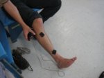
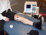
Biofeedback (BFB) has been practiced in clinical settings since the 1970’s, and has become a commonly used treatment in stroke rehabilitation. Normal regulation of muscle tone can be disrupted by central nerve damage caused by a stroke. This can prevent your muscles from functioning adequately. With the help of electromyographic biofeedback (EMG-BFB), you can get feedback concerning when your muscles are tense or relaxed. Electromyography or EMG is when a set of electrodes is placed on the skin over the chosen muscle or muscle group to detect the electrical signals that occur when a muscle is tense or contracted. This electrical signal will provide you with visual or auditory feedback on whether or not your muscle is contracting and the amount of force in the contraction.
Does it work for stroke?
Research studies have shown that biofeedback of the lower extremity can lead to improvements in the ability to walk, move your lower extremity to their full range, as well as improve the quality of lower extremity movements while walking. This intervention may also improve the ability to walk in a more natural, functional setting, such as on a sidewalk or street. However, these improvements do not seem to impact performance in daily activities or the muscle stiffness in your lower extremity that is commonly associated with a stroke. These studies did not mention if there are any adverse or harmful effects of biofeedback for the lower extremity in clients who have experienced a stroke, such that this therapy seems to be safe.
Who provides the treatment?
Biofeedback for the lower extremity is typically performed by a physiotherapist. Most rehabilitation centers and private clinics are equipped with EMG equipment.
Clinician Information
Note: When reviewing the findings, it is important to note that they are always made according to randomized clinical trial (RCT) criteria – specifically as compared to a control group. To clarify, if a treatment is “effective” it implies that it is more effective than the control treatment to which it was compared. Non-randomized studies are no longer included when there is sufficient research to indicate strong evidence (level 1a) for an outcome.
Ten RCTs have investigated the efficacy of biofeedback in the lower extremity as a treatment intervention post-stroke. Specifically, biofeedback in the lower extremity has been examined in relation to gait recovery, range of motion (ROM), performance of activities of daily living (ADLs), functional ambulation, dorsiflexion strength, spasticity, and postural control. In eight of nine randomized controlled trials there were significant differences found for most of these outcome measures in favour of biofeedback therapy.
One high quality study investigated the relationship between biofeedback interventions and the ability to perform activities of daily living (ADL) post-stroke (Intiso et al. 1994). Using the Barthel Index as a measure of ADL performance, this study found no significant differences between groups. A recently published fair quality RCT explored the use of biofeedback for standing balance training and its impact on ADL, as assessed by the Functional Independence Measure (Heller et al. 2005). There were significant improvements for both groups however no observed differences between the treatment and control group.
Conclusion: There is moderate (Level 1b) evidence from one high quality RCT and one fair quality RCT that biofeedback interventions in the lower extremity are not effective in the recovery of functional performance in activities of daily living (ADL) post-stroke.
Dorsiflexion strength
Effective
1b
Two RCT have investigated the efficacy of biofeedback interventions for improving dorsiflexion strength post-stroke. One high quality study (Burnside et al. 1982) found a significant difference between groups, suggesting that biofeedback interventions in the lower extremity help to improve dorsiflexion strength post-stroke. One fair quality RCT (Basmajian et al. 1975) also tested strength of dorsiflexion and observed significant differences between groups.
Conclusion: There is moderate (Level 1b) evidence from one high quality RCT that dorsiflexion strength can be improved as a result of biofeedback treatment in the upper extremity post-stroke.
Functional ambulation
Effective
1b
Two RCT studies investigated the relationship between biofeedback interventions and functional ambulation post-stroke. One fair quality study (Mandel et al. 1990) found that walking speeds increased more rapidly for patients treated with a combination of biofeedback and conventional physical therapy. One high quality study (Intiso et al. 1994) also noted a significant improvement in walking ability for those who received biofeedback treatment. A recently published fair quality RCT explored the use of biofeedback for standing balance training and its impact on functional ambulation, as assessed by the Functional Ambulation Categories and walking speed (Heller et al. 2005). There were significant improvements for both groups on these assessments however no observed differences between the treatment and control group.
Conclusion: There is moderate (Level 1b) evidence from one high quality RCT that biofeedback therapy in the lower extremity improves functional ambulation post-stroke.
Conclusion: There is strong evidence from three high quality RCTs (Level 1a) that biofeedback improves gait recovery post-stroke.
Postural control
Effective
2a
One fair quality study (Engardt et al. 1993) investigated the effect of biofeedback interventions for improving postural control post-stroke, using measures of sit-to-stand and rising to sit-down as primary outcomes. Significant improvements were noted in favour of biofeedback treatment. Another fair quality study (Wong et al. 1997) found that biofeedback improved the ability to maintain stance post-stroke. A recently published fair quality RCT explored the use of biofeedback for standing balance training and its impact on postural control, as assessed by the Postural Assessment Scale for Stroke (PASS) (Heller et al. 2005). There were significant improvements for both groups on these assessments however no observed differences between the treatment and control group. One important finding was that the experimental group showed significant improvements in the duration of reception double stance on the paretic limb as compared to the control group.
Conclusion: There is limited (Level 2a) evidence to suggest that biofeedback interventions are effective in improving postural control post-stroke as noted in three fair quality studies.
Range of motion (ROM)
Effective
1A
Two high quality RCT studies investigated the effect of biofeedback on range of motion in the lower extremity and all observed significant differences between groups. Burnside et al. (1982) noted significant improvements in range of motion for those who received biofeedback treatment. In a similar investigation, Bradley et al. (1998) found that active movement was significantly increased in patients that had received a combination of standard physiotherapy and biofeedback. One fair quality RCT (Basmajian et al. 1975) found a significant improvement in the range of motion of dorsiflexion in favour of the treatment group that received combined biofeedback with standard physical therapy. A recently published fair quality RCT explored the use of biofeedback for standing balance training and its impact on motor recovery, as assessed by the Fugl-Meyer Motor Recovery Scale (Heller et al. 2005). There were significant improvements for both groups however no observed differences between the treatment and control group.
Conclusion: There is strong evidence (Level 1a) from two high quality RCTs and one fair quality RCT that biofeedback interventions improve range of motion (ROM) post-stroke.
Spasticity
Not effective
1B
One high quality study (Intiso et al. 1994) investigated the relationship between biofeedback interventions in the lower extremity and spasticity post-stroke. While the primary measure used to assess spasticity was the Ashworth Scale, no significant differences were noted between groups. Another recently published fair quality RCT explored the use of biofeedback for standing balance training and its impact on spasticity, as assessed by the Ashworth Scale (Heller et al. 2005). There were no significant improvements for both groups, as well as no observed differences between the treatment and control group.
Conclusion: There is moderate (Level 1b) evidence from one high quality RCT and one fair quality RCT that spasticity is not improved as a result of biofeedback interventions in the lower extremity.
References
Basmajian JV, Kukulka CG, Narayan MG, Takebe K. (1975). Biofeedback treatment of foot-drop after stroke compared with standard rehabilitation technique: effects on voluntary control and strength. Arch Phys Med Rehabil, 56, 231-236.
Bradley L, Hart BB, Mandana S, Flowers K, Riches M, Sanderson P. (1998). Electromyographic biofeedback for gait training after stroke. Clin Rehabil, 12, 11-22.
Burnside IG, Tobias HS, Bursill D. (1982). Electromyographic feedback in the remobilization of stroke patients: a controlled trial. Arch Phys Med Rehabil, 63, 217-222.
Cozean CD, Pease WS, Hubbell SL. Biofeedback and functional electric stimulation in stroke rehabilitation. Arch Phys Med Rehabil 1988; 69: 401-405
Engardt M, Ribbe T, Olsson E. (1993). Vertical ground reaction force feedback to enhance stroke patients’ symmetrical body-weight distribution while rising/sitting down. Scand J Rehabil Med, 25(1), 41-8.
Heller F., Beuret-Blanquart F., & Weber, J. (2005). [Postural biofeedback and locomotion reeducation in stroke patients]. Ann Readapt Med Phys, 48(4), 187-195.
Intiso D, Santilli V, Grasso MG, Rossi R, Caruso I. (1994). Rehabilitation of walking with electromyographic biofeedback in foot-drop after stroke. Stroke, 25, 1189-1192.
Mandel AR, Nymark JR, Balmer SJ, Grinnell DM, O’Riain MD. (1990). Electromyographic versus rhythmic positional biofeedback in computerized gait retraining with stroke patients. Arch Phys Med Rehabil 71, 649-654.
Morris ME, Matyas TA, Bach TM, Goldie PA. (1992). Electrogoniometric feedback: its effect on genu recurvatum in stroke. Arch Phys Med Rehabil, 73, 1147-1154.
Wong AM, Lee MY, Kuo JK, Tang FT.(1997).The development and clinical evaluation of a standing biofeedback trainer. J Rehabil Res Dev, 34, 322-327.
Biofeedback – Upper Extremity
Evidence Reviewed as of before: 26-10-2010
Author(s)*: Robert Teasell, MD; Norine Foley, BASc; Sanjit Bhogal, MSc; Jamie Bitensky, MSc OT; Mark Speechley, MD; Nicol Korner-Bitensky, PhD OT
Biofeedback (BFB) is commonly used as a treatment intervention for stroke rehabilitation. Following a stroke, the main central motor pathways that regulate normal muscle tone and functioning can be disrupted or even damaged. However, some motor pathways that are often unused remain relatively unaffected by the stroke. Individuals may learn how to activate these unused pathways with the help of electromyographic biofeedback (EMG-BFB) and this may lead to improvements in their muscle tone and functioning. Given that hemiparesis of the upper extremity can result in functional disability following stroke and can affect important aspects of daily living (i.e. feeding and dressing), the use of EMG-BFB as an effective means of treatment for upper extremity hemiparesis has been carefully studied. Specifically, studies have examined the use of biofeedback to improve hand function as well as upper extremity range of motion and function.
Patient/Family Information
Author: Marc-André Roy, MSc.
What is biofeedback for the upper extremity?
A stroke can damage the central nervous system and disrupt normal regulation of muscle tone. This can prevent your muscles from functioning adequately. With the help of electromyographic biofeedback (EMG-BFB), you can receive feedback to know when your muscles are tense or relaxed. Electromyography (EMG) is when a set of electrodes is placed on the skin over the chosen muscle (or muscle group) to detect the electrical signals that occur when a muscle is tense (or contracted). This electrical signal will provide you with a visual or auditory feedback to know whether or not your muscle is contracting and indicate the amount of contraction. This biofeedback can help you re-educate your muscles to contract or relax at your own will in order to increase voluntary muscle control.
Does it work for stroke?
Research has shown that the main reason for functional impairment following a stroke is upper extremity hemiparesis which can affect important activities of daily living (e.g. feeding and dressing). Biofeedback (BFB) is commonly used as a treatment intervention for stroke rehabilitation. Following a stroke, the main central motor pathways that regulate normal muscle tone and functioning can be disrupted or even damaged. However, some motor pathways that are often unused remain relatively unaffected by the stroke. Individuals may learn how to activate these unused pathways with the help of electromyographic biofeedback (EMG-BFB) and this may lead to improvements in their muscle tone and functioning. Specifically, studies have examined the use of biofeedback to improve hand function as well as upper extremity range of motion and function. There is conflicting evidence that biofeedback interventions are effective for improving upper extremity function post-stroke. But, they are not effective for improving manual dexterity and for improving range of motion in the upper extremity post-stroke. At the follow-up evaluation, biofeedback interventions were not effective for improving upper extremity function.
Who provides the treatment?
Biofeedback for the upper extremity is typically performed by a physiotherapist. Most rehabilitation centers and private clinics are equipped with EMG equipment.
Clinician Information
Note: When reviewing the findings, it is important to note that they are always made according to randomized clinical trial (RCT) criteria – specifically as compared to a control group. To clarify, if a treatment is “effective” it implies that it is more effective than the control treatment to which it was compared. Non-randomized studies are no longer included when there is sufficient research to indicate strong evidence (level 1a) for an outcome.
Six studies, four high quality RCTs and two of fair quality , have investigated the effect of biofeedback interventions on stroke rehabilitation. Specifically, studies have investigated the effectiveness of biofeedback to improve manual dexterity, upper extremity function and range of motion (ROM).
Three high quality RCTs and one fair quality RCT have investigated the relationship between biofeedback treatment upper extremity function post-stroke. Two studies, Basmajian et al. (1982) and Basmajian et al. (1987) investigated the effect of biofeedback compared to physical therapy using the biofeedback approach. No significant differences were observed in upper extremity function, as assessed using the Upper Extremity Function Test. Similar results were found in a similar study by Prevo et al. (1982) as assessed using a non-standardized functional test.
One high quality RCT (Crow et al. 1989) compared the use of EMG biofeedback therapy to sham biofeedback. The treatment group scored significantly higher on the Action Research Arm Test and on the Brunnstrom-Fugl Meyer Test. However, these results were not maintained at follow up.
Conclusion: There is conflicting evidence (Level 4) from three high quality RCTs and one fair quality RCT, that biofeedback interventions are effective for improving upper extremity function post-stroke. However, at follow-up there is strong evidence that biofeedback interventions are not effective for improving upper extremity function post-stroke.
Upper extremity manual dexterity
Not Effective
1A
Two high quality RCTs investigated the relationship between biofeedback treatment and manual dexterity post-stroke. In the first, Basmajian et al. (1982) examined manual dexterity and biofeedback using the Minnesota Rate of Manipulation test. No improvements in manual dexterity were noted for either group. This finding was substantiated by another high quality RCTs Basmajian et al. (1987) that also investigated biofeedback interventions and manual dexterity. Results were similar to those found in the previous study, such that no significant differences were noted when the Finger Oscillation Test was used as a measure of manual dexterity.
Conclusion: There is strong (level 1a) evidence from two high quality RCTs, that biofeedback interventions are not effective for improving manual dexterity post-stroke.
Upper extremity range of motion
Not Effective
1b
Two RCTs have investigated the efficacy of biofeedback treatment for improving range of motion (ROM) in the upper extremity post-stroke. One high quality RCT (Hurd et al. 1980) investigated the relationship between biofeedback and range of motion using measures of active range of motion and muscle activity in the upper extremity. No significant differences were noted. One fair quality RCT (Greenberg and Fowler, 1980) also examined the use of biofeedback methods for improving the active elbow extension ROM. No significant improvements were reported in either group.
Conclusion: There is moderate evidence (Level 1b) from one high and one fair quality RCT, that biofeedback interventions are not effective for improving range of motion in the upper extremity post-stroke.
References
Basmajian JV, Gowland CA, Finlayson MA, Hall AL, Swanson LR, Stratford PW, Trotter JE, Brandstater ME. (1987). Stroke treatment: comparison of integrated behavioral-physical therapy vs traditional physical therapy programs. Arch Phys Med Rehabil, 68 (5 Pt 1), 267-272.
Basmajian, Gowland, Brandstater, Swanson, Trotter (1982). EMG feedback treatment of upper limb in hemiplegic stroke patients: a pilot study. Arch Phys Med Rehabil, 63(12), 613-616.
Crow, Lincoln, Nouri, De Weerdt (1989). The effectiveness of EMG biofeedback in the treatment of arm function after stroke. Int Disabil Stud, 11, 155-160.
Greenberg, Fowler (1980). Kinesthetic biofeedback: a treatment modality for elbow range of motion in hemiplegia. Am J Occup Ther, 34(11), 738-743.
Hurd, Pegram, Nepomuceno. (1980). Comparison of actual and simulated EMG biofeedback in the treatment of hemiplegic patients. Am J Phys Med, 59(2), 73-82.
Prevo, Visser, Vogelaar (1982). Effect of EMG feedback on paretic muscles and abnormal co-contraction in the hemiplegic arm, compared with conventional physical therapy. Scand J Rehabil Med, 14(3), 121-131.
Functional electrical stimulation (FES), also sometimes called functional neuromuscular stimulation (FNS), is a technique used to elicit a voluntary muscle contraction during a functional task by applying low-level electrical current to the nerves that control muscles or directly over the motor end-plate of the muscle (just like a pacemaker makes a heart beat).
Neuromuscular electrical stimulation, or simply ‘electrical stimulation’ (ES), is a modality used primarily for strengthening muscles, without the purpose of integrating a functional task as done with FES. All three (FES, FNS, ES) basically focus on eliciting muscular contractions.
This module summarizes the scientific evidence on FES effectiveness in the treatment of the lower extremities post-stroke (FES of the shoulder and FES of the upper extremities are reviewed in separate modules). TENS and other therapeutic electrical stimulation that do not elicit muscular contraction are reviewed in other modules.
NOTE: Studies involving the use of medications such as Botox have not been included in our analyses.
Functional electrical stimulation (FES) is a technique that causes a muscle to contract through the use of an electrical current. This might sound strange, but actually, the body naturally uses electrical currents to make muscles move. Normally, when a part of the body needs to move, the brain sends electrical signals through the nervous system. The nerves, acting like electrical wires, relay these signals to the muscles, directing them to contract. This contraction causes the body part – for example, the ankles, wrists, elbows – to move in a controlled, deliberate fashion. After a stroke, some of these electrical signals do not function as well as they should, causing patients to have trouble walking and coordinating movements. Pain and stiffness can result as well.
When using FES as an intervention after a stroke, the therapist applies an electrical current to either the skin over the nerve, or over the bulk of the muscle, and this will cause a muscle contraction.
The idea behind FES is that it allows the muscles that are paralyzed or partially paralyzed by stroke to move again. The electrical stimulation applied to the muscle is controlled so that the movement produced will provide useful function, and not random movement. FES devices translate input controlled by the patient into patterns that will produce the desired motion in the paralyzed muscles.
This module will look at the use of FES for loss of function, pain, or spasticity (stiffness) of the legs and feet. There is also an intervention using electrical stimulation that does not cause muscle contraction. This is called Transcutaneous Electrical Neuromuscular Stimulation (TENS) and it is described in another module of StrokEngine (soon to come).
Are there different kinds of FES?
Yes, and you will see different names, including functional electrical stimulation, functional neuromuscular stimulation, and electrical stimulation. But, they all have the same goal: to stimulate muscle contraction which in turn may lead to an increase in function, strength, and movement as well as a decrease in pain and spasticity. FES can be used on different parts of the body (arms, legs, shoulders, etc) and also on specific muscles in order to achieve different goals. For example, in FES of the lower extremities, a therapist may apply the stimulation to the quadriceps (muscles of the thighs) in order to help the patient to walk. Two other modules on StrokEngine focus on other kinds of FES, namely Functional Electrical Stimulation of the Hemiplegic Shoulder and Functional Electrical Stimulation of the Upper Extremities.
Why use FES on the legs after stroke?
Loss of leg function, movement, and strength are common after a stroke, and thus impair walking and standing. This occurs because muscles become paralyzed, and cannot receive electrical impulses from the brain. Pain and spasticity are also common after a stroke. FES may be useful for increasing leg function as well as for preventing pain and dysfunction after a stroke.
Does it work for stroke?
Researchers have studied how FES can help patients with stroke through its effects on the muscles in the legs and feet:
Strength of muscle contraction: In individuals 1-6 months post-stroke, FES was shown to strengthen muscle contractions.
Walking: In patients1-6 months post-stroke , FES showed some improvement in walking. However, FES is more useful in people more than 6 months post-stroke.
Perceived health status: Studies showed that FES for the lower extremities was not effective in improving the perceived health of patients more than 6 months post-stroke.
Spasticity (stiffness): Research showed that when combined with other therapy, FES caused a reduction in spasticity when used for patients more than 6 months post-stroke.
Range of motion (movement of joints): FES was shown to be moderately effective in improving range of motion for individuals more than 6 months post-stroke, when combined with other therapy.
Functional Ambulation (mobility): There was some evidence that FES improved the ability of patients more than 6 months post-stroke to move around more easily.
Lower extremity coordination: There was improvement in knee coordination in patients more than 6 months post stroke, however coordination of the legs and feet in general did not show improvement.
Activity level: Studies did not show improvement in level of activity of patients more than 6 months post-stroke after using FES.
Balance: Research showed some evidence that FES did not improve the balance of individuals more than 6 months post stroke.
What can I expect?
Small square stickers (electrodes) are placed over the centre of the bulk of the muscle to be stimulated. Wires connect the electrodes to a stimulator, a small machine that produces the electrical current. The stimulation is usually started at a very low level, causing a tingling “pins and needles” feeling on the skin. The current will then slowly be increased after each stimulation until it is strong enough to make the muscle contract. This level (the smallest current necessary to make the muscle contract) will be used for the FES treatment.
Although some people find the treatment uncomfortable, it is usually well tolerated. FES may give some discomfort, but it is virtually painless. Treatment times may vary, however, the time is usually divided into a number of daily sessions. FES treatments are usually done for 30 – 45 minutes, but once you are set up, you can typically perform the treatments on your own or with a family member.
Are there any side effects/risks?
There are few risks that come with the use of functional electrical stimulation. The electrodes can irritate the skin they contact, but this is uncommon. Using non-latex hypoallergenic electrodes can often solve this problem. Some people may find that certain types of electrical stimulations are irritating, but this can be easily fixed by changing the level of the current. After the treatment, there may be pink marks left on the skin where the electrodes were placed, but these usually fade within an hour. Although very rare, this type of therapy can increase spasticity (muscle tightness).
NOTE: People with epilepsy, poor skin condition, hypersensitivity to the electrical stimulation, cancer, and cardiac pacemakers should not receive FES treatment.
Who provides the treatment?
Physical therapists or occupational therapists will usually provide the FES treatment. However, due to the long duration of the stimulation, it is possible for the treatment to be done at home after discharge from the hospital. This will require having a stimulator at home. If you are provided with a home stimulator, family members or friends will be given instructions on how to assist with treatments. Usually, once the electrodes are placed, the rest of the procedure is very simple. To operate an FES machine, you simply switch it on and increase (slowly and gradually) the intensity of the current on a knob – just like switching on a radio and increasing the volume.
NOTE: Consult with your therapist or medical professional on the exact use of specific models of FES equipment.
How many treatments?
Some patients continue to use FES for many years. To maximize the benefits after stroke, it should be used for at least 6 weeks.
How much does it cost? Does insurance pay for it?
Although the cost of an FES machine varies, some systems are relatively inexpensive. Rental or lease options bring the cost down to the equivalent of 1 or 2 clinical visits per month. Some insurance plans cover the purchase or rental of such equipment. Check with your insurance company.
Is FES for me?
There is clear evidence that there are benefits to using functional electrical stimulation in comparison to regular therapy. These benefits include increased force of contraction and improved walking. However, in terms of general activity level after stroke, balance, and perceived health status, FES was not shown to be more effective than conventional therapy. So, overall, FES is an effective treatment you may want to consider after a stroke. If you are interested in learning more about FES, speak to your rehabilitation provider about the possibility of using this treatment.
Clinician Information
Note: When reviewing the findings, it is important to note that they are always made according to randomized clinical trial (RCT) criteria – specifically as compared to a control group. To clarify, if a treatment is “effective” it implies that it is more effective than the control treatment to which it was compared. Non-randomized studies are no longer included when there is sufficient research to indicate strong evidence (level 1a) for an outcome.
Thirty three studies (of which 13 high quality RCTs, 13 fair quality RCTs, two quasi-experimental design studies and two pre-post design studies) as well as two meta-analyses and one systematic review have examined the effectiveness of functional electrical stimulation (FES) as a means to improve lower extremity function post-stroke.
The first fair quality RCT (MacDonell et al., 1994) investigated the effectiveness of FES for improving performance in activities of daily living in 38 patients with acute stroke with weakness of dorsiflexion Grade 4 or less measured by the Medical Research Council Scale (MRC). The treatment group received FES five times a week for four weeks producing ankle dorsiflexion of the affected leg in addition to standard physical therapy, while the control group received standard physical therapy alone. Outcomes were measured at four weeks (post-treatment) and at eight weeks. No significant between-group difference was found on performance of activities of daily living at post-treatment (four weeks) or at the eight week follow-up as measured by the Barthel Index.
The second fair quality RCT (Kojovic et al., 2009) investigated the effectiveness of FES for improving ADL in 13 patients with acute stroke who were able to ambulate with a single cane or hand support. The patients were randomly assigned to either the functional electrical therapy (FET) group, or a control group. Both FET and control groups participated in a standard rehabilitation program and walking sessions. During the walking sessions, patients used the tripod cane or were physically assisted by the therapist in addition to instructions given by the therapist on how to improve their walking pattern. The FET group used a sensor-driven electrical stimulator device to stimulate four muscle groups: quadriceps, hamstring, soleus and tibialis anterior of the paretic leg in order to improve knee flexion/extension and ankle flexion/extension during walking. Outcomes were assessed at post-treatment (four weeks). At the end of the four week intervention, the Barthel Index score was significantly higher (reflecting better functioning) in the FET group compared to the control group.
The quasi-experimental design study (Solopova et al., 2011) pseudo-randomized patients with acute stroke to receive FES with assisted passive/active locomotor-like leg movements and progressive limb loading (FES group) or conventional rehabilitation (control group). FES was applied to the soleus, tibialis anterior, hamstring, quadriceps, hip adductors, gluteus maximus and medial gastrocnemius muscles as participants performed leg movements while semi reclined on a tilt table. ADL function was measured by the Barthel Index. At post-treatment (2 weeks) a significant between-group difference in ADL function was seen in favour of the FES group compared to the control group.
Conclusion: There is limited evidence (level 2a) from 1 fair quality RCT and 1 quasi-experimental design study that FES is more effective than conventional rehabilitation in improving ADL performance in patients with acute stroke.
Note: However, another fair quality RCT found that FES was not more effective than conventional rehabilitation alone in improving ADL performance among patients with acute stroke. Both fair quality RCTs had small sample sizes, which may in part contribute to different results between studies.
Electrophysiology
Not Effective
2A
One fair quality RCT (MacDonell et al., 1994) has investigated the effectiveness of FES for improving electrophysiological functioning in patients with acute stroke.
The fair quality RCT (MacDonell et al., 1994) investigated the effectiveness of FES for improving electrophysiological functioning in 38 patients with acute stroke and weakness of dorsiflexion Grade 4 or less as measured by the Medical Research Council Scale (MRC). The treatment group received FES producing ankle dorsiflexion of the affected leg in addition to standard physical therapy and the control group received standard physical therapy alone. Outcomes were measured at post-treatment (four weeks) and at eight weeks. No significant between-group difference was found on any electrophysiological measure at post-treatment or at the eight week follow-up as measured by foot tap frequency, duration and frequency of electromyographic (EMG) burst activity in tibialis anterior, H max/ M max ratio in gastocnemius, degree of vibratory inhibition of the H reflex, and F mean/ M max ratio using responses from the flexor hallucis brevis.
Conclusion: There is limited evidence (Level 2a) from one fair quality RCT that FES in combination with standard physical therapy is not more effective than standard physical therapy alone for improving electrophysiological functioning in patients with acute stroke.
MIVC and EMG co-contraction
Effective
1a
Two high quality RCTs (Ferrante et al., 2008, Yan et al., 2005) and one quasi-experimental design study (Solopova et al., 2011) investigated the effectiveness of FES for improving maximum isometric voluntary contraction in patients with acute stroke.
The first high quality RCT (Ferrante et al., 2008) investigated the use of FES for improving MIVC in 20 patients with acute stroke. The participants were randomized to receive either (1) standard rehabilitation and FES-cycling (5 minutes of passive cycling, 10 minutes of FES, 5 minutes of passive cycling, 10 minutes of FES and 5 minutes of passive cycling); or (2) standard rehabilitation alone. Outcomes were measured at post-treatment (four weeks). Following four weeks of treatment, a significant between-group difference in favour of the group receiving FES-cycling was found on the quadriceps isometric maximum force as measured by maximal voluntary contraction.
The second high quality RCT (Yan et al., 2005) investigated the effectiveness of FES on maximum isometric voluntary contraction (MIVC) of ankle dorsiflexors and planter flexors in 46 patients with acute stroke. Patients received either (1) standard rehabilitation and FES-cycling; (2) standard rehabilitation and placebo stimulation (placebo); or (3) standard rehabilitation alone (control) for three weeks. All participants were assessed on MIVC and EMG co-contraction ratio during ankle dorsiflexion and plantarflexion at one, two, three and 8 weeks. During ankle dorsiflexion, percent increases in MIVC torques and integrated EMG of the FES group were significantly larger than those of the control group from week one onward, and significantly larger than the placebo group at week three only. Also, the EMG co-contraction ratio during dorsiflexion of the affected ankle showed a significantly greater reduction in the FES group compared to the other groups from week one or two onward, reflecting greater improvement. For ankle plantarflexion, a significant difference was found in favour of the FES group only at week three only compared to the other groups. No significant between group difference was found on either measure at the 8 week follow-up.
The quasi-experimental design study (Solopova et al., 2011) pseudorandomized patients with acute stroke to receive FES with assisted passive/active locomotor-like leg movements and progressive limb loading (FES group) or conventional rehabilitation (control group). FES was applied to the soleus, tibialis anterior, hamstring, quadriceps, hip adductors, gluteus maximus and medial gastrocnemius muscles as participants performed leg movements while semi reclined on a tilt table. At post-treatment (2 weeks) there were significant between-group differences in maximum isometric voluntary contraction of the paretic flexor and extensor muscles and the nonparetic extensors, in favour of the FES group compared to the control group. There was also a significant between-group difference in EMG patterns of the rectus femoris and biceps femoris muscles during knee flexion of the ipsilateral leg, in favour of the intervention group compared to the control group.
Conclusion: There is strong evidence (Level 1a) from two high quality RCTs and one quasi-experimental study that FES in addition to standard rehabilitation is more effective than standard rehabilitation alone for improving maximal isometric voluntary contraction in patients with acute stroke.
Note:mprovements were not maintained beyond follow-up in one of the high quality RCT.
Mobility
Not Effective
1b
One high quality RCT (Ferrante et al., 2008) has investigated the effectiveness of FES for improving mobility in patients with acute stroke.
The high quality RCT (Ferrante et al., 2008) examined the effectiveness of FES on mobility in 20 patients with acute stroke. Patients were randomized to receive either (1) standard rehabilitation and FES-cycling (5 minutes of passive cycling, 10 minutes of FES, 5 minutes of passive cycling, 10 minutes of FES and 5 minutes of passive cycling; or (2) standard rehabilitation alone. Outcomes were measured at post-treatment (four weeks). No significant between-group differences were found for mobility of the ankle, or knee and hip joint during voluntary movement, as measured by the Motricity Index (MI) following 4 weeks of treatment. Due to the small sample size, failure to find group differences may have occurred because of type 2 error.
Conclusion: There is moderate evidence (Level 1b) from one high quality RCT that FES in addition to standard rehabilitation is not more effective than standard rehabilitation alone for improving mobility in patients with acute stroke.
Motor control
Effective
1B
One high quality RCT (Ferrante et al., 2008) investigated the use of FES for improving motor control in patients with acute stroke.
The high quality RCT (Ferrante et al., 2008), examined the effectiveness of FES on motor control in 20 patients with acute stroke. The participants were randomized to receive either (1) standard rehabilitation and FES-cycling (5 minutes of passive cycling, 10 minutes of FES, 5 minutes of passive cycling, 10 minutes of FES and 5 minutes of passive cycling); or (2) standard rehabilitation alone. Patients were asked to perform sit to stand trials at 3 different speeds (patient selected, one slower than patient selected, and one faster than patient selected) to test motor control. Outcomes were measured at post-treatment (four weeks). Following four weeks of treatment, a significant between-group difference in favour of the FES-cycling group was found on the percentage ratio between the slow and self selected speeds. The percentage ratio between fast and self selected speed failed to reach significance.
Conclusion: There is moderate evidence (Level 1b) from one high quality RCT that FES in addition to standard rehabilitation is more effective than standard rehabilitation alone for improving motor control in patients with acute stroke.
The first fair quality RCT (MacDonell et al., 1994) investigated the effectiveness of FES for improving motor function in 38 patients with acute stroke with weakness of dorsiflexion Grade 4 or less measured by the Medical Research Council Scale (MRC). The treatment group received FES five times a week for four weeks producing ankle dorsiflexion of the affected leg in addition to standard physical therapy, while the control group received standard physical therapy alone. Outcomes were measured at post-treatment (four weeks) and at eight weeks. No significant between-group difference was found in motor function at post-treatment or at eight week follow-up measured by the Fugl-Meyer Lower Extremity Motor Assessment Scale.
The second fair quality RCT (Kojovic et al., 2009) investigated the effectiveness of FES for improving motor function in 13 patients with acute stroke who were able to ambulate with a single cane or hand support. The patients were randomly assigned to either the functional electrical therapy (FET) group, or a control group. Both FET and control groups participated in a standard rehabilitation program and walking sessions for four weeks. During the walking sessions, patients used the tripod cane or were physically assisted by the therapist in addition to instructions given by the therapist on how to improve their walking pattern. The FET group used a sensor-driven electrical stimulator device to stimulate four muscle groups: quadriceps, hamstring, soleus and tibialis anterior of the paretic leg in order to improve knee flexion/extension and ankle flexion/extension during walking. At the end of the 4 week intervention, the FET group had significantly higher scores on the Fugl-Meyer Lower Extremity Motor Assessment Scale compared to the control group.
The quasi-experimental design study (Solopova et al., 2011) pseudorandomized patients with acute stroke to receive FES with assisted passive/active locomotor-like leg movements and progressive limb loading (FES group) or conventional rehabilitation (control group). FES was applied to the soleus, tibialis anterior, hamstring, quadriceps, hip adductors, gluteus maximus and medial gastrocnemius muscles as participants performed leg movements while semi reclined on a tilt table. Motor function was measured by the Fugl Meyer Assessment. At post-treatment (2 weeks) a significant between-group difference in motor function was seen in favour of the FES group compared to the control group. Significant between-group differences in motor function were also seen among subgroups of patients with paralysis, severe hemiparesis and pronounced hemiparesis, in favour of the FES group compared to the control group.
Conclusion: There is limited evidence (level 2a) from 1 fair quality RCT and 1 quasi-experimental design study that FES is more effective than conventional rehabilitation in improving motor function in patients with acute stroke.
Note: However, another fair quality RCT found that FES was not more effective than conventional rehabilitation alone in patients with acute stroke. Both fair quality RCTs had small sample sizes, which may in part contribute to different results between studies.
Muscle strength
Not Effective
1B
One high quality RCT (Ferrante et al., 2008) has investigated the use of FES for improving muscle strength in patients with acute stroke.
The high quality RCT (Ferrante et al., 2008) examined the effectiveness of FES on muscle strength in 20 patients with acute stroke. Patients were randomized to receive either (1) standard rehabilitation and FES cycling FES-cycling (5 minutes of passive cycling, 10 minutes of FES, 5 minutes of passive cycling, 10 minutes of FES and 5 minutes of passive cycling); or (2) standard rehabilitation alone for four weeks. No significant between group difference was found for muscle strength of the hemiplegic limb, as measured by the Upright Motor Control Test following 4 weeks of treatment. Due to the small sample size, failure to find group differences may have occurred because of type 2 error.
Conclusion: There is moderate evidence (Level 1b) from one high quality RCT that FES in addition to standard rehabilitation is not more effective than standard rehabilitation alone for improving muscle strength of the hemiplegic limb patients with acute stroke.
Severity of stroke
Effective
2b
One quasi-experimental design study (Solopova et al., 2011) has investigated the effectiveness of FES on severity of stroke in patients with acute stroke.
The quasi-experimental design study (Solopova et al., 2011) pseudorandomized patients with acute stroke to receive FES with assisted passive/active locomotor-like leg movements and progressive limb loading (FES group) or conventional rehabilitation (control group). FES was applied to the soleus, tibialis anterior, hamstring, quadriceps, hip adductors, gluteus maximus and medial gastrocnemius muscles as participants performed leg movements while semi reclined on a tilt table. Stroke severity was measured by the National Institutes of Health Stroke Scale (NIHSS) and European Stroke Scale (ESS). At post-treatment (2 weeks) a significant between-group difference in scores was seen in favour of the FES group compared to the control group.
Conclusion: There is limited evidence (level 2b) from 1 quasi-experimental study that FES is more effective than conventional rehabilitation in improving severity of stroke in patients with acute stroke.
Spasticity
Effective
1A
Two high quality RCTs (Yan et al., 2005, Yeh et al., 2010) have investigated the effectiveness of FES for improving spasticity in patients with acute stroke.
The first high quality RCT (Yan et al., 2005) randomized patients to receive (1) standard rehabilitation and FES (FES group); (2) standard rehabilitation and placebo stimulation (placebo group); or standard rehabilitation alone (control group) for 3 weeks. Spasticity was measured by the Composite Spasticity Scale (CSS). At 3 weeks (post-treatment) a significant between-group difference was found in favour of the FES group compared to the placebo group and the control group. No significant between-group difference was found at the 8 week follow-up.
A high quality cross-over design study (Yeh et al., 2010) randomized patients to one 20-minute session of cycling with FES or one 20-minute session of cycling without FES. The groups performed the opposite treatment on the second intervention day. Hypertonicity was measured by the Modified Ashworth Scale (MAS) and the pendulum test (relaxation index and peak velocity). Both treatments were found to improve hypertonia, as seen by significantly reduced MAS scores and significantly improved pendulum test scores from pre- to post-testing. After the second intervention day there were significant between-group differences in all measures of hypertonia, in favour of the FES group compared to the non-FES group.
Conclusion: There is strong evidence (level 1a) from one high quality RCT and one high quality randomized cross-over design study that FES is more effective than standard rehabilitation alone for improving spasticity in patients with acute stroke in the short term.
Trunk movements and balance
Not Effective
1B
One high quality RCT (Ferrante et al., 2008) has investigated the effectiveness of FES for improving trunk movement and balance in patients with acute stroke.
The high quality RCT (Ferrante et al., 2008), examined the effectiveness of FES on trunk movements and balance in 20 patients with acute stroke. Patients were randomized to receive either (1) standard rehabilitation and FES-cycling (5 minutes of passive cycling, 10 minutes of FES), 5 minutes of passive cycling, 10 minutes of FES and 5 minutes of passive cycling; or (2) standard rehabilitation alone for four weeks. No significant between group differences were found for trunk movement and balance, as measured by the Trunk Control Test (TCT) following four weeks of treatment. Due to the small sample size failure to find group differences may have occurred because of type 2 error.
Conclusion: There is moderate evidence (Level 1b) from one high quality RCT that FES in addition to standard rehabilitation is not more effective than standard rehabilitation alone for improving trunk movement and balance in patients with acute stroke.
The first high quality RCT (Ferrante et al., 2008) investigated the use of FES for improving walking ability in 20 patients with acute stroke. The participants were randomized to receive either (1) standard rehabilitation and FES-cycling (5 minutes of passive cycling, 10 minutes of FES, 5 minutes of passive cycling, 10 minutes of FES and 5 minutes of passive cycling); or (2) standard rehabilitation alone for four weeks. Following four weeks of treatment, the group that received FES – cycling showed more improvement in a 50m walk test than the control group, however, the between group difference failed to reach statistical significance.
The second high quality RCT (Yan et al., 2005) investigated the effectiveness of FES for improving walking ability in 46 patients with acute stroke. Patients received either (1) standard rehabilitation or FES; (2) standard rehabilitation and placebo stimulation (placebo); or (3) standard rehabilitation alone (control) for three weeks. Outcomes were measured at one, two, three and 8 weeks. No significant between group difference was found for walking ability, measured by the Timed Up and Go (TUG) test at all periods of assessment.
The fair quality RCT (MacDonell et al., 1994) investigated the effectiveness of FES for improving walking ability in 38 patients with acute stroke and weakness in dorsiflexion Grade 4 or less as measured by the Medical Research Council Scale (MRC). The treatment group received FES producing ankle dorsiflexion of the affected leg in addition to standard physical therapy and the control group received standard physical therapy alone for four weeks. Outcomes were measured at post-treatment and at eight weeks. No significant between-group difference was found in walking ability at post-treatment or at eight week follow-up as measured by the Massachusetts General Hospital Functional Ambulation Categories (MGH FAC) scale.
The quasi-experimental design study (Solopova et al., 2011) pseudorandomized patients with acute stroke to receive FES with assisted passive/active locomotor-like leg movements and progressive limb loading (FES group) or conventional rehabilitation (control group). FES was applied to the soleus, tibialis anterior, hamstring, quadriceps, hip adductors, gluteus maximus and medial gastrocnemius muscles as participants performed leg movements while semi reclined on a tilt table. Walking ability was measured by movement in the knee and ankle joints. At post-treatment (2 weeks) a significant difference in ankle range of movement was found in favour of the FES group compared to the control group, however there were no differences between groups in knee joint motion.
Conclusion: There is strong evidence (Level 1a) from two high quality RCTs, one fair quality RCT and and one quasi-experimental study that FES in addition to standard rehabilitation is not more effective than standard rehabilitation alone for improving walking ability in patients with acute stroke.
Walking efficiency
Effective
2A
One fair quality RCT (Kojovic et al., 2009) has investigated the effectiveness of FES for improving walking efficiency in patients with acute stroke.
In the fair quality RCT, (Kojovic et al., 2009) investigated the effectiveness of FES for improving walking efficiency in 13 patients with acute stroke who were able to ambulate with a single cane or hand support. The patients were randomly assigned to either the functional electrical therapy (FET) group, or a control group. Both FET and control groups participated in a standard rehabilitation program and walking sessions for four weeks. During the walking sessions, patients used the tripod cane or were physically assisted by the therapist in addition to instructions given by the therapist on how to improve their walking pattern. The FET group used a sensor-driven electrical stimulator device to stimulate four muscle groups: quadriceps, hamstring, soleus and tibialis anterior of the paretic leg in order to improve knee flexion/extension and ankle flexion/extension during walking. At the end of the 4 week intervention, a statistically significant decrease in the Physiological Cost Index (PCI) was observed between groups in favour of the FET group, reflecting more efficient walking in the treatment group.
Conclusion: There is limited evidence (Level 2a) from one fair quality RCT that FES in addition to standard therapy is more effective than standard therapy alone for improving walking efficiency in patients with acute stroke.
Walking speed
Effective
2A
One fair quality RCT (Kojovic et al., 2009) has investigated the effectiveness of FES for improving walking speed in patients with acute stroke.
In the fair quality RCT, (Kojovic et al., 2009) investigated the effectiveness of FES for improving walking efficiency in 13 patients with acute stroke who were able to ambulate with a single cane or hand support. The patients were randomly assigned to either the functional electrical therapy (FET) group, or a control group. Both FET and control groups participated in a standard rehabilitation program and walking sessions for four weeks. During the walking sessions, patients used the tripod cane or were physically assisted by the therapist in addition to instructions given by the therapist on how to improve their walking pattern. The FET group used a sensor-driven electrical stimulator device to stimulate four muscle groups: quadriceps, hamstring, soleus and tibialis anterior of the paretic leg in order to improve knee flexion/extension and ankle flexion/extension during walking. At the end of the 4 week intervention, a statistically significant increase in favour of the FES group was found on mean walking velocity over a 6 meter walkway.
Conclusion: There is limited evidence (Level 2a) from one fair quality RCT that FES in addition to standard therapy is more effective than standard therapy alone for improving walking speed in patients with acute stroke.
Subacute Phase
Activities of Daily Living
Not Effective
1b
One high quality RCT (Tong et al., 2006) and one fair quality RCT (Granat et al., 1996) have investigated the effectiveness of FES on ADL in patients with subacute stroke.
The high quality RCT (Tong et al., 2006) randomized patients with subacute stroke to receive (1) gait training using an electromechanical gait trainer with functional electric stimulation (EGT-FES), (2) gait training using an electromechanical gait trainer (EGT), or (3) conventional gait training (CGT, control) for four weeks. Performance of ADLs was measured using the Barthel Index. At 4 weeks (post-treatment) no significant between-group differences in ADLs were found.
One fair quality crossover RCT (Granat et al., 1996) assigned patients with subacute and chronic stroke to receive four weeks of standard rehabilitation followed by four weeks of FES intervention. ADLs were measured using the Barthel Index. A significant improvement in ADLs was seen following the intervention period but not following the control period.
Conclusion : There is moderate evidence (Level 1b) from one high quality RCT that FES is not more effective than conventional or electromechanical gait training for improving performance of ADLs in patients with subacute stroke.
Note: However, there is limited evidence (level 2a) from one fair quality RCT that FES is more effective than standard rehabilitation in improving performance of ADLs in patients with subacute and chronic stroke.
Balance
Not Effective
1B
One high quality RCT (Tong et al., 2006) has investigated the effectiveness of FES on balance in patients with subacute stroke.
In the high quality RCT (Tong et al., 2006) has investigated the effectiveness of gait training using an electromechanical gait trainer with and without functional electric stimulation for improving balance in 46 patients with sub-acute stroke. The patients were randomly assigned to receive either: (1) gait training using an electromechanical gait trainer with functional electric stimulation (EGT-FES); (2) gait training using an electromechanical gait trainer (EGT), or (3) conventional gait training (CGT, control) for four weeks. At the end of the four week intervention, no significant between group difference was found on the Berg Balance Scale.
Conclusion: There is moderate evidence (level 1b) from one high quality RCT that FES not more effective than electromechanical or conventional gait training alone for improving balance in patients with sub-acute stroke.
Contraction force
Effective
1B
One high quality cross-over RCT (Bogataj et al., 1995) and 2 fair quality RCTs (Newsam et al., 2004, Winchester et al., 1983) has investigated the effectiveness of FES on maximum isometric voluntary contraction (MIVC) and contraction force in patients with subacute stroke.
The high quality cross-over RCT (Bogataj et al., 1995) randomized patients with subacute stroke and severe hemiplegia to receive either (1) 3 weeks of multi-channel functional electrical stimulation (MFES) followed by 3 weeks of conventional physiotherapy; or (2) 3 weeks of conventional therapy followed by 3 weeks of MFES. Gait stability was measured by the vertical components of ground reaction force and the trajectories of center of pressure (TCP) for each foot in the stance phase. A significant between-group difference in TCP was found, in favour of FES compared to conventional therapy.
The first fair quality RCT (Newsam et al., 2004) assigned patients with subacute stroke to receive physical therapy and an electric stimulation facilitation program of the quadriceps (FES group), or physical therapy alone (control group). A significant between-group difference in supramaximal contraction torque of the quadriceps was found at 3 weeks (post-treatment) in favour of the FES group compared to the control group. There was no significant difference in MIVC of the quadriceps.
The second fair quality RCT (Winchester et al., 1983) randomized patients with subacute stroke to receive either standard physiotherapy and FES (FES group) or standard physiotherapy alone (control group). Contraction force was measured by knee extension torque. A significant between-group difference in contraction force was seen at 3 weeks (during training) and 4 weeks (post-treatment) in favour of the FES group compared to the control group.
Conclusion: There is moderate evidence (level 1b) from 1 high quality cross-over RCT and 2 fair quality RCTs that FES is more effective than standard rehabilitation for increasing contraction force in patients with subacute stroke.
Note: However, there is limited evidence (level 2a) from 1 fair quality RCT that FES is not more effective than standard rehabilitation for improving MIVC in patients with subacute stroke.
The first high quality RCT (Yavuzer et al., 2006) randomly assigned patients with subacute stroke to receive either (1) conventional rehabilitation and neuromuscular electric stimulation applied to the tibialis anterior for 10 minutes per session (NMES group) or (2) conventional rehabilitation alone (control group). Gait kinematics were measured by walking velocity, step length, percentage of stance phase at the paretic side, sagittal plane kinematics of the pelvis, hip, knee and ankle, maximum ankle dorsiflexion at swing and maximum ankle plantar flexion angle at initial contact. No significant between-group differences in gait kinematics were found at 4 weeks (post-intervention).
The second high quality RCT (Yavuzer et al., 2007) randomly assigned patients with subacute stroke to receive (1) conventional rehabilitation and sensory-amplitude electric stimulation (SES) to the paretic leg, or (2) standard rehabilitation alone (control group). Gait kinematics were measured by walking velocity, step length, percentage of stance phase at the paretic side, sagittal plane kinematics of the pelvis, hip, knee and ankle, maximum ankle dorsiflexion at swing and maximum ankle plantar flexion angle at initial contact. No significant between-group differences in gait kinematics were found at 4 weeks (post-treatment).
The first fair quality RCT (Sabut et al., 2010b) randomized patients with subacute stroke to receive physical therapy and FES (FES group) or physical therapy alone (control) for 12 weeks. Gait kinematics and parameters were taken as a measure of cadence, step length, step width, and toe-in toe-out. No significant between-group difference in gait kinematics and parameters were seen at 12 weeks (post-treatment).
The second fair quality crossover RCT (Granat et al., 1996) assigned patients with subacute or chronic stroke to receive four weeks of conventional rehabilitation followed by four weeks of FES intervention. Gait kinematics and parameters were taken as a measure of swing symmetry, heel strike and foot inversion. Group results at 11 weeks (post-treatment) revealed a significant improvement in foot inversion on linoleum, carpet and uneven ground, and swing symmetry on linoleum when receiving FES as compared to when not receiving FES.
Conclusion: There is strong evidence (Level 1a) from 2 high quality RCTs and 1 fair quality RCT that FES is not more effective than standard rehabilitation alone for improving gait kinematics and parameters in patients with subacute stroke.
NOTE: However, 1 fair quality crossover RCT found that FES was more effective than conventional rehabilitation in improving some aspects of gait kinematics and parameters in patients with subacute and chronic stroke.
Joint position sense
Not Effective
2A
One fair quality RCT (Winchester et al., 1983) has investigated the effect of FES on joint position sense in patients with subacute stroke.
The fair quality RCT (Winchester et al., 1983) investigated the effects of standard physiotherapy and FES (FES group) compared to standard physiotherapy alone (control group) in patients with subacute stroke. No significant between-group difference in joint position sense was found at 4 weeks (post-treatment).
Conclusion: There is limited evidence (level 2a) from 1 fair quality RCT that FES and standard rehabilitation is not more effective than standard rehabilitation alone for improving joint position sense in patients with subacute stroke.
Metabolic function
Effective
2B
One pre-post design study (Sabut et al., 2010a) has investigated the effect of FES on metabolic function in patients with subacute and chronic stroke.
The pre-post design study (Sabut et al., 2010a) assigned patients with subacute or chronic stroke to receive FES and standard physical therapy. Metabolic function was measured by oxygen consumption, carbon dioxide production, heart rate and energy cost. A significant improvement in metabolic function was seen at 12 weeks (post-treatment).
Conclusion: There is limited evidence (level 2b) from one pre-post design study that FES and standard therapy is effective for improving measures of metabolic response in patients with subacute and chronic stroke.
Mobility
Effective
1B
One high quality RCT (Tong et al., 2006) investigated the effectiveness of FES on mobility in patients with sub-acute stroke.
The high quality RCT (Tong et al., 2006) investigated the effectiveness of gait training using an electromechanical gait trainer with and without functional electric stimulation for improving mobility in 46 patients with sub-acute stroke. The patients were randomly assigned to receive either: (1) gait training using an electromechanical gait trainer with functional electric stimulation (EGT-FES), (2)gait training using an electromechanical gait trainer (EGT), or (3) conventional gait training (CGT, control) for four weeks. At the end of the four week intervention, there was a significant between-group difference on the Elderly Mobility Scale (EMS) and on the Five-meter Walking Speed Test in the EGT and EGT-FES groups compared to controls, however no significant between-group differences were found on the Functional Independence Measure (FIM).
Conclusion: There is moderate evidence (level 1b) from one high quality RCT that FES in addition to gait training is more effective than gait training alone for improving mobility in patients with sub-acute stroke.
The first high quality RCT (Ambrosini et al., 2011) randomized patients with subacute stroke or traumatic brain injury to either an FES-induced cycling group (FES group) or a placebo FES cycling group (control group). Function of the paretic limb was measured by the Upright Motor Control Test and by pedaling unbalance between the paretic and non-paretic legs, and motor power of the paretic limb was measured by the Motricity Index leg subscale. Significant between-group differences in all measures were seen at 4 weeks (post-treatment), in favour of the FES group compared to the control group. Results remained significant for the Upright Motor Control Test and the Motricity Index at follow-up (3-5 months post-treatment), in favour of the FES group.
The second high quality cross-over RCT (Bogataj et al., 1995) randomized patients with subacute stroke and severe hemiplegia to receive: (1) 3 weeks of multi-channel functional electrical stimulation (MFES) followed by 3 weeks of conventional physiotherapy; or (2) 3 weeks of conventional physiotherapy followed by 3 weeks of MFES. Functional motor status was assessed using the Fugl-Meyer Assessment. A significant improvement in motor function was seen at the middle of intervention (week 3), in favour of FES compared to conventional physiotherapy. However, no differences were seen at week 6.
The third high quality RCT (Tong et al., 2006) investigated the effectiveness of gait training using an electromechanical gait trainer with and without functional electric stimulation for improving motor impairment in 46 patients with sub-acute stroke. The patients were randomly assigned to receive either: (1) gait training using an electromechanical gait trainer with functional electric stimulation (EGT-FES), (2) gait training using an electromechanical gait trainer (EGT), or (3) conventional gait training (CGT, control) for four weeks. At the end of the four week intervention, there was a significant between group difference on the Motricity Index Leg subscale (MI) in the EGT and EGT-FES groups compared to control group.
One fair quality RCT (Sabut et al., 2010b) randomized patients with subacute stroke to receive physical therapy and FES (FES group) or physical therapy alone (control group) for 12 weeks. Motor function was measured by the Fugl-Meyer Assessment. No significant between-group differences were reported at 12 weeks (post-treatment), although the authors reported “better” improvement was made with FES.
Conclusion: There is strong evidence (level 1a) from 3 high quality RCTs that FES is more effective than control therapies (e.g. placebo intervention, conventional rehabilitation) in improving motor function in patients with subacute stroke. A fair quality RCT also reported improved motor function following FES, although between-group differences were not reported.
The first high quality RCT (Yavuzer et al., 2007) randomized patients with subacute stroke to receive either sensory amplitude electrical stimulation (SES group) or sham stimulation (control group). Motor recovery was measured using the Brunnstrom Stages of Motor Recovery. No significant between-group difference in motor recovery was seen at 4 weeks (post-treatment).
The second high quality RCT (Yavuzer et al., 2006) randomly assigned patients with subacute stroke to receive either conventional stroke rehabilitation and neuromuscular electric stimulation to the tibialis anterior (NMES group) or conventional therapy alone (control group). Motor function was measured using the Brunnstrom Stages of Motor Recovery. No significant between-group difference in motor recovery was seen at 4 weeks (post-treatment).
Conclusion: There is strong (level 1a) evidence from 2 high quality RCTs that FES is not more effective than standard rehabilitation in improving motor recovery in patients with subacute stroke.
Motor unit recruitment
Effective
2a
One fair quality RCT (Newsam et al., 2004) has investigated the effectiveness of FES on motor unit recruitment in patients with sub-acute stroke.
In the fair quality RCT (Newsam et al., 2004) investigated the effectiveness of FES on motor unit recruitment in 20 patients with sub-acute stroke. The treatment group received an electric stimulation facilitation program of the quadriceps during gait training and weight-bearing activities as well as standard physical therapy. The control group received standard physical therapy alone for three weeks. To measure the interpolated twitch and determine the motor unit recruitment, a short-duration, supramaximal electric stimulus was delivered at approximately three seconds into each of the maximal tests. After the three week intervention there was a significant between group difference in motor unit recruitment in favour of the patients that received FES.
Conclusion: There is limited evidence (Level 2a) from one fair quality RCT that FES of the quadriceps in combination with standard physical therapy is more effective than standard physical therapy alone for increasing motor unit recruitment in the quadriceps in patients with sub-acute stroke.
Muscle girth
Not Effective
2A
One fair quality RCT (Winchester et al., 1983) has investigated the effects of FES on muscle girth in patients with subacute stroke.
The fair quality RCT (Winchester et al., 1983) investigated the effects of standard physiotherapy and FES (FES group) compared to standard physiotherapy alone in patients with subacute stroke. No significant between-group difference in quadriceps muscle girth was seen at 4 weeks (post-treatment).
Conclusion: There is limited evidence (level 2a) from 1 fair quality RCT that FES is not more effective than standard rehabilitation in improving muscle girth of the quadriceps in patients with subacute stroke.
Muscle strength
Effective
2B
One fair quality RCT (Sabut et al., 2010b) and 1 pre-post design study (Sabut et al., 2010a) have investigated the effect of FES on muscle strength in patients with subacute stroke.
The fair quality RCT (Sabut et al., 2010b) randomized patients with subacute stroke to receive physical therapy and FES (FES group) or physical therapy alone (control group) for 12 weeks. Muscle output was measured by the maximum value of the root mean square on electromyography analysis. No between-group comparisons in muscle strength were reported, however the authors state that the FES group showed a “better” improvement than the control group at 12 weeks (post-treatment).
The pre-post design study (Sabut et al., 2010a) assigned patients with subacute or chronic stroke to receive FES and standard physical therapy. Muscle strength was measured by the mean absolute value, root mean square, median frequency and median amplitude of the tibialis anterior muscle EMG signal. A significant effect of FES treatment on muscle strength was found at 12 weeks (post-treatment).
Conclusion: There is limited evidence (level 2b) from 1 fair quality RCT and 1 pre-post design study that FES is effective improving muscle strength in patients with subacute stroke.
Note: Neither study reported between-group differences so it cannot be determined whether FES is more effective than control therapy in improving muscle strength in patients with subacute stroke.
The first fair quality RCT (Sabut et al., 2010b) randomized patients with subacute stroke to receive physical therapy and FES (FES group) or physical therapy alone (control group) for 12 weeks. No between-group comparisons on ankle range of motion were made, however the authors state that the FES group showed “better” improvement than the control group at 12 weeks (post-treatment).
The second fair quality RCT (Winchester et al., 1983) randomized patients with subacute stroke to receive either standard physiotherapy and FES (FES group) or standard physiotherapy alone (control group). Range of motion was measured weekly during the 4-week intervention. A significant between-group difference in active range of motion was found at week 2 (during treatment) in favour of the FES group compared to the control group. No significant differences were found at week 1 or 3 of treatment, or at week 4 (post-treatment).
Conclusion: There is limited evidence (level 2a) from 1 fair quality RCT that FES is not more effective than standard rehabilitation in improving range of motion in patients with subacute stroke, although temporary gains were seen mid-treatment.
Note: One fair quality RCT found improvement in ankle range of motion following FES, however these results are not used to determine level of evidence as between-group differences were not reported.
The first fair quality RCT (Sabut et al., 2010b) randomized patients with subacute stroke to receive physical therapy and FES (FES group) or physical therapy alone (control group) for 12 weeks. Spasticity was measured by the Modified Ashworth Scale. No between-group comparison in spasticity was provided, however, the authors state that the FES group showed a “better” improvement than the control group at 12 weeks (post-treatment).
The second fair quality RCT (Winchester et al., 1983) randomized patients with subacute stroke to receive either standard physiotherapy and FES (FES group) or standard physiotherapy alone (control group). Spasticity was measured on a scale of “none,” “minimal,” “moderate,” or “severe”. No significant between-group difference in spasticity was seen at 4 weeks (post-treatment).
Conclusion: There is limited (level 2a) from 1 fair quality RCT that FES and standard rehabilitation is not more effective than standard rehabilitation alone for decreasing spasticity in patients with subacute stroke.
Note: One fair quality RCT reported reduced spasticity following FES, however these results are not used to determine level of evidence as between-group differences were not reported.
Trunk control
Effective
1B
One high quality RCT (Ambrosini et al., 2011) has investigated the effectiveness of FES on trunk control in patients with subacute stroke.
In the high quality RCT (Ambrosini et al., 2011) randomized patients with subacute stroke or traumatic brain injury to either an FES-induced cycling group (FES group) or a placebo FES cycling group (control group). Trunk control was measured by the Trunk Control Test. A significant between-group difference on trunk control was seen at 4 weeks (post-treatment) and follow-up, in favour of the FES group compared to the control group.
Conclusion: There is moderate evidence (level 1b) from 1 high quality RCT that FES is more effective than placebo intervention in improving trunk control in patients with sub-acute stroke.
Walking efficiency
Effective
1B
One high quality RCT (Tong et al., 2006) ), 1 fair quality RCT (Sabut et al., 2010b) and 1 pre-post design study (Sabut et al., 2010a) have investigated the effectiveness of FES on walking efficiency in patients with subacute and chronic stroke.
The high quality RCT (Tong et al., 2006) randomly assigned patients with subacute stroke to receive: (1) gait training using an electromechanical gait trainer with functional electric stimulation (EGT-FES); (2) gait training using an electromechanical gait trainer (EGT); or (3) conventional gait training (CGT, control) for four weeks. Walking efficiency was measured using the Functional Ambulatory Category (FAC). At four weeks (post-intervention), there was a significant between-group difference in walking efficiency in favour of the EGT and EGT-FES groups compared to the control group.
One fair quality RCT (Sabut et al., 2010b) randomized patients with subacute stroke to receive physical therapy and FES (FES group) or physical therapy alone (control group) for 12 weeks. Walking efficiency was measured using the Physiological Cost Index. No significant between-group difference in walking efficiency was seen at 12 weeks (post-treatment).
One pre-post design study (Sabut et al., 2010a) assigned patients with subacute or chronic stroke to receive FES and standard physical therapy. Walking efficiency was measured using the Physiological Cost Index. A significant effect of FES treatment on walking efficiency was seen at 12 weeks (post-treatment).
Conclusion: There is moderate evidence (level 1b) from 1 high quality RCT that FES is more effective than conventional gait training or standard rehabilitation in improving walking efficiency in patients with subacute stroke. One pre-post study also reported improved walking efficiency following FES in patients with subacute and chronic stroke.
NOTE: However, 1 fair quality RCT did not find FES to be more effective than standard rehabilitation in improving walking efficiency.
The high quality RCT (Ambrosini et al., 2011) randomized patients with subacute stroke or traumatic brain injury to either an FES-induced cycling group (FES group) or a placebo FES cycling group (control group). Self-selected gait speed was measured by the 50m walking test. No significant between-group difference in gait speed was seen at 4 weeks (post-treatment) or follow-up. However, a subgroup analysis of patients with ischaemic stroke revealed a significant between-group difference in favour of the FES group compared to the control group.
The fair quality RCT (Sabut et al., 2010b) randomized patients with subacute stroke to receive physical therapy and FES (FES group) or physical therapy alone (control group) for 12 weeks. Walking speed was measured using the 10 Meter Walk-Way Test. No significant between-group difference in walking speed was found at 12 weeks (post-treatment).
The fair quality crossover RCT (Granat et al., 1996) assigned patients with subacute or chronic stroke to receive four weeks of standard rehabilitation followed by four weeks of FES intervention. Walking speed was measured by the walk-path length divided by the time to traverse the walk-path. No between-group comparisons in walking speed were made, however the authors reported improved speed following 4 weeks of FES intervention.
One pre-post design study (Sabut et al., 2010a) assigned patients with subacute or chronic stroke to receive FES and standard physical therapy. Walking speed was measured over 10 metres. A significant effect of FES treatment on walking speed was found at 12 weeks (post-treatment).
Conclusion: There is moderate evidence (level 1b) from 1 high quality RCT that FES is more effective than placebo intervention in improving walking speed in patients with subacute stroke. One pre-post design study also reported improved walking speed following FES.
NOTE: However, one fair quality RCT reported that FES is not more effective than standard rehabilitation alone in improving walking speed. Further, a second fair quality crossover RCT did not find significant improvement in walking speed following FES, although these results are not used to determine level of evidence as between-group differences were not reported.
Chronic Phase
Activities of Daily Living and participation
Effective
2A
Two fair quality crossover RCTs (Embrey et al., 2010; Granat et al., 1996) have investigated the effect of FES on Activities of Daily Living (ADLs) and participation in patients with chronic stroke.
The first fair quality crossover RCT (Embrey et al., 2010) randomized patients with chronic stroke and hemiplegia to receive FES or no FES for 3 months (phase I). The groups performed the opposite treatment for the following 3 months (phase II). Function and participation was measured using the Stroke Impact Scale. Patients who received FES in phase I showed significantly improved function and participation compared with patients who received no FES, and retained these improvements following no FES in phase II. Patients who received FES in phase II demonstrated significantly improved function and participation at 6 months (post-treatment) compared to baseline scores.
The second fair quality crossover RCT (Granat et al., 1996) assigned patients with sub-acute or chronic stroke to receive 4 weeks of standard rehabilitation followed by 4 weeks of FES intervention. Performance of ADLs was measured using the Barthel Index. A significant difference in ADLs was seen at 4 weeks (post-treatment) in favour of FES compared to standard rehabilitation.
Conclusion: There is limited evidence (level 2a) from 2 fair quality crossover RCTs that FES is more effective than standard rehabilitation for improving ADLs in patients with chronic stroke.
Activity level
Not Effective
2a
One fair quality RCT (Kottink et al., 2007) investigated the effectiveness of FES on physical activity in patients with chronic stroke.
The fair quality RCT (Kottink et al., 2007) investigated the effectiveness of using a 2-channel implantable peroneal nerve stimulator for improving physical activity in 29 patients with chronic stroke and foot drop. Participants were randomly assigned to receive either: (1) a 2-channel implantable peroneal nerve stimulator, (stimulation treatment group) or (2) a control group. Participants in the treatment group had the stimulator implanted under the epineurium of the peroneal nerve under general or spinal anesthesia. The control group was able to continue using their regular walking devices (ankle-foot orthosis, orthopedic shoes, or no device). Each participant was assessed on physical activity at baseline and at week 26. After 26 weeks, there was no significant between group difference in physical activity measured by the percentage of time spent stepping or standing, however there was a significant difference between the groups on time spent sitting/lying in favour of the stimulation group.
Conclusion: There is limited evidence (Level 2a) from one fair quality RCT that FES is not more effective than standard therapy for increasing physical activity in patients with chronic stroke.
Aerobic capacity
Not Effective
1B
One high quality RCT (Janssen et al., 2008) has investigated the effectiveness of FES on aerobic capacity in patients with chronic stroke.
The high quality RCT (Janssen et al., 2008) compared the effectiveness of cycling with and without FES on aerobic capacity in 12 patients with chronic stroke. Subjects were randomly assigned to either the treatment or control group in which both groups performed a cycling exercise. During cycling, the treatment group received electrical stimulation evoking muscle contractions and the control group received sensible stimulation not evoking muscle contractions for six weeks. After six weeks of treatment, there was no significant difference between the groups on VO2 peak scores.
Conclusion: There is moderate evidence (Level 1b) from one high quality RCT that FES in addition to cycling is not more effective than cycling alone in improving aerobic capacity in patients with chronic stroke.
Balance
Not Effective
1A
Two high quality RCTs (Daly et al., 2006, Janssen et al., 2008) have investigated the effectiveness of FES on improving balance in patients with chronic stroke.
The first high quality RCT (Daly et al., 2006) investigated the effectiveness of functional neuromuscular stimulation using intramuscular electrodes (FNS-IM) on balance in 32 patients with chronic stroke. The treatment group received functional neuromuscular stimulation using intramuscular electrodes, body-weight support treadmill training, coordination exercises, over ground walking (OG) and a home exercise program. The control group received the same treatment as the intervention group but without FNS for 12 weeks. No significant between group difference was reported for balance as measured by the Tinetti Balance (TB) after 12 weeks of treatment.
The second high quality RCT (Janssen et al., 2008) compared the effectiveness of cycling with and without FES on balance in 12 patients with chronic stroke. Participants were randomly assigned to either the treatment or control group in which both groups performed a cycling exercise. During cycling, the treatment group received electrical stimulation of the paretic leg evoking muscle contractions and the control group received sensible stimulation not evoking muscle contractions for six weeks. After the six week intervention, no significant difference between the groups was found on the Berg Balance Scale (BBS).
Conclusion: There is strong evidence (Level 1a) from two high quality RCTs that FES in addition to a gait training or a cycling program compared to a gait training or cycling program alone does not improve balance in patients with chronic stroke.
Coordination
Not Effective
1A
Two high quality RCTs (Daly et al., 2011, Daly et al., 2006) has investigated the effectiveness of FES for improving coordination in patients with chronic stroke.
The first high quality RCT (Daly et al., 2011) randomized patients with chronic stroke to receive intramuscular FES or no FES. Participants performed strengthening exercises, overground gait training and body-weight supported treadmill training for 1.5 hours 4 times/week for 12 weeks. Lower extremity coordination was measured by the Fugl-Meyer Lower Limb Scale and by the Functional Independence Measure (FIM), Locomotion and Mobility subscales. Both groups demonstrated significant improvements in lower extremity coordination at post-treatment (12 weeks), however between-group differences were not reported for these measures.
The second high quality RCT (Daly et al., 2006) investigated the effectiveness of functional neuromuscular stimulation using intramuscular electrodes (FNS-IM) on coordination in 32 patients with chronic stroke. The treatment group received functional neuromuscular stimulation using intramuscular electrodes, body-weight support treadmill training, coordination exercises, over ground walking (OG) and a home exercise program. The control group received the same treatment as the intervention group except without FNS for 12 weeks. After 12 weeks of treatment there was a significant between-group difference in favour of the treatment group on the Fugl-Meyer knee flexion coordination measure (FMKnFx). No statistically significant between group difference was found on the Fugl-Meyer lower extremity coordination (FMLE).
Conclusion: There is strong evidence (level 1a) from one high quality RCTs that FES is more effective than no FES in improving gait training program alone improves knee coordination but not general lower extremity coordination in patients with chronic stroke. Another high quality RCT also reported significantly improved coordination following FES, however these results are not used to determine level of evidence as between-group differences were not reported.
Functional ambulation
Effective
2A
One fair quality crossover study (Sheffler et al., 2006) has investigated the effectiveness of FES for improving functional ambulation in patients with chronic stroke.
The fair quality crossover study (Sheffler et al., 2006) investigated the effect of a transcutaneous peroneal nerve stimulation device on functional ambulation in 14 patients with chronic stroke and a drop foot. The modified Emory Functional Ambulation Profile was used to measure functional ambulation while the 14 patients underwent the test with the Odstock Dropped-Foot Stimulator (ODFS), with an Ankle-Foot Orthosis (AFO), or with no device. The order of use of the device was randomized, and all evaluations and training were completed over two days. After two days, there was a significant difference in favour of using the ODFS compared to no device as well as in favour of using an AFO compared to no device but there was no significant difference between using an AFO and the ODFS as measured by the modified Emory Functional Ambulation Profile.
Conclusion: There is limited evidence (Level 2a) from one fair quality crossover study that FES (via the ODFS) is more effective than using no device for improving functional ambulation in patients with chronic stroke.
Gait component execution
Effective
1A
Two high quality RCTs (Daly et al., 2011; Daly et al., 2006) have investigated the effectiveness of FES for improving gait component execution in patients with chronic stroke.
The first high quality RCT (Daly et al., 2011) randomized patients with chronic stroke to receive intramuscular FES or no FES. Participants performed strengthening exercises, overground gait training and body-weight supported treadmill training for 1.5 hours 4 times/week for 12 weeks. Gait component analysis was performed using the Gait Assessment and Intervention Tool (GAIT). A significant between-group difference was found in favour of the FES group compared to the no-FES group at post-treatment (12 weeks) and follow-up (6 months).
The second high quality RCT (Daly et al., 2006) investigated the effectiveness of functional neuromuscular stimulation using intramuscular electrodes (FNS-IM) on gait component execution in 32 patients with chronic stroke. The treatment group received functional neuromuscular stimulation using intramuscular electrodes, body-weight support treadmill training, coordination exercises, over ground walking (OG) and a home exercise program. The control group received the same treatment as the intervention group except without FNS for 12 weeks. There was a significant between-group difference in favour of the treatment group after 12 weeks of treatment on the Tinetti Gait scale (TG) which measured: (1) gait initiation; (2) walking path; (3) trunk alignment; (4) swing phase limb trajectory; (5) step continuity; (6) step symmetry; and (7) swing limb floor clearance and forward swing limb execution.
Conclusion: There is strong evidence (level 1a) from two high quality RCTs that FES is more effective than no FES in improving gait component execution in patients with chronic stroke.
Gait kinematics and parameters
Effective
2A
Two fair quality RCTs (Daly et al., 2004, Granat et al., 1996) and 1 quasi-experimental design study (Kesar et al., 2011) have investigated the effectiveness of FES for improving gait kinematics in patients with chronic stroke.
The first fair quality RCT (Daly et al., 2004) assigned patients with chronic stroke to: (1) a treatment group that received functional neuromuscular stimulation (FNS) with intramuscular electrodes during strengthening and coordination exercises, ground gait training, and weight supported treadmill training, or (2) a control group received the same treatment except for the FNS. Data was collected in three sessions: pre-treatment, post-treatment and follow-up (six months after the end of treatment). At each session gait kinematics were measured during volitional over-ground walking at a self-selected speed. Three limb flexion gait components were chosen as indicators of swing phase limb advancement in the sagittal plane: peak swing hip flexion, peak swing knee flexion and mid-swing ankle dorsiflexion. Although the stimulation treatment group showed a significant gain in volitional peak swing knee flexion and volitional mid-swing ankle dorsiflexion after treatment, no group comparisons were reported.
The second fair quality crossover RCT (Granat et al., 1996) assigned patients with sub-acute or chronic stroke to receive four weeks of standard rehabilitation followed by four weeks of FES intervention. Gait parameters were measured by swing symmetry and foot inversion in stance. A significant difference in gait parameters in patients was seen at 4 weeks (post-treatment) in favour of FES compared to standard rehabilitation.
One quasi-experimental design study (Kesar et al., 2011) measured gait kinematics of patients with chronic stroke during four walking conditions: 1) self-selected speed without FES (SS); 2) self-selected speed with FES (SS-FES); 3) faster than self-selected speed without FES (FAST); and 4) faster than self-selected speed with FES (FAST-FES). Gait kinematics were measured by peak anterior ground reaction force during paretic stance (AGRF) and trailing limb angle. Comparison of walking faster than self-selected speed conditions revealed significant differences on the AGRF in favour of FES (condition 4) compared to no FES (condition 3). Comparison of FES conditions at post-evaluation revealed significant differences on the AGRF, trailing limb angle and peak knee flexion in favour of condition 4 (walking faster than self selected speed with FES) compared to condition 2 (walking at a self selected speed with FES). Comparison of non-FES conditions revealed a significant difference in gait kinematics in favour of condition 3 (walking faster than self selected speed) compared to condition 1 (walking at self selected speed). No differences were found when comparing non-FES conditions (condition 3: walking at a faster than self selected speed vs. condition 1: walking at a self selected speed) on peak knee flexion. No differences were found when comparing FES conditions (condition 4: walking faster than self selected speed with FES vs. condition 2: walking at a self selected speed with FES) on trailing limb angle and peak knee flexion. No significant differences were found on any comparison on the percent paretic propulsion.
Conclusion: There is limited evidence (level 2a) from 1 fair quality RCT and 1 quasi-experimental designs study that FES is more effective than standard rehabilitation or no FES in improving gait parameters and kinematics in patients with chronic stroke. A second fair quality RCT reported improved gait parameters following FES, however these results are not used to determine level of evidence as between-group differences were not reported.
Maximal power output
Not Effective
1b
One high quality RCT (Janssen et al., 2008) has investigated the effectiveness of FES on maximal power output in patients with chronic stroke.
The high quality RCT (Janssen et al., 2008) compared the effectiveness of cycling with and without FES on maximal power output in 12 patients with chronic stroke. Subjects were randomly assigned to either the treatment or control group in which both groups performed a cycling exercise. During cycling, the treatment group received electrical stimulation evoking muscle contractions and the control group received sensible stimulation not evoking muscle contractions for six weeks. After six weeks of treatment, there was no significant difference between the groups on PO2max scores.
Conclusion: There is moderate evidence (Level 1b) from one high quality RCT that FES in addition to cycling is not more effective than cycling alone in improving maximal power output in patients with chronic stroke.
Metabolic responses
Effective
2b
One pre-post design study (Sabut et al., 2010a) has investigated the effect of FES on metabolic responses in patients with sub-acute and chronic stroke.
The pre-post design study (Sabut et al., 2010a) assigned patients with sub-acute or chronic stroke to receive FES and standard physical therapy. Metabolic responses were measured according to oxygen consumption, carbon dioxide production, heart rate and energy cost. A significant effect of FES treatment on metabolic responses was seen at 12 weeks (post-treatment) in patients with sub-acute and chronic stroke.
Conclusion: There is limited evidence (level 2b) from one pre-post design study that FES is effective in improving metabolic responses in patients with sub-acute and chronic stroke.
MIVC
Not Effective
1B
One high quality RCT (Janssen et al., 2008) has investigated the effectiveness of FES on maximum isometric voluntary contraction (MIVC) in patients with chronic stroke.
The high quality RCT (Janssen et al., 2008) compared the effectiveness of cycling with and without FES on maximum isometric voluntary contraction in 12 patients with chronic stroke. Subjects were randomly assigned to either the treatment or control group in which both groups performed a cycling exercise. During cycling, the treatment group received electrical stimulation evoking muscle contractions and the control group received sensible stimulation not evoking muscle contractions for six weeks. After six weeks of treatment, no significant difference was found between the groups on maximal voluntary knee extension.
Conclusion: There is moderate evidence (Level 1b) from one high quality RCT that FES in addition to cycling is not more effective than cycling alone in improving maximum isometric voluntary contraction in patients with chronic stroke.
The first high quality RCT (Daly et al., 2011) randomized patients with chronic stroke to receive intramuscular FES or no FES. Participants performed strengthening exercises, overground gait training and body-weight supported treadmill training for 1.5 hours 4 times/week for 12 weeks. Muscle strength was measured by manual muscle testing. Both groups demonstrated significant improvements in muscle strength at post-treatment. Between-group differences were not reported for this outcome.
The second high quality RCT (Janssen et al., 2008) randomized patients to an intervention group that received FES to evoke muscle contractions during cycling exercises, or a control group that received sensible stimulation that did not evoke muscle contractions during cycling exercises. Muscle strength was measured by maximal knee extension torque. No significant difference in muscle strength was seen at 6 weeks (post-intervention).
A fair quality crossover RCT (Embrey et al., 2010) randomized patients with chronic stroke and hemiplegia to receive FES or no FES for 3 months (phase I). The groups performed the opposite treatment for the following 3 months (phase II). Isometric muscle strength was measured by dynamometer. Significant between-group differences in dorsiflexion strength in the paretic leg were seen in favour of the FES group compared to the no-FES group following phase I (3 months). Similar results were found in favour of FES compared to no-FES following phase II (6 months). There were no significant between-group differences in strength of plantar flexor muscles at either time point.
In the meta-analysis (Glanz et al., 1996), four randomized controlled trials were analyzed to assess the effectiveness of functional electrical stimulation (FES) therapy for improving muscular strength in chronic patients post-stroke. The range of mean time since onset of symptoms for the individuals in the four studies included was 1.5 months to 29.2 months. The treatment group received FES for a muscle in their hemiparetic extremity (ankle, knee or wrist), along with standard physical therapy. The control group received physical therapy alone for all studies except one, where these patients received a sham treatment. All four studies generated a positive effect size (0.63), where patients who received FES had significant improvements in muscle strength of the hemiparetic extremity (ankle, knee or wrist) at post-treatment, in comparison to those who received standard physical therapy or sham treatment.
One pre-post design study (Sabut et al., 2010a) assigned patients with subacute or chronic stroke to receive FES and standard physical therapy. Muscle strength was measured by the mean absolute value, root mean square, median frequency and median amplitude of the tibialis anterior muscle EMG signal. A significant effect of FES treatment on muscle strength was seen at 12 weeks (post-treatment).
Conclusion: There is strong evidence (level 1a) from one meta-analysis and one fair quality RCT that FES is more effective than no FES, standard therapy, or no therapy for improving muscle strength in patients with chronic stroke. One high quality RCT and one pre-post study also found significant improvement in muscle strength following FES, although between-group differences were not reported.
NOTE: One high quality RCT, published after the meta-analysis, found no significant difference between FES treatment and sensible stimulation during cycling.
Quality of life
Effective
1B
One high quality RCT (Kottink et al., 2010) has investigated the effectiveness of FES on quality of life in patients with chronic stroke.
The high quality RCT (Kottink et al., 2010) randomized patients with stroke and chronic hemiplegia with foot drop to a group that received FES by an implantable two-channel peroneal nerve stimulator for correction of their foot drop, or to a control group who continued using conventional walking devices (e.g. ankle-foot orthoses, orthopedic shoes or no device). Health status was measured using the Short Form-36 (SF-36), Disability Impact Profile (DIP), and mean preference-based summary indexes of the SF-36 and EuroQoL (EQ-5D). At 26 weeks there was a significant between-group difference in favour of the intervention group compared to the control group on the SF-36 (physical functioning, general health and physical component summary scores), the DIP (mobility, self-care and psychological status scores), and the EQ-5D preference-based summary index. There were no differences in other domains of the DIP (symptoms, social activities, communication), the SF-36 (physical role functioning, bodily pain, social functioning, mental health, emotional role functioning, vitality, mental component summary) or the SF-36 mean preference-based summary index.
Conclusion: There is moderate evidence (level 1b) from one high quality RCT that FES is more effective than control therapies (no FES, orthotic devices) in improving aspects of quality of life in patients with chronic stroke.
Range of motion
Effective
1B
One high quality RCT (Cozean et al., 1988) has investigated the effectiveness of FES for improving range of motion in patients with chronic stroke.
The high quality RCT (Cozean et al., 1988) examined the effectiveness of FES on range of motion in 32 patients with chronic stroke who demonstrated a dynamic gait problem of spastic equinus posturing of the affected leg. Patients received either (1) a combination of electromyographic biofeedback (BFB) and functional electrical stimulation (FES), (2) BFB only, (3) FES only, or (4) control therapy (standard physical therapy only). At four weeks post-treatment, statistically significant differences were seen in the improvement indices of knee flexion and ankle dorsiflexion in the combined treatment group compared to the control group who received standard physical therapy only.
Conclusion: There is moderate evidence (Level 1b) from one high quality RCT that FES combined with BFB and physiotherapy compared to physiotherapy alone improves range of motion (knee flexion and ankle dorsiflexion) in patients with chronic stroke.
Self-reported functional mobility
Not Effective
1B
One high quality RCT (Janssen et al., 2008) has investigated the effectiveness of FES on self-reported functional mobility in patients with chronic stroke.
One high quality RCT (Janssen et al., 2008) compared the effectiveness of cycling with and without FES on self-reported functional mobility in 12 patients with chronic stroke. Subjects were randomly assigned to either the treatment or control group in which both groups performed a cycling exercise. During cycling, the treatment group received electrical stimulation evoking muscle contractions and the control group received sensible stimulation not evoking muscle contractions for six weeks. After six weeks of treatment, no significant difference was found between the groups on the Rivermead Mobility Index (RMI).
Conclusion: There is moderate evidence (Level 1b) from one high quality RCT that FES in addition to cycling is not more effective than cycling alone in improving self-reported functional mobility in patients with chronic stroke.
Spasticity
Not Effective
2A
One high quality RCT (Chen et al., 2005) and one fair quality RCT (Embrey et al., 2010) have investigated the effect of FES on spasticity in patients with chronic stroke.
The high quality RCT (Chen et al., 2005) randomized patients with chronic stroke to receive electrical stimulation (ES) or placebo ES. Spasticity was measured using the Modified Ashworth Scale and electrophysiological evaluation of lower limb responses (tibial Fmax/Mmax ratio, H-reflex latency and H-reflex recovery curve). Patients who received ES demonstrated a significant decrease in electrophysiology measures of spasticity (Fmax/Mmax ratio, H-reflex latency). More patients from the ES group than the placebo group demonstrated decreased spasticity on the modified Ashworth scale, although between-group differences were not reported.
A fair quality crossover RCT (Embrey et al., 2010) randomized patients with chronic stroke and hemiplegia to receive FES or no FES for 3 months (phase I). The groups performed the opposite treatment for the following 3 months (phase II). Spasticity was measured by the Modified Ashworth Scale. No significant between-group differences in spasticity were seen following phase I or phase II.
Conclusion: There is limited evidence (level 2a) from 1 fair quality crossover RCT that FES is not more effective than no stimulation in reducing spasticity in patients with chronic stroke.
Note: 1 high quality RCT reported significantly reduced spasticity following FES, although between-group differences were not reported.
Walking distance
Not Effective
1a
Two high quality RCTs (Daly et al., 2006, Janssen et al., 2008) have investigated the effectiveness of FES on walking distance in patients with chronic stroke.
The first high quality RCT (Daly et al., 2006) investigated the effectiveness of functional neuromuscular stimulation using intramuscular electrodes (FNS-IM) on walking distance in 32 patients with chronic stroke. The treatment group received functional neuromuscular stimulation using intramuscular electrodes, body-weight support treadmill training, coordination exercises, over ground walking (OG) and a home exercise program. The control group received the same treatment as the intervention group but without FNS for 12 weeks. No significant between group difference was found for walking distance measured by the 6 minute walking test (6MWT) after 12 weeks of treatment.
The second high quality RCT (Janssen et al., 2008) compared the effectiveness of cycling with and without FES on walking distance in 12 patients with chronic stroke. Subjects were randomly assigned to either treatment or control group in which both groups performed a cycling exercise. During cycling, the treatment group received electrical stimulation evoking muscle contractions and the control group received sensible stimulation not evoking muscle contractions for six weeks. After six weeks of treatment, no significant difference was found between the groups on the 6 minute walking test (6MWT).
Conclusion: There is strong evidence (Level 1a) from two high quality RCTs that FES in addition to a gait training or cycling program is not more effective than a gait training or cycling program alone for improving walking distance in patients with chronic stroke.
The first fair quality RCT (Burridge and McLellan, 2000) assigned patients with chronic stroke and age-matched controls to receive FES to the common peroneal nerve during swing phase of walking, for a period of 3 months. Walking efficiency was measured by the Physiological Cost Index. Subjects were assessed with and without stimulation. Comparison of scores from baseline to post-intervention revealed a significant improvement in walking efficiency, both with and without stimulation. Note: this study did not provide between-group differences, so is not used to determine level of evidence in the conclusion below.
The second fair quality RCT (Burridge et al., 1997)randomized patients to a group that received FES and physiotherapy (FES group) or a group that received physiotherapy alone (control group). Assessment of the Physiological Cost Index (PCI) was done at the start of the trial, between four and five weeks, and between 12 and 13 weeks. Subjects in the treatment group were assessed with and without stimulation. A statistically significant difference was found between the change in PCI in the control group and change in PCI in the FES group (measured without stimulation at the start of the trial and with stimulation at the end).
The first pre-post design study (Sabut et al., 2010a) assigned patients with subacute or chronic stroke to receive FES and standard physical therapy. Walking efficiency was measured by the Physiological Cost Index. A significant effect of FES treatment on walking efficiency was seen at 12 weeks (post-treatment).
The second pre-post design study (Stein et al., 2006) investigated the use of FES to the peroneal nerve and tibialis anterior for improving walking efficiency in 26 individuals with drop foot as a result of various central nervous system disorders, including 12 patients with stroke.. Physiological Cost Index (PCI) was measured before the subjects took the device home and at approximately monthly intervals for at least three months. At the end of treatment, a trend was seen in the PCI toward lower values (from 1.06 to 1.01) however these results were not significant.
The systematic review (Kottink et al., 2004) examined the literature on the effectiveness of FES for improving walking efficiency in eight studies of patients with stroke. Only one of these studies was an RCT and has been reviewed above (Burridge et al., 1997). A second pre-post designed study by the same authors reported a significant decrease in PCI with and without stimulation after three months in 56 individuals with stroke. Overall this systematic review suggests a positive impact of FES on improving walking efficiency in individuals with chronic stroke.
Conclusion: There is limited evidence (level 2a) from 1 fair quality RCT that FES is more effective than standard rehabilitation alone for improving walking efficiency in patients with sub-acute and chronic stroke. A second fair quality RCT, a pre-post design study and a systematic review also reported significant improvements in walking efficiency following FES, and a second pre-post design study reported a non-significant trend towards improved walking efficiency.
The meta-analysis (Robbins et al., 2006) which included three controlled trials examined changes in gait speed that had occurred after a series of FES treatments over a period of weeks or months. The main outcome was gait speed. Time since stroke was 3.9 to 59 months with a mean of 34 months. All studies examined differences in gait speed in subjects who had received training with FES for weeks or months compared to those that did not receive FES training. A fixed-effects model of three studies produced a mean difference that was significant, indicating the effectiveness of FES treatment at increasing gait speed in subjects post-stroke.
Conclusion: There is strong evidence (Level 1a) from one meta-analysis that FES is more effective even after the sessions have ended for improving walking speed compared to conventional therapy.
Note:Due to the small number of controlled trials, the authors also included in their discussion the results of 1 pre-post design and 1 crossover study to provide a more comprehensive review. In total three of the five FES studies showed a real improvement in gait speed (defined in this article as a minimal improvement of 7.9% in gait speed). This meta-analysis showed that previous training using FES produces sustained improvements in gait speed even after the FES is turned off (therapeutic effect).
The first high quality RCT (Cozean et al., 1988) randomized (1) a combination of electromyographic biofeedback (BFB) and functional electrical stimulation (FES), (2) BFB only, (3) FES only, or (4) control therapy (standard physical therapy only). At four weeks post-treatment a statistically significant improvement in the velocity of gait was observed in the combined treatment group, however no group comparisons were made.
The second high quality RCT (Chen et al., 2005) randomized patients with chronic stroke to receive electrical stimulation (ES) for 20 minutes/day, 6 days/week for 1 month, or placebo ES. Walking speed was measured by a 10m walking time assessment. Patients who received ES demonstrated significant improvements in walking time at post-treatment, while patients who received placebo ES did not demonstrate a significant improvement. Between-group differences were not reported.
The third high quality RCT (Daly et al., 2011) randomized patients with chronic stroke to receive intramuscular FES or no FES. Participants performed strengthening exercises, overground gait training and body-weight supported treadmill training for 1.5 hours 4 times/week for 12 weeks. Walking speed was measured using the 6-Minute Walk Test (6MWT). Both groups demonstrated significant improvements in walking speed at post-treatment.
The first fair quality RCT (Burridge et al., 1997) examined the effectiveness of FES and physical therapy on walking speed in patients with chronic stroke whose walking was impaired by a drop foot. The participants were randomized into a treatment group that received FES and physiotherapy or to a control group which received physiotherapy alone. The Odstock Dropped Foot Stimulator provided FES. Walking speed was measured over a 10-meter walkway. Treatment group subjects were asked to perform the walk three times with stimulation and three times without. Control subjects completed the walk three times without stimulation. At the first assessment there was no significant difference between the walking speed of the control and the FES group with stimulation. However, there was a significant difference between the groups when the mean change in walking speed was compared (measured without stimulation at the start of the trial and with stimulation at the end for the FES group).
The second fair quality RCT (Kottink et al., 2007) investigated the effectiveness of using a 2-channel implantable peroneal nerve stimulator for improving walking speed in patients with chronic stroke and foot drop. Participants were randomly assigned to receive either: (1) a 2-channel implantable peroneal nerve stimulator, (stimulation treatment group) or (2) a control group. Participants in the treatment group had the stimulator implanted under the epineurium of the peroneal nerve under general or spinal anesthesia. The implanted device communicates with an external transmitter, and applied continual stimulation to the nerve. The control group was able to continue using their regular walking devices (ankle-foot orthosis, orthopedic shoes, or no device). Each participant was assessed on this measure at baseline, week 12 and week 26. After 12 weeks, there was no significant difference between the groups on the 6MWT or 10-meter walkway test, however, after 26 weeks, there was a significant improvement in walking speed in the stimulation group compared to the control group measured by the 6MWT and the 10-meter walkway test.
The third fair quality RCT (Alon and Ring, 2003) assigned patients with chronic stroke to receive either FES (electrically augmented stimulation training) or a control treatment consisting of the same exercises without stimulation. Walking speed was measured by the 10 Meter Walk Test: time and cadence. A significant between-group difference in walking speed was seen at 2 months (post-treatment) in favour of FES and standard rehabilitation compared to standard rehabilitation only.
The fourth fair quality RCT (Burridge and McLellan, 2000) assigned patients with stroke and age-matched controls to receive 3 months of FES to the common peroneal nerve. Walking speed was measured by the 10 Meter Walk Test. Comparison of scores from baseline to post-intervention revealed a significant improvement in walking speed when the stimulator was turned on.
The first fair quality crossover RCT (Granat et al., 1996) assigned patients with sub-acute or chronic stroke to receive four weeks of standard rehabilitation followed by four weeks of FES intervention. Walking speed was measured by the walk-path length divided by the time to traverse the walk-path. A significant improvement in walking speed was seen after 4 weeks of FES intervention, although between-group comparisons were not reported.
A second fair quality crossover RCT (Embrey et al., 2010) randomized patients with chronic stroke and hemiplegia to receive FES or no FES for 3 months (phase I). The groups performed the opposite treatment for the following 3 months (phase II). Walking speed was measured by the 6-Minute Walk Test (6MWT) and the Emory Functional Ambulatory Profile (EFAP). At 3 months (phase I), participants who had received FES demonstrated a statistically significant improvement in walking speed. At 6 months (phase II) the group that received FES in phase I retained significant improvements from baseline and the group that received FES in phase II demonstrated significantly improved walking speed compared to baseline scores.
The first pre-post design study (Sabut et al., 2010a) assigned patients with sub-acute or chronic stroke to receive FES and standard physical therapy. Walking speed was measured over 10 metres. A significant effect of FES treatment on walking speed was found at 12 weeks (post-treatment).
The second pre-post designed study (Stein et al., 2006) investigated the use of a peroneal nerve and tibialis anterior FES for improving walking efficiency in individuals with drop foot as a result of various central nervous system disorders including 12 patients with stroke. Walking speed was measured over a straight distance of 10 meters as well as by walking around a continuous 10 meter figure of eight. Measurements were taken before the subject took the device home and at approximately monthly intervals for at least three months. Subjects were asked to perform the 10 meter straight walking test twice with FES and twice without FES at each subsequent visit. The average increase in walking speed from the initial measure without FES to the final measure with FES was 12%, which was not a significant difference.
The systematic review (Kottink et al., 2004), investigated the results of eight studies (including only one RCT, Burridge et al., 1997) on the relationship between FES and walking speed. Walking speed was measured in six of the eight studies analyzed for the review, and it was concluded that FES interventions improved walking speed post-stroke.
Conclusion: There is limited evidence (level 2a) from 5 fair quality RCTs (one which using a cross-over design) that FES is more effective than control therapies (physiotherapy, conventional rehabilitation, no FES) in improving walking speed in patients with chronic stroke. Two high quality RCTs, one fair quality cross-over RCT, one pre-post design study and one systematic review found significant improvements in walking speed following FES, although between-group differences were not reported.
Note: One high quality RCT reported significant improvements in walking speed at post-treatment in both groups whereas 1 pre-post design study reported no significant improvement in walking speed following FES.
References
Alon, G., and Ring, H. (2003). Gait and hand function enhancement following training with a multi-segment hybrid-orthosis stimulation system in stroke patients. Journal of stroke and cerebrovascular diseases, 12(5), 209-216.
Ambrosini, E., Ferrante, S., Pedrocchi, A., Ferrigno, G., & Molteni, F. (2011). Cycling induced by electrical stimulation improves motor recovery in postacute hemiparetic patients: A randomized controlled trial. Stroke, 42, 1068-1073.
Bogataj U, Gros N, Kljajic M, Acimovic R, Malezic M. The rehabilitation of gait in patients with hemiplegia: a comparison between conventional therapy and multichannel functional electrical stimulation therapy. Phys Ther 1995; 75: 490-502.
Burridge, J.H., & McLellan, D.L. (2000). Relation between abnormal patterns of muscle activation and response to common peroneal nerve stimulation in hemiplegia. Journal of Neurosurgery and Psychiatry, 69, 353-361.
Burridge JH, Taylor PN, Hagan SA, Wood DE, Swain ID. The effects of common peroneal stimulation on the effort and speed of walking: a randomized controlled trial with chronic hemiplegic patients. Clin Rehabil 1997;11:201-210.
Chen, S.C., Chen, Y.L., Chen, C.J., Lai, C.H., Chiang, W.H., & Chen, W.L. (2005). Effects of surface electrical stimulation on the muscle-tendon junction of spastic gastrocnemius in stroke patients. Disability and Rehabilitation, 27(3), 105-110.
Cozean CD, Pease WS, Hubbell SL. Biofeedback and functional electric stimulation in stroke rehabilitation. Arch Phys Med Rehabil 1988; 69: 401-405.
Daly J.J., Roenigk K., Holcomb J., Rogers J.M., Butler K., Gansen, J. & al. (2006). A Randomized controlled Trial of Functional Neuromuscular Stimulation in Chronic Stroke Subjects. Stroke, 37, 172-8.
Daly JJ, Roenigk KL, Butler KM, Gansen JL, Frederickson E, Marsolais B, Rogers J, Ruff, RL. Response of sagittal plane gait kinematics to weight-supported treadmill training and functional neuromuscular stimulation following stroke. Journal of Rehabilitation Research and Development. 2004 ; 41 (6A); 807-820.
Daly, J.J., Zimbelman, J., Roenigk, K.L., McCabe, J.P., Rogers, J.M., Butler, K., Burdsall, R., Holcomb, J.P., Marsolais, E.B., & Ruff, R.L. (2011). Recovery of Coordinated Gait: Randomized Controlled Stroke Trial of Functional Electrical Stimulation (FES) Versus No FES, With Weight-Supported Treadmill and Over- Ground Training. Neurorehabilitation and Neural Repair, 25(7), 588-596.
Embrey, D.G., Holtz, S.L., Alon, G., Brandsma, B.A., & McCoy, S.W. (2010). Functional electrical stimulation to dorsiflexors and plantar flexors during gait to improve walking in adults with chronic hemiplegia. Archives of Physical Medicine and Rehabilitation, 91, 687-696.
Ferrante S., Pedrocchi A., Ferrigno G., Molteni, F.(2008). Cycling induced by functional electrical stimulation improves the muscular strength and the motor control of individuals with post-acute stroke. European Journal of Physical & Rehabilitation Medicine. 44 (2):159-67.
Glanz M., Klawansky S., Stason W., Berkey C., & Chalmers, T. C. (1996). Functional electrostimulation in poststroke rehabilitation: a meta-analysis of the randomized controlled trials. Arch Phys Med Rehabil, 77(6), 549-553.
Granat, M.H., Maxwell, D, J., Ferguson, A.C.B., Lees, K.R., & Barbenel, J.C. (1996). Peroneal stimulator: Evaluation for the correction of spastic drop foot in hemiplegia. Archives in Physical and Medical Rehabilitation, 77, 19-24.
Janssen T., Beltman J., Elich P., Koppe P., Konijnenbelt H., de Haan A., Gerrits K. (2008). Effects of Electric Stimulation- Assisted Cycling Training in People With Chronic Stroke. Archives of physical medicine and rehabilitation, 89(3), 463-469.
Johnson CA, Burridge JH, Strike PW, Wood DE, Swain ID. The effect of combined use of botulinum toxin type A and functional electrical stimulation in the treatment of spastic drop foot after stroke: a preliminary investigation. Arch Phys Med Rehabil 2004; 85(6): 902-909.
Kesar, T.M., Reisman, D.S., Perumal, R., Jancosko, A.M., Higginson, J.S., Rudolph, K.S., & Binder-Macleod, S.A. (2011). Combined effects of fast treadmill walking and functional electrical stimulation on post-stroke gait. Gait and Posture, 33, 309-313.
Kottink A.I., Hermens H.J., Nene A.V., Tenniglo M.J., vander Aa H.E., Hendrik P. Buschman,H.P., Maarten J. Jzerman, I.(2007). A Randomized Controlled Trial of an Implantable 2-Channel Peroneal Nerve Stimulator on Walking Speed and Activity in Postroke Hemiplegia. Archives of Physical Medicine and Rehabilitation; 88: 971-8.
Kottink AI, Oostendorp LJ, Buurke JH, Nene AV, Hermens HJ, IJzerman MJ. (2004). The orthotic effect of functional electrical stimulation on the improvement of walking in stroke patients with a dropped foot: a systematic review. Artif Organs. Jun;28(6):577-86.
Kottink, A.I., IJzerman, M.J., Groothuis-Oudshoorn, C.G., & Hermens, H.J. (2010). Measuring quality of life in stroke subjects receiving an implanted neural prosthesis for drop foot. Artificial Organs, 34, 366-376.
MacDonell RA, Triggs WJ, Leikauskas J, Bourque M, Robb K, Day BJ, Shahani BT. Functional electrical stimulation to the affected lower limb and recovery after cerebral infarction. J. Stroke Cerebrovasc Dis 1994; 4: 155-160.
Newsam, C.J Baker, L.L.. Effect of an Electric Stimulation Facilitation Program on Quadriceps Motor Unit Recruitment After Stroke. Arch Phys Med Rehabil 2004; 85: 2040-5.
Robbins SM, Houghton PE, Woodbury MG, Brown JL. (2006). The therapeutic effect of functional and transcutaneous electric stimulation on improving gait speed in stroke patients: a meta-analysis. Arch Phys Med Rehabil. Jun;87(6):853-9.
Sabut, S.K., Lenka, P.K., Kumar, R., & Mahadevappa, M. (2010). Effect of functional electrical stimulation on the effort and walking speed, surface electromyography activity, and metabolic responses in stroke subjects. Journal of Electromyography and Kinesiology, 20, 1770-1177.
Sabut, S.K., Sikdar, C., Mondal, R., Kumar, R., & Mahadevappa, M. (2010). Restoration of gait and motor recovery by functional electrical stimulation therapy in persons with stroke. Disability and Rehabilitation, 32(19), 1594-1603.
Solopova, I.A., Tihonova, D.Y., Grishin, A.A., & Ivanenko, Y.P. (2011). Assisted leg displacements and progressive loading by a tilt table combined with FES promote gait recovery in acute stroke. NeuroRehabilitation, 29, 67-77.
Sheffler L, Hennessey M, Naples G, Chae J. (2006). Peroneal Nerve Stimulation versus an ankle foot orthosis for Correction of footdrop in stroke: impact on functional ambulation. Neurorehabilitation and Neural Repain, 20, 355-360.
Stein B, Phil D, Chong S, Everaert D, Rolf R, Thompson A, Whittaker M, Robertson J, Fung J,Preuss R, Momose K, Ihashi K. (2006) A Multicenter Trial of a Footdrop Stimulator Controlled by a Tilt Sensor. Neurorehabil Neural Repair; 20:371-379.
Tong R.K., Ng M.F., Li L,S. (2006). Effectiveness of gait training using an electromechanical gair trainer, with and without functional electric stimulation, in subacute stroke: A randomized controlled trial. Archives of Physical Medicine and Rehabilitation, 87, 1298-1304.
Winchester, P., Montgomery, J., Bowman, B., & Hislop, H. (1983). Effects of feedback stimulation training and cyclical stimulation on knee extension in hemiparetic patients. Physical Therapy, 63(7), 1096-1103.
Yan T, Hui-Chan CW, Li LS. (2005) Functional electrical stimulation improves motor recovery of the lower extremity and walking ability of subjects with first acute stroke: a randomized, placebo-controlled trial. Stroke; 36(1): 80-85.
Yavuzer G, Geler-Kulcu D, Sonel-Tur B, Kutlay S, Ergin S, Stam HJ. (2006). Neuromuscular electric stimulation effect on lower-extremity motor recovery and gait kinematics of patients with stroke: A randomized controlled trial. Arch Phys Med Rehabil, 87: 536-40.
Yavuzer G., Öken Ö., Atay M., Stam H. (2007). Effect of Sensory-Amplitude Electric Stimulation on Motor Recovery and Gait Kinematics After Stroke: A Randomized Controlled Study. Archives of Physical Medicine and Rehabilitation, 88(6), 710-714.
Yeh, C-Y., Tsai, K-H., Su, F-C., Lo, H-C. (2010). Effect of a bout of leg cycling with electrical stimulation on reduction of hypertonia in patients with stroke. Archives in Physical and Medical Rehabilitation, 91, 1731-1736.
Mirror Therapy – Lower Extremity
Evidence Reviewed as of before: 01-11-2018
Author(s)*: Annabel McDermott, OT; Adam Kagan, B.Sc; Samuel Harvey-Vaillancourt, PT U3; Shahin Tavakol, PT U3; Dan Moldoveanu, PT U3; Phonesavanh Cheang, PT U3; Elissa Sitcoff, BA BSc; Nicol Korner-Bitensky, PhD OT
Mirror therapy is a type of motor imagery whereby the patient moves his unaffected limb while watching the movement in a mirror; this in turn sends a visual stimulus to the brain to promote movement in the affected limb. Some of the effects of mirror therapy on the brain have already been demonstrated. A crossover study on healthy individuals by Garry, Loftus & Summers (2004) showed that viewing the mirror image of an individual’s active hand increased the excitability of neurons in the ipsilateral primary motor cortex significantly more than viewing the inactive hand directly (no mirror). The study also found a trend toward significance in favour of viewing a mirror image of the active hand compared to viewing the active hand directly (no mirror).
While numerous studies have investigated the use of mirror therapy on the upper extremity following stroke, there is a limited body of evidence regarding lower extremity mirror therapy. As more studies become available, the benefits and use of mirror therapy with the lower extremity can be better understood. In order to gain a clearer appreciation for the effect of mirror therapy on lower extremity outcomes, this review includes studies where mirror therapy is provided to the intervention group in isolation rather than as a combined treatment (e.g. mirror therapy with repetitive transcranial magnetic stimulation).
Please also see our Mirror Therapy – Upper Extremity module for studies that have investigated the use of mirror therapy with the upper limbs.
Clinician Information
Note: When reviewing the findings, it is important to note that they are always made according to randomized clinical trial (RCT) criteria – specifically as compared to a control group. To clarify, if a treatment is “effective” it implies that it is more effective than the control treatment to which it was compared. Non-randomized studies are no longer included when there is sufficient research to indicate strong evidence (level 1a) for an outcome.
Nine randomized trials (four high quality RCTs, four fair quality RCTs and one poor quality RCT) have studied the use of lower extremity mirror therapy following stroke. Of these:
One fair quality study investigated the effect of mirror therapy on balance, mobility and motor function in the acute phase of stroke recovery;
Three high quality RCTs involved patients in the subacute phase of recovery and outcomes included functional ambulation, gait and mobility, functional independence, motor recovery, spasticity and range of motion;
One high quality RCT and one fair quality RCT involved patients in the chronic phase of recovery and included outcomes of gait and walking speed, motor function and motor recovery, range of motion and spasticity; and
The remaining two fair quality RCTs and one poor quality RCT included patients across the recovery continuum and outcomes included balance, gait and walking, motor function and motor recovery.
Comparison interventions included sham mirror therapy, conventional rehabilitation, electrical stimulation and facilitated movement/exercises.
One fair quality RCT (Mohan et al., 2013) investigated the effect of lower extremity mirror therapy on balance in patients with acute stroke. This fair quality RCT randomized patients to receive lower extremity mirror therapy or sham mirror therapy; both groups also received conventional stroke rehabilitation. Balance was measured by the Brunnel Balance Assessment at post-treatment (2 weeks). No significant between-group difference was found.
Conclusion: There is limited evidence (Level 2a) from one fair quality RCT that lower extremity mirror therapy is not more effective than a comparison therapy (sham mirror therapy) for improving balance in patients with acute stroke.
Mobility
Effective
2A
One fair quality RCT (Mohan et al., 2013) investigated the effect of lower extremity mirror therapy on mobility in patients with acute stroke. This fair quality RCT randomized patients to receive lower extremity mirror therapy or sham mirror therapy; both groups also received conventional stroke rehabilitation. Mobility was measured by the Functional Ambulation Categories at post-treatment (2 weeks). A significant between-group difference was found, in favour of mirror therapy vs. sham therapy.
Conclusion: There is limited evidence (Level 2a) from one fair quality RCT that lower extremity mirror therapy is more effective than a comparison therapy (sham mirror therapy) for improving mobility in patients with acute stroke.
Motor function
Not Effective
2A
One fair quality RCT (Mohan et al., 2013) investigated the effect of lower extremity mirror therapy on motor function in patients with acute stroke. This fair quality RCT randomized patients to receive lower extremity mirror therapy or sham mirror therapy; both groups also received conventional stroke rehabilitation. Lower extremity motor function was measured by the Fugl-Meyer Assessment (Lower Extremity score) at post-treatment (2 weeks). No significant between-group difference was found.
Conclusion: There is limited evidence (Level 2a) from one fair quality RCT that lower extremity mirror therapy is not more effective than a comparison therapy (sham therapy) for improving lower extremity motor function in patients with acute stroke.
Subacute Phase
Functional ambulation
Not Effective
1B
One high quality RCT (Sutbeyaz et al., 2007) investigated the effect of lower extremity mirror therapy on functional ambulation in patients with subacute stroke. This high quality RCT randomized patients to receive lower extremity mirror therapy or sham mirror therapy; both groups received conventional rehabilitation. Functional ambulation was measured by the Functional Ambulation Categories at post-treatment (4 weeks) and follow-up (6 months). No significant between-group difference was found at follow-up. Note: Post-treatment results were not reported.
Conclusion: There is moderate evidence (Level 1b) from one high quality RCT that lower extremity mirror therapy is not more effective than a comparison intervention (sham mirror therapy) for improving functional ambulation in patients with subacute stroke.
Functional independence
Effective
1B
One high quality RCTs (Sutbeyaz et al., 2007) investigated the effect of lower extremity mirror therapy on functional independence in patients with subacute stroke. This high quality RCT randomized patients to receive lower extremity mirror therapy or sham mirror therapy; both groups received conventional rehabilitation. Functional independence was measured by the Functional Independence Measure (Motor score) at post-treatment (4 weeks) and follow-up (6 months). A significant difference was seen at follow-up, in favour of mirror therapy vs. sham mirror therapy. Note: Post-treatment results were not reported.
Conclusion: There is moderate evidence (Level 1b) from one high quality RCT that lower extremity mirror therapy is more effective than a comparison intervention (sham mirror therapy) for improving functional independence in patients with subacute stroke.
Gait
Effective
1B
One high quality RCT (Ji & Kim, 2014) investigated the effect of lower extremity mirror therapy on gait in patients with subacute stroke. This high quality RCT randomized patients to receive lower extremity mirror therapy or sham mirror therapy; both groups received conventional rehabilitation. Temporospatial gait characteristics (single stance, step length, stance phase, swing phase, velocity, cadence, stride length, step width) were measured by a motion analysis device at post-treatment (4 weeks). Significant between-group differences were found in 3 measures (single stance, step length, stride length), in favour of mirror therapy vs. sham mirror therapy.
Conclusion: There is moderate evidence (Level 1b) from one high quality RCT that lower extremity mirror therapy is more effective than a comparison intervention (sham mirror therapy) for improving some gait measures in patients with subacute stroke.
Mobility
Effective
1B
One high quality RCT (Xu et al., 2017) investigated the effect of lower extremity mirror therapy on mobility in patients with subacute stroke. This high quality RCT randomized patients with foot drop to receive lower extremity mirror therapy, mirror therapy and neuromuscular electrical stimulation (NMES), or sham mirror therapy. Mobility was measured by the 10 meter Walk Test at post-treatment (4 weeks). Significant between-group difference was found, in favour of lower extremity mirror therapy vs. sham mirror therapy.
Note: Significant between-group differences were also found in favour of mirror therapy + NMES vs. lower extremity mirror therapy and vs. sham mirror therapy.
Conclusion: There is moderate evidence (Level 1b) from one high quality RCT that lower extremity mirror therapy is more effective than a comparison intervention (sham mirror therapy) for improving mobility in patients with subacute stroke.
Motor recovery
Effective
1A
Two high quality RCTs (Sutbeyaz et al., 2007; Xu et al., 2017) investigated the effect of lower extremity mirror therapy on lower extremity motor recovery in patients with subacute stroke.
The first high quality RCT (Sutbeyaz et al., 2007) randomized patients to receive lower extremity mirror therapy or sham mirror therapy; both groups received conventional rehabilitation. Lower extremity motor recovery was measured by Brunnstrom stages of motor recovery (Lower extremity score) at post-treatment (4 weeks) and follow-up (6 months). A significant between-group difference was found at follow-up, in favour of mirror therapy vs. sham mirror therapy.
Note: post-treatment results were not reported.
The second high quality RCT (Xu et al., 2017) randomized patients with foot drop to receive lower extremity mirror therapy, mirror therapy and neuromuscular electrical stimulation (NMES), or sham mirror therapy. Lower extremity motor recovery was measured by Brunnstrom stages of motor recovery (Lower extremity score) at post-treatment (4 weeks). A significant between-group difference was found, in favour of lower extremity mirror therapy vs. sham mirror therapy.
Note: A significant between-group difference was found in favour of mirror therapy + NMES vs. sham mirror therapy. There were no significant differences between lower extremity mirror therapy vs. mirror therapy + NMES.
Conclusion: There is strong evidence (Level 1a) from two high quality RCTs that lower extremity mirror therapy is more effective than a comparison intervention (sham mirror therapy) for improving lower extremity motor recovery in patients with subacute stroke.
Range of motion
Effective
1B
One high quality RCT (Xu et al., 2017) investigated the effect of lower extremity mirror therapy on range of motion in patients with subacute stroke . This high quality RCT randomized patients with foot drop to receive lower extremity mirror therapy, mirror therapy and neuromuscular electrical stimulation (NMES), or sham mirror therapy. Passive range of motion on ankle dorsiflexion was measured by a goniometer at post-treatment (4 weeks). A significant between-group difference was found, in favour of lower extremity mirror therapy vs. sham mirror therapy.
Note: A significant between-group difference was found in favour of mirror therapy + NMES vs. sham mirror therapy. There were no significant differences between lower extremity mirror therapy vs. mirror therapy + NMES.
Conclusion: There is moderate evidence (Level 1b) from one high quality RCT that lower extremity mirror therapy is more effective than a comparison intervention (sham mirror therapy) for improving range of motion (ankle dorsiflexion) in patients with subacute stroke.
Spasticity
Not Effective
1A
Two high quality RCTs (Sutbeyaz et al., 2007; Xu et al., 2017) investigated the effect of lower extremity mirror therapy lower extremity spasticity in patients with subacute stroke.
The first high quality RCT (Sutbeyaz et al., 2007) randomized patients to receive lower extremity mirror therapy or sham mirror therapy; both groups received conventional rehabilitation. Lower extremity spasticity was measured by the Modified Ashworth Scale at post-treatment (4 weeks) and follow-up (6 months). No significant between-group difference was found at follow-up. Note: Post-treatment results were not reported.
The second high quality RCT (Xu et al., 2017) randomized patients with foot drop to receive lower extremity mirror therapy, mirror therapy and neuromuscular electrical stimulation (NMES), or sham mirror therapy. Lower extremity spasticity was measured by the Modified Ashworth Scale (plantar flexion) at post-treatment (4 weeks). No significant between-group difference was found when comparing lower extremity mirror therapy vs. sham mirror therapy. Note: Significant between-group difference was found in favour of mirror therapy + NMES vs. sham mirror therapy. There were no significant differences between lower extremity mirror therapy vs. mirror therapy + NMES.
Conclusion: There is strong evidence (Level 1a) from two high quality RCTs that mirror therapy is not more effective than a comparison intervention (sham mirror therapy) for reducing lower extremity spasticity in patients with subacute stroke.
Chronic Phase
Gait
Effective
1b
One high quality RCT (Arya, Pandian & Kumar, 2017) investigated the effect of lower extremity mirror therapy on gait in patients with chronic stroke. This high quality RCT randomized patients to receive lower extremity mirror therapy or time-matched conventional rehabilitation. Gait was measured by the Rivermead visual gait assessment at post-treatment (3 months). A significant between-group difference was found, in favour of mirror therapy vs. conventional rehabilitation.
Conclusion: There is moderate evidence (Level 1b) from one high quality RCT that lower extremity mirror therapy is more effective than a comparison intervention (conventional rehabilitation) for improving gait in patients with chronic stroke.
Motor function
Effective
1B
One high quality RCT (Arya, Pandian & Kumar, 2017) investigated the effect of lower extremity mirror therapy on lower extremity motor function in patients with chronic stroke. This high quality RCT randomized patients to receive lower extremity mirror therapy or time-matched conventional rehabilitation. Lower extremity motor function was measured by the Fugl-Meyer Assessment (Lower extremity score) at post-treatment (3 months). A significant between-group difference was found, in favour of mirror therapy vs. conventional rehabilitation.
Conclusion: There is moderate evidence (Level 1b) from one high quality RCT that lower extremity mirror therapy is more effective than a comparison intervention (conventional rehabilitation) for improving lower extremity motor function in patients with chronic stroke.
Motor recovery
Not Effective
1B
One high quality RCT (Arya, Pandian & Kumar, 2017) and one fair quality RCT (Abo Salem & Huang, 2015) investigated the effect of lower extremity mirror therapy on motor recovery in patients with chronic stroke.
The high quality RCT (Arya, Pandian & Kumar, 2017) randomized patients to receive lower extremity mirror therapy or time-matched conventional rehabilitation. Lower extremity motor recovery was measured by the Brunnstrom stages of motor recovery (Lower extremity score) at post-treatment (3 months). No significant between-group difference was found.
The fair quality RCT (Abo Salem & Huang, 2015) randomized patients to receive lower extremity mirror therapy or sham mirror therapy; both groups received conventional rehabilitation. Lower extremity motor recovery was measured by the Brunnstrom stages of motor recovery (Lower extremity score) at post-treatment (4 weeks). A significant between-group difference was found, in favour of lower extremity mirror therapy vs. sham mirror therapy.
Conclusion: There is moderate evidence (Level 1b) from one high quality RCT that lower extremity mirror therapy is not more effective than a comparison intervention (conventional rehabilitation) for improving motor recovery in the chronic phase of stroke recovery.
Note: However, a fair quality RCT found that lower extremity mirror therapy was more effective than sham mirror therapy for improving motor recovery. The studies differed in the intensity and duration of intervention, as well as the type of mirror therapy and comparison intervention provided.
Range of motion
Effective
2a
One fair quality RCT (Abo Salem & Huang, 2015) investigated the effect of lower extremity mirror therapy on range of motion in patients with chronic stroke. This fair quality RCT randomized patients to receive lower extremity mirror therapy or sham mirror therapy; both groups received conventional rehabilitation. Passive range of motion on ankle dorsiflexion was measured by a goniometer at post-treatment (4 weeks). A significant between-group difference was found, in favour of lower extremity mirror therapy vs. sham mirror therapy.
Conclusion: There is limited evidence (Level 2a) from one fair quality RCT that lower extremity mirror therapy is more effective than a comparison intervention (sham mirror therapy) for improving range of motion (ankle dorsiflexion) in patients with chronic stroke.
Spasticity
Not Effective
2A
One fair quality RCT (Abo Salem & Huang, 2015) investigated the effect of lower extremity mirror therapy on spasticity in patients with chronic stroke. This fair quality RCT randomized patients to receive lower extremity mirror therapy or sham mirror therapy; both groups received conventional rehabilitation. Spasticity (ankle plantarflexion) was measured by the Modified Ashworth Scale at post-treatment (4 weeks). No significant between-group difference was found.
Conclusion: There is limited evidence (Level 2a) from one fair quality RCT that lower extremity mirror therapy is not more effective than a comparison intervention (sham mirror therapy) for reducing spasticity (ankle plantarflexion) in patients with chronic stroke.
Walking speed
Not Effective
1B
One high quality RCT (Arya, Pandian & Kumar, 2017) and one fair quality RCT (Abo Salem & Huang, 2015) investigated the effect of lower extremity mirror therapy on walking speed in patients with chronic stroke.
The high quality RCT (Arya, Pandian & Kumar, 2017) randomized patients to receive mirror therapy or time-matched conventional rehabilitation. Walking speed was measured by the 10 Meter Walk Test (10MWT -Comfortable speed, Maximum speed) at post-treatment (3 months). No significant between-group differences were found.
The fair quality RCT (Abo Salem & Huang, 2015) randomized patients to receive lower extremity mirror therapy or sham mirror therapy; both groups received conventional rehabilitation. Walking speed was measured by the 10MWT at post-treatment (4 weeks). A significant between-group difference was found, in favour of lower extremity mirror therapy vs. sham mirror therapy.
Conclusion: There is moderate evidence (Level 1b) from one high quality RCT that lower extremity mirror therapy is not more effective than a comparison intervention (conventional rehabilitation) for improving walking speed in patients with chronic stroke.
Note: However, a fair quality RCT found that lower extremity mirror therapy was more effective than sham mirror therapy for improving walking speed. The studies differed in the intensity and duration of intervention, as well as the type of mirror therapy and comparison intervention provided.
Phase of stroke recovery not specific to one period
Balance
Not Effective
2A
One fair quality RCT (Wang et al., 2017) investigated the effect of lower extremity mirror therapy on balance in patients with stroke. This fair quality RCT randomized patients with acute/subacute stroke to receive lower extremity mirror therapy or sham mirror therapy; both groups received conventional rehabilitation. Balance was measured by the Berg Balance Scale at post-treatment (6 weeks). No significant between-group difference was found.
Conclusion: There is limited evidence (Level 2a) from one fair quality RCT that lower extremity mirror therapy is not more effective than a comparison intervention (sham mirror therapy) for improving balance in patients with stroke.
Gait
Not Effective
2A
One fair quality RCT (Ji et al., 2014) investigated the effect of lower extremity mirror therapy on gait in patients with stroke. This fair quality RCT randomized patients with subacute / chronic stroke to receive lower extremity mirror therapy, lower extremity mirror therapy + functional electrical stimulation (FES), or sham mirror therapy; all participants received additional rehabilitation. Gait was measured by a three-dimensional motion capture system (velocity, cadence, step length and stride length) at post-treatment (6 weeks). A significant between-group difference in one measure (velocity) was found, in favour of lower extremity mirror therapy vs. sham mirror therapy. Note: There were significant between-group differences in three measures of gait (velocity, step length, stride length), in favour of mirror therapy + FES vs. sham mirror therapy. There were no significant differences between lower extremity mirror therapy vs. mirror therapy + FES.
Conclusion: There is limited evidence (Level 2a) from one fair quality RCT that lower extremity mirror therapy is not more effective than a comparison intervention (sham mirror therapy) for improving gait in patients with stroke.
Note: Mirror therapy with FES was more effective than sham mirror therapy for improving gait following stroke.
Motor function
Not Effective
2B
One poor quality RCT (Kawakami et al., 2015) investigated the effect of mirror therapy on lower extremity motor function in patients with stroke. This poor quality RCT randomized patients with acute/subacute stroke to receive (i) lower extremity mirror therapy, (ii) integrated volitional-control electrical stimulation, (iii) therapeutic electrical stimulation, (iv) repetitive facilitative exercises, or (v) facilitated movement. Lower extremity motor function was measured by the Stroke Impairment Assessment Set (Hip flexion test, Knee extension test, Foot pad test) at post-treatment (4 weeks). No significant between-group differences were found.
Conclusion: There is limited evidence (Level 2b) from one poor quality RCT that mirror therapy is not more effective than comparison interventions (integrated volitional-control electrical stimulation, therapeutic electrical stimulation, repetitive facilitative exercises, facilitated movement) for improving lower extremity motor function in patients with stroke.
Motor recovery
Effective
2A
One fair quality RCT (Wang et al., 2017) investigated the effect of lower extremity mirror therapy on motor recovery in patients with stroke. This fair quality RCT randomized patients with acute/subacute stroke to receive lower extremity mirror therapy or lower extremity sham mirror therapy; both groups received conventional rehabilitation. Motor recovery was measured by the Brunnstrom stages of motor recovery at post-treatment (6 weeks). A significant between-group difference was found, in favour of mirror therapy vs. sham mirror therapy.
Conclusion: There is limited evidence (Level 2a) from one fair quality RCT that lower extremity mirror therapy is more effective than a comparison intervention (sham mirror therapy) for improving lower extremity motor recovery in patients with stroke.
Walking
Effective
2A
One fair quality RCT (Wang et al., 2017) investigated the effect of lower extremity mirror therapy on walking in patients with stroke. This fair quality RCT randomized patients with acute/subacute stroke to receive lower extremity mirror therapy or lower extremity sham mirror therapy; both groups received conventional rehabilitation. Walking was measured by the Functional Ambulation Categories and the Functional Independence Measure (Locomotion subtest) at post-treatment (6 weeks). Significant between-group differences were found on both measures, in favour of mirror therapy vs. sham mirror therapy.
Conclusion: There is limited evidence (Level 2a) from one fair quality RCT that lower extremity mirror therapy is more effective than a comparison intervention (sham mirror therapy) for improving walking in patients with stroke.
References
Abo Salem, H.M. & Huang, X. (2015). The effects of mirror therapy on clinical improvement in hemiplegic lower extremity rehabilitation in subjects with chronic stroke. International Journal of Biomedical and Biological Engineering, 9(2), 163-6. DOI: 10.1177/0269215518766642
Arya, K.N., Pandian, S., & Kumar, S.P. (2017, September 26). Effect of activity-based mirror therapy on lower limb motor-recovery and gait in stroke: a randomised controlled trial. Neuropsychological Rehabilitation, DOI: 10.1080/09602011.2017.1377087.
Ji, S.G., Cha, H.-G., Kim, M.-K., & Lee, C.-R. (2014). The effect of mirror therapy integrating functional electrical stimulation on the gait of stroke patients. Journal of Physical Therapy Science, 26(4), 497-9. DOI: 10.1589/jpts.26.497
Ji, S.G. & Kim, M.K. (2014). The effects of mirror therapy on the gait of subacute stroke patients: a randomized controlled trial. Clinical Rehabilitation, 29(4), 348-54. DOI: 10.1177/0269215514542356.
Kawakami, K., Miyasaka, H., Nonoyama, S., Hayashi, K., Tonogai, Y., Tanino, G., Wada, Y., Narukawa, A., Okuyama, Y., Tomita, Y., & Sonoda, S. (2015). Randomized controlled comparative study on effect of training to improve lower limb motor paralysis in convalescent patients with post-stroke hemiplegia. Journal of Physical Therapy Science, 27(9), 2947-50. DOI: 10.1589/jpts.27.2947
Mohan, U., Babu, S.K., Kumar, K.V., Suresh, B.V., Misri, Z.K., Chakrapani, M. (2013). Effectiveness of mirror therapy on lower extremity motor recovery, balance and mobility in patients with acute stroke: a randomized sham-controlled pilot trial. Annals of Indian Academy of Neurology, 16(4), 634-9. DOI: 10.4103/0972-2327.120496
Sütbeyaz S., Yavuzer G., Sezer N., Koseoglu B. F. (2007). Mirror Therapy Enhances Lower-Extremity Motor Recovery and Motor Functioning After Stroke: A Randomized Controlled Trial. Archives of Physical Medicine and Rehabilitation, 88, 555-559. DOI: 10.1016/j.apmr.2007.02.034
Wang, H., Zhao, Z., Jiang, P., Li, X., Lin, Q., & Wu, Q. (2017). Effect and mechanism of mirror therapy on rehabilitation of lower limb motor function in patients with stroke hemiplegia. Biomedical Research, 28(22), 10165-70.
Xu, Q., Guo, F., Salem, H.M.A., Chen, H., & Huang, X. (2017). Effects of mirror therapy combined with neuromuscular electrical stimulation on motor recovery of lower limbs and walking ability of patients with stroke: a randomized controlled study. Clinical Rehabilitation, 31(12), 1583-91. DOI: 10.1177/0269215517705689
Excluded Studies
Cha, H.-G. & Kim, M.K. (2015). Therapeutic efficacy of low frequency transcranial magnetic stimulation in conjunction with mirror therapy for sub-acute stroke patients. Journal of Magnetics, 20(1), 52-6. Reason for exclusion: Study compared mirror therapy + rTMS vs. sham mirror therapy + sham rTMS, limiting the ability to compare mirror therapy vs. sham mirror therapy alone.
Cha, H.G. & Oh, D.W. (2016). Effects of mirror therapy integrated with task-oriented exercise on the balance function of patients with poststroke hemiparesis: a randomized-controlled pilot trial. International Journal of Rehabilitation Research, 39(1), 70-6. DOI: 10.1097/MRR.0000000000000148 Reason for exclusion: Participants performed movements in front of mirrors with full view of both sides of the body simultaneously.
Motor Imagery / Mental Practice
Evidence Reviewed as of before: 01-06-2017
Author(s)*: Tatiana Ogourtsova, MSc BSc OT; Annabel McDermott, OT; Angela Kim, B.Sc.; Adam Kagan, B.Sc.; Emilie Belley B.A. Psychology, B.Sc PT; Mathilde Parent-Vachon Bsc PT; Josee-Anne Filion; Alison Nutter; Marie Saulnier; Stephanie Shedleur, Bsc PT; Tsz Ting Wan, BSc PT; Elissa Sitcoff, BA BSc; Nicol Korner-Bitensky, PhD OT
Motor imagery or mental practice/mental imagery/mental rehearsal involves activation of the neural system while a person imagines performing a task or body movement without actually physically performing the movement. Motor imagery has been used after a stroke to attempt to treat loss of arm, hand and lower extremity movement, to help improve performance in activities of daily living, to help improve gait, and to minimize the effects of unilateral spatial neglect. Motor imagery can be used in the acute phase, subacute phase or chronic phase of rehabilitation. It has been shown that while motor imagery is beneficial by itself, it is most effective when used in addition to physical practice. In fact, many of the first studies on motor imagery were designed to investigate whether motor imagery improved motor performance in athletes. Brain scanning techniques have shown that similar areas of the brain are activated during motor imagery and physical movement. In addition, motor imagery has been shown in one study to help the brain reorganize its neural pathways, which may help promote learning of motor tasks after a stroke.
Motor imagery is a form of therapy that can be used to strengthen the arms, hands, feet and legs which may be weakened by stroke. In motor imagery, we mentally rehearse the movement of the affected body parts, without ever actually attempting to perform the movement. In other words, you imagine doing the motion in your mind. For example, you may imagine hitting a golf ball or drinking a cup of tea. Researchers have shown that this “mental rehearsal” actually works, as it stimulates the brain areas responsible for making the weaker arm or leg move. Courtesy of Dr. Stephen Page and his team at Drake Center and University of Cincinnati
What is motor imagery used for?
It has been used to improve strength, increase hip movements, and improve postural control in the elderly, as well as treat people who have health problems, including injury to the spinal cord, Parkinson’s disease, or fibromyalgia (general muscle pain). It is especially useful for people with problems with the arms, legs, and hands.
Are there different types of motor imagery?
There are two distinct types of motor imagery:
Kinaesthetic motor imagery – imagining the feeling associated with performing a movement.
Visual motor imagery – imagining the movement itself.
What can I expect from a motor imagery session?
An example of a motor imagery session for a person with a weakened arm might include:
5 minutes of listening to a tape recording of relaxation techniques
20 minutes of exercises related to motor imagery. In week one the mental imagery training involves using computer images and movies to analyze steps and sequences required to successfully complete a task ie. reaching for a cup or turning a page in a book. In week two, patients are trained to identify problems they are having with the tasks and correct them using mental imagery. In the third week, they practice the corrected tasks mentally as well as perform the actual tasks.
The session concludes with time given to the individual to refocus on the room around them.
Does it Work for Stroke?
Experts have done experiments to compare mental imagery with other treatments, to see if mental imagery helps people who have had a stroke.
In individuals with ACUTE stroke (up to 1 month after stroke), 1 high quality study and one fair quality study found that mental imagery:
Was more helpful than the usual treatment alone for improving self-care skills (e.g. dressing and shopping);
Was as helpful as other treatments for improving thinking skills (e.g. attention) and motor function of the arms and legs.
In individuals with SUBACUTE stroke (1 month to 6 months after stroke), 2 high quality studies and 1 fair quality study found that mental imagery:
Was more helpful than the usual treatment alone for improving walking speed;
Was as helpful as other treatments for improving self-care skills (e.g. dressing) and physical skills of the arms and legs, including mobility, dexterity and grip strength.
In individuals with CHRONIC stroke (more than 6 months after stroke), 10 high quality studies, 6 fair quality studies in 1 poor quality study found that mental imagery:
Was more helpful than the usual treatment alone for improving balance, walking speed, and motor function of the arms and legs;
Was as helpful as other treatments for improving self-care skills (e.g. dressing and shopping) and spasticity.
When can motor imagery be used after stroke?
Motor imagery techniques can be started at any time following a stroke. However, it is believed that the treatments would be most useful in the first 6 to 18 months after a stroke when the majority of post-stroke recovery occurs.
Are there any risks to me?
There are no specific risks involved in participating in motor imagery. Motor imagery is actually quite easy to do at home, and many people find it a fun and relaxing way of having additional therapy.
How do I begin?
Your rehabilitation therapist should be able to provide you with a program to meet your individual needs. She/He can guide you as to:
how many times a week you should do motor imagery exercises,
what specific activities and movements you should do,
what activities you should not do,
how long each motor imagery session should be,
how to change activities as you improve.
How much does it cost? Do I need special equipment?
Motor imagery is inexpensive and accessible. Insurance will cover the services that you will receive in the hospital or rehabilitation centre. Once you are home you can continue this treatment on your own. No special equipment is required.
Clinician Information
Note: When reviewing the findings, it is important to note that they are always made according to randomized clinical trial (RCT) criteria – specifically as compared to a control group. To clarify, if a treatment is “effective” it implies that it is more effective than the control treatment to which it was compared. Non-randomized studies are no longer included when there is sufficient research to indicate strong evidence (level 1a) for an outcome.
Studies were excluded if: (1) they were not RCTs and outcomes within those studies could be found in RCTs; (2) both groups were receiving a form of mental imagery training; and/or (3) no between-group analyses were performed.
Studies included in this review used mental imagery across all stages of stroke recovery, although most studies included individuals in the chronic phase or mixed phases of recovery (acute/subacute/chronic). Overall, mental imagery was often provided in combination with other interventions (e.g. conventional rehabilitation, physical therapy, occupational therapy, electrical stimulation or modified-Constraint Induced Movement Therapy – mCIMT). While in many instances it was found to achieve similar results to other interventions, mental imagery was shown to be more effective than comparison interventions in improving outcomes such as:
Acute stroke – functional independence and instrumental activities of daily living;
Subacute stroke – gait speed;
Chronic stroke – balance, gait speed, lower extremity motor function, mobility and stroke outcomes.
Note: Mental imagery, motor imagery or mental rehearsal are used interchangeably in this module.
One high quality RCT (Liu et al., 2004) investigated the effect of mental imagery on functional independence in patients with acute stroke. This high quality RCT randomized patients to receive mental imagery + activity of daily living (ADL) training or ADL training alone. Functional independence of trained and untrained tasks was measured by a 7-point Likert Scale at post-treatment (3 weeks) and at follow-up (1 month). Significant between-group differences in functional independence (trained and untrained tasks) were found at post-treatment, favoring mental imagery + ADL training vs. ADL training alone. Significant between-group differences in functional independence (trained tasks only) were found at follow-up, favoring mental imagery + ADL training vs. ADL training alone. Note: In this study, mental imagery training was aimed at creating a strategy to correct ADLs in general, rather than to improve a particular movement.
Conclusion: There is moderate evidence (Level 1b) from one high quality RCT that mental imagery + ADL training is more effective than a comparison intervention (ADL training alone) in improving functional independence in patients with acute stroke.
Instrumental activities of daily living (IADLs)
Effective
2a
One fair quality RCT (Liu et al., 2009) investigated the effect of mental imagery on instrumental activities of daily living (IADLs) in patients with acute stroke. This fair quality RCT randomized patients to receive mental imagery training or conventional functional rehabilitation. IADLs (trained: sweeping, tidying, cooking, going outdoors, going to a shop; untrained: cooking, cleaning, visiting a resource center) were measured at post-treatment (3 weeks). There were significant between-group differences in performance of 3/5 trained tasks (tidying, cooking, going outdoors) and 2/3 untrained tasks (cleaning, visiting a resource center) at post-treatment, favoring mental imagery training vs. conventional functional rehabilitation.
Conclusion: There is limited evidence (Level 2a) from one fair quality RCT that mental imagery training is more effective than a comparison intervention (conventional functional rehabilitation) in improving IADLs in patients with acute stroke.
Motor function - lower extremity
Not effective
1b
One high quality RCT (Liu et al., 2004) investigated the effect of mental imagery on lower extremity motor function in patients with acute stroke. This high quality RCT randomized patients to receive mental imagery + activity of daily living (ADL) training or ADL training alone. Lower extremity motor function was measured by the Fugl-Meyer Assessment – Lower Extremity at post-treatment (3 weeks). No significant between-group differences were found.
Conclusion: There is moderate evidence (Level 1b) from one high quality RCT that mental imagery + ADL training is not more effective than a comparison intervention (ADL training alone) in improving lower extremity motor function in patients with acute stroke.
Motor function - upper extremity
Not effective
1b
One high quality RCT (Liu et al., 2004) investigated the effects of mental imagery on upper extremity motor function in patients with acute stroke. This high quality RCT randomized patients to receive mental imagery + activity of daily living (ADL) training or ADL training alone. Upper extremity motor function was measured by the Fugl-Meyer Assessment – Upper Extremity at post-treatment (3 weeks). No significant between-group differences were found.
Conclusion: There is moderate evidence (Level 1b) from one high quality RCT that mental imagery + ADL training is not more effective than a comparison intervention (ADL training alone) in improving upper extremity motor function in patients with acute stroke.
Sensation
Not effective
1b
One high quality RCT (Liu et al., 2004) investigated the effect of mental imagery on sensation in patients with acute stroke. This high quality RCT randomized patients to receive mental imagery + activity of daily living (ADL) training or ADL training alone. Sensation was measured by the Fugl-Meyer Assessment – Sensation subtest at post-treatment (3 weeks). No significant between-group differences were found.
Conclusion: There is moderate evidence (Level 1b) from one high quality RCT that mental imagery + ADL training is not more effective than a comparison intervention (ADL training) in improving sensation in patients with acute stroke.
Sustained visual attention
Not effective
1b
One high quality RCT (Liu et al., 2004) investigated the effects of mental imagery on sustained visual attention in patients with acute stroke. This high quality RCT randomized patients to receive mental imagery + activity of daily living (ADL) training or ADL training alone. Sustained attention was measured by the Color Trails Test at post-treatment (3 weeks). No significant between-group differences were found.
Conclusion: There is moderate evidence (Level 1b) from one high quality RCT that mental imagery + ADL training is not more effective than a comparison intervention (ADL training alone) in improving sustained attention in patients with acute stroke.
Subacute phase
Dexterity
Not effective
1b
One high quality RCT (Ietswaart et al., 2011) investigated the effect of mental imagery on dexterity in patients with subacute stroke. This high quality RCT randomized patients to receive mental rehearsal training, non-motor mental rehearsal training or conventional rehabilitation. Dexterity was measured by a timed manual dexterity task at post-treatment (4 weeks). No significant between-group differences were found.
Conclusion: There is moderate evidence (Level 1b) from one high quality RCT that mental rehearsal training is not more effective than comparison interventions (non-motor mental rehearsal training, conventional rehabilitation) in improving dexterity in patients with subacute stroke.
Functional independence
Not effective
1b
One high quality RCT (Ietswaart et al., 2011) investigated the effect of mental imagery on functional independence in patients with subacute stroke. This high quality RCT randomized patients to receive mental rehearsal training, non-motor mental rehearsal training or conventional rehabilitation. Functional independence was measured by the Barthel Index and the Modified Functional Limitations Profile at post-treatment (4 weeks). No significant between-group differences were found on any of the measures.
Conclusion: There is moderate evidence (Level 1b) from one high quality RCT that mental rehearsal training is not more effective than comparison interventions (non-motor mental rehearsal training, conventional rehabilitation) in improving functional independence in patients with subacute stroke.
Gait speed
Effective
1b
One high quality RCT (Oostra et al., 2015) investigated the effect of mental imagery on gait speed in patients with subacute stroke. This high quality RCT randomized patients to receive lower extremity mental imagery practice or muscle relaxation. Gait speed was measured by the 10 Meter Walking Test at post-treatment (6 weeks). Significant between-group differences were found at post-treatment, favoring lower extremity mental imagery practice vs. muscle relaxation.
Conclusion: There is moderate evidence (Level 1b) from one high quality RCT that lower extremity mental imagery practice is more effective than a comparison intervention (muscle relaxation) in improving gait speed in patients with subacute stroke.
Grip strength
Not effective
1b
One high quality RCT (Ietswaart et al., 2011) investigated the effect of mental imagery on grip strength in patients with subacute stroke. This high quality RCT randomized patients to receive mental rehearsal training, non-motor mental rehearsal training or conventional rehabilitation. Grip strength was measured with a dynamometer at post-treatment (4 weeks). No significant between-group differences were found.
Conclusion: There is moderate evidence (Level 1b) from one high quality RCT that mental rehearsal training is not more effective than comparison interventions (non-motor mental rehearsal training, conventional rehabilitation) in improving grip strength in patients with subacute stroke.
Motor function - lower extremity
Not effective
1b
One high quality RCT (Oostra et al., 2015) investigated the effect of mental imagery on lower extremity motor function in patients with subacute stroke. This high quality RCT randomized patients to receive lower extremity mental imagery practice or muscle relaxation. Lower extremity motor function was measured by the Fugl-Meyer Assessment – Lower Extremity (far transfer) at post-treatment (6 weeks). No significant between-group differences were found.
Conclusion: There is moderate evidence (Level 1b) from one high quality RCT that lower extremity mental imagery practice is not more effective than a comparison intervention (muscle relaxation) in improving lower extremity motor function in patients with subacute stroke.
Motor function - upper extremity
Not effective
1b
One high quality RCT (Ietswaart et al., 2011) and one fair quality RCT (Riccio et al., 2010) investigated the effect of mental imagery on upper extremity motor function in patients with subacute stroke.
The high quality RCT (Ietswaart et al., 2011) randomized patients to receive mental rehearsal training, non-motor mental rehearsal training or conventional rehabilitation. Upper extremity motor function was measured by the Action Research Arm Test at post-treatment (4 weeks). No significant between-group differences were found.
The fair quality RCT (Riccio et al., 2010) randomized patients to receive mental rehearsal training + conventional rehabilitation or conventional rehabilitation alone, in a cross-over design study. Upper extremity motor function was measured by the Motricity Index – Upper Extremity subscale (MI-UE) and the Arm Functional Test – Functional Ability Scale and Time score (AFT-FAS, AFT-T) score at post-treatment of Phase 1 (3 weeks) and post-treatment of Phase 2 (6 weeks). Significant between-group differences were found on all measures of upper extremity motor function at both time points, in favour of the group that had just undergone mental rehearsal training + conventional rehabilitation vs. conventional rehabilitation alone.
Conclusion: There is moderate evidence (Level 1b) from one high quality RCT that mental rehearsal training is not more effective than comparison interventions (non-motor mental rehearsal training, conventional rehabilitation) in improving upper extremity motor function in patients with subacute stroke. Note: However, one cross-over fair quality RCT found that mental rehearsal training + conventional rehabilitation was more effective than conventional rehabilitation alone in improving upper extremity motor function in patients with subacute stroke.
Motor imagery ability
Not effective
1b
One high quality RCT (Oostra et al., 2015) investigated the effect of mental imagery on motor imagery ability in patients with subacute stroke. This high quality RCT randomized patients to receive lower extremity mental imagery practice or muscle relaxation. Motor imagery ability was measured by the Movement Imagery Questionnaire Revised – Visual and Kinesthetic scales, and the Walking Trajectory Test (imagery/actual walking time) at post-treatment (6 weeks). There was a significant between-group difference on only one measure (Movement Imagery Questionnaire Revised – kinesthetic scale) at post-treatment, favoring lower extremity mental imagery practice vs. muscle relaxation.
Conclusion: There is moderate evidence (Level 1b) from one high quality RCT that lower extremity mental imagery practice is not more effective than a comparison intervention (muscle relaxation) in improving motor imagery ability in patients with subacute stroke. Note: However, there was a significant difference in kinaesthetic motor imagery, in favour of lower extremity mental imagery practice vs. muscle relaxation.
The first high quality RCT (Hwang et al., 2010) randomized patients to receive videotape-based locomotor imagery training or sham imagery training. Balance was measure by the Berg Balance Scale (BBS) at post-treatment (4 weeks). Significant between-group differences were found in balance, favoring videotape-based locomotor imagery training vs. sham imagery training.
The second high quality RCT (Cho et al., 2012) randomized patients to receive mental imagery + gait training or gait training alone. Balance was measured by the Functional Reach Test (FRT) at post-treatment (6 weeks). Significant between-group differences were found in balance, favoring mental imagery + gait training vs. gait training alone.
The third high quality RCT (Hosseini et al., 2012) randomized patients to receive mental imagery + occupational therapy or occupational therapy alone. Balance was measured by the BBS at post-treatment (5 weeks) and at follow-up (7 weeks). Significant between-group differences were found in balance at post-treatment, favoring mental imagery + occupational therapy vs. occupational therapy alone. Differences did not remain significant at follow-up.
The forth high quality RCT (Kim & Lee, 2013) randomized patients to receive mental imagery + physical therapy, action observation training + physical therapy or physical therapy alone. Balance was measured by the FRT at post-treatment (4 weeks). No significant between-group differences were found.
Conclusion: There is strong evidence (Level 1a) from three high quality RCTs that mental imagery training is more effective than comparison interventions (sham imagery training, gait training alone, occupational therapy alone) in improving balance in patients with chronic stroke. However, a fourth high quality RCT reported no significant between-group differences when comparing mental imagery + physical therapy, action observation training + physical therapy or physical therapy alone in improving balance in patients with chronic stroke.
Balance confidence
Conflicting
4
Two high quality RCTs (Hwang et al., 2010Dickstein et al., 2013) investigated the effect of mental imagery on balance confidence in patients with chronic stroke.
The first high quality RCT (Hwang et al., 2010) randomized patients to receive videotape-based locomotor imagery training or sham imagery training. Balance confidence was measure by the Activities Specific Balance Confidence Scale at post-treatment (4 weeks). Significant between-group differences were found, favoring videotape-based locomotor imagery training vs. sham imagery training.
The second high quality RCT (Dickstein et al., 2013) randomized patients to receive mental imagery training or physical therapy. Balance confidence was measured by the Falls Efficacy Scale at post-treatment (4 weeks) and at follow-up (6 weeks). No significant between-group differences were found at either time point.
Conclusion: There is conflicting evidence (Level 4) regarding the effect of mental imagery on balance confidence in patients with chronic stroke. While one high quality RCT found that videotape-based locomotor imagery training was more effective than sham mental imagery training, one second high quality RCT found that mental imagery training was not more effective than physical therapy in improving balance confidence in patients with chronic stroke. Note: Studies used different measures of balance confidence.
Functional independence
Not effective
1a
Two high quality RCTs (Bovend’Eerdt et al., 2010; Hong et al., 2012) investigated the effect of mental imagery on functional independence in patients with chronic stroke.
The first high quality RCT (Bovend’Eerdt et al., 2010) randomized patients to receive mental imagery + conventional rehabilitation or conventional rehabilitation alone. Functional independence was measured by the Barthel Index (BI) at post-treatment (6 weeks). No significant between-group differences were found.
The second high quality RCT (Hong et al., 2012) randomized patients to receive mental imagery with electromyogram-triggered electric stimulation or functional electric stimulation to the affected forearm. Functional independence was measured by the modified BI at post-treatment (4 weeks). No significant between-group differences were found.
Conclusion: There is strong evidence (Level 1a) from two high quality RCTs that mental imagery is not more effective than comparison interventions (conventional rehabilitation alone, functional electric stimulation) in improving functional independence in patients with chronic stroke.
The first high quality RCT (Hwang et al., 2010) randomized patients to receive videotape-based locomotor imagery training or sham imagery training. Gait parameters (cadence, joint motion, stride length) were measured by a 3D motion capture system at post-treatment (4 weeks). Significant between-group differences in some gait parameters (joint motion, stride length) were found, favoring videotape-based locomotor imagery training vs. sham imagery training.
The second high quality RCT (Kim & Lee, 2013) randomized patients to receive mental imagery + physical therapy, action observation training + physical therapy or physical therapy alone. Gait parameters (cadence, speed, single/double limb support, step/stride length) were measured by the GAITRite system at post-treatment (4 weeks). There were significant between-group differences in three gait parameters (cadence, speed, single limb support) at post-treatment, favoring action observation training + physical therapy vs. physical therapy alone.
The fair quality RCT (Lee et al., 2011) randomized patients to receive mental imagery + treadmill training or treadmill training alone. Gait parameters (cadence, speed, single/double limb support, step/stride length) were measured at post-treatment (2 weeks following a 6-week treatment block). No significant between-group differences were found.
Conclusion: There is conflicting evidence (Level 4) regarding the effect of mental imagery training on gait parameters in patients with chronic stroke. While one high quality RCT found that videotape-based locomotor imagery training is more effective than a comparison intervention (sham mental imagery training) in improving some gait parameters in patients with chronic stroke, another high quality RCT and one fair quality RCT found that mental imagery training is not more effective than comparison interventions (action observation training with physical therapy, physical therapy alone, treadmill training alone) in improving gait parameters in patients with chronic stroke.
The first high quality RCT (Hwang et al., 2010) randomized patients to receive videotape-based locomotor imagery training or sham imagery training. Gait speed was measured by the 10 Meter Walk Test (10MWT) at post-treatment (4 weeks). Significant between-group differences were found, favoring videotape-based locomotor imagery training vs. sham imagery training.
The second high quality RCT (Cho et al., 2012) randomized patients to receive mental imagery + gait training or gait training alone. Gait speed was measured by the 10MWT at post-treatment (6 weeks). Significant between-group differences were found in gait speed at post-treatment, favoring mental imagery + gait training vs. gait training alone.
The third high quality RCT (Dickstein et al., 2013) randomized patients to receive mental imagery training or physical therapy. Gait speed was measured by the 10MWT at post-treatment (4 weeks) and at follow-up (6 weeks). Significant between-group differences were found at both time points, favoring mental imagery training vs. physical therapy. Note: Further, all participants who received physical therapy crossed-over to receive mental imagery training for 4 weeks. A significant improvement in gait speed was reported among those participants at both time points.
Conclusion: There is strong evidence (Level 1a) from three high quality RCTs that mental imagery training is more effective than comparison interventions (sham imagery training, gait training alone, physical therapy) in improving gait speed in patients with chronic stroke.
Goal attainment
Not effective
1b
One high quality RCT (Bovend’Eerdt et al., 2010) investigated the effect of mental imagery training on goal attainment in patients with chronic stroke. This high quality RCT randomized patients to receive mental imagery + conventional rehabilitation or conventional rehabilitation alone. Goal attainment was measured by the Goal Attainment Scale at post-treatment (6 weeks). No significant between-group differences were found.
Conclusion: There is moderate evidence (Level 1b) from one high quality RCT that mental imagery is not more effective than a comparison intervention (conventional rehabilitation alone) in improving goal attainment in patients with chronic stroke.
Instrumental activities of daily living (IADLs)
Not effective
1b
One high quality RCT (Bovend’Eerdt et al., 2010) investigated the effect of mental imagery training on instrumental activities of daily living (IADLs) in patients with chronic stroke. This high quality RCT randomized patients to receive mental imagery + conventional rehabilitation or conventional rehabilitation alone. IADLs were measured by the Nottingham Extended Activities of Daily Living at post-treatment (6 weeks). No significant between-group differences were found.
Conclusion: There is moderate evidence (Level 1b) from one high quality RCT that mental imagery is not more effective than a comparison intervention (conventional rehabilitation alone) in improving IADLs in patients with chronic stroke.
The first high quality RCT (Malouin et al., 2009) randomized patients to receive mental imagery + physical practice, cognitive training + physical practice, or no training. Mobility was measured by the change scores in leg loading of the affected leg as a percent of body weight during the rising-to-sitting action at baseline, post-treatment (4 weeks) and follow-up (7 weeks). Significant between-group differences in change scores from baseline to post-treatment were found, favoring mental imagery training + physical practice vs. cognitive training + physical practice; and favoring mental imagery training + physical practice vs. no training. Significant between-group differences were not maintained at follow-up.
The second high quality RCT (Bovend’Eerdt et al., 2010) randomized patients to receive mental imagery + conventional rehabilitation or conventional rehabilitation alone. Mobility was measured by the Timed Up and Go Test (TUGT) and the Rivermead Mobility Index at post-treatment (6 weeks). No significant between-group differences were found on any of the measures.
The third high quality RCT (Hwang et al., 2010) randomized patients to receive videotape-based locomotor imagery training or sham imagery training. Mobility was measured by the Dynamic Gait Index and the Modified Emory Functional Ambulation Profile at post-treatment (4 weeks). Significant between-group differences in both measures of mobility were found, favoring videotape-based locomotor imagery training vs. sham imagery training.
The forth high quality RCT (Cho et al., 2012) randomized patients to receive mental imagery + gait training or gait training alone. Mobility was measured by the TUGT at post-treatment (6 weeks). Significant between-group differences were found, favoring mental imagery + gait training vs. gait training alone.
The fifth high quality RCT (Hosseini et al., 2012) randomized patients to receive mental imagery + occupational therapy or occupational therapy alone. Mobility was measured by the TUGT at post-treatment (5 weeks) and at follow-up (7 weeks). Significant between-group differences were found at post-treatment, favoring mental imagery + occupational therapy vs. occupational therapy alone. Significant between-group differences were not maintained at follow-up.
The sixth high quality RCT (Dickstein et al., 2013) randomized patients to receive mental imagery training or physical therapy. Mobility was measured by step activity monitor (community ambulation) and number of steps/minute at post-treatment (4 weeks) and at follow-up (6 weeks). There were no significant between-group differences in both measures of mobility at either time point.
The seventh high quality RCT (Kim & Lee, 2013) randomized patients to receive mental imagery + physical therapy, action observation training + physical therapy or physical therapy alone. Mobility was measured by the TUGT, Walking Ability Questionnaire, and Functional Ambulation Category at post-treatment (4 weeks). A significant between-group difference in one measure of mobility (TUGT) was found at post-treatment, favoring action observation training + physical therapy vs. physical therapy alone.
Conclusion: There is conflicting evidence (Level 4) regarding the effect of mental imagery on mobility in patients with chronic stroke. While four high quality RCTs found that mental imagery training is more effective than comparison interventions (cognitive training + physical practice, no training, sham imagery training, gait training alone, occupational therapy alone) in improving mobility in patients with chronic stroke; three other high quality RCTs found that mental imagery is not more effective than comparison interventions (conventional rehabilitation alone, physical therapy, action observation training + physical therapy) in improving mobility in patients with chronic stroke.
Motor activity - upper extremity
Not effective
1b
One high quality RCT (Hong et al., 2012) and one fair quality RCT (Page et al., 2005) investigated the effect of mental imagery on upper extremity motor activity among patients with chronic stroke.
The high quality RCT (Hong et al., 2012) randomized patients to receive mental imagery + electromyogram-triggered electric stimulation or functional electric stimulation to the affected forearm. Upper extremity motor activity was measured by the Motor Activity Log – Amount of Use and Quality of Movement (MAL-AOU, MAL-QOM) at post-treatment (4 weeks). No significant between-group differences were found.
The fair quality RCT (Page et al., 2005) randomized patients to receive mental imagery training or relaxation training. Upper extremity motor activity was measured by the MAL at post-treatment (6 weeks). No significant between-group differences were found.
Conclusion: There is moderate evidence (Level 1b) from one high quality RCT and one fair quality RCT that mental imagery training is not more effective than comparison interventions (functional electrical stimulation to the affected forearm, relaxation training) in improving upper extremity motor activity in patients with chronic stroke.
Motor function - lower extremity
Effective
1b
One high quality RCT (Cho et al., 2012) investigated the effect of mental imagery on lower extremity motor function in patients with chronic stroke. This high quality RCT randomized patients to receive mental imagery + gait training or gait training alone. Lower extremity motor function was measured by the Fugl-Meyer Assessment – Lower Extremity at post-treatment (6 weeks). Significant between-group differences were found, favoring mental imagery + gait training vs. gait training alone.
Conclusion: There is moderate evidence (Level 1b) from one high quality RCT that mental imagery + gait training is more effective than a comparison intervention (gait training alone) in improving lower extremity motor function in patients with chronic stroke.
The first high quality RCT (Bovend’Eerdt et al., 2010) randomized patients to receive mental imagery + conventional rehabilitation or conventional rehabilitation alone. Upper extremity motor function was measured by the Action Research Arm Test (ARAT) at post-treatment (6 weeks). No significant between-group differences were found.
The second high quality RCT (Page et al., 2011) randomized patients to receive mental imagery or sham audio therapy. Upper extremity motor function was measured by the Fugl-Meyer Assessment – Upper Extremity (FMA-UE) and the ARAT at post-treatment (10 weeks). No significant between-group differences were found on any of the measures.
The third high quality RCT (Hong et al., 2012) randomized patients to receive mental imagery + electromyogram-triggered electric stimulation or functional electric stimulation to the affected forearm. Upper extremity motor function was measured by the FMA-UE at post-treatment (4 weeks). Significant between-group differences in upper extremity motor function were found at post-treatment, favoring mental imagery + electromyogram-triggered electric stimulation vs. functional electric stimulation to the affected forearm.
The forth high quality RCT (Nilsen et al., 2012) randomized patients to receive mental imagery training using an internal perspective (internal group), mental imagery training using an external perspective (external group), or relaxation imagery; all groups received occupational therapy. Upper extremity motor function was measured by the FMA-UE and the Jebsen-Taylor Test of Hand Function at post-treatment (6 weeks). Significant between-group differences were found on both measures, favoring both styles of mental imagery training (internal group, external group) vs. relaxation imagery.
The first fair quality RCT (Page, 2000) randomized patients to receive mental imagery training + occupational therapy or occupational therapy alone. Upper extremity motor function was measured by the FMA-UE at post-treatment (4 weeks). Significant between-group differences were found at post-treatment, favoring mental imagery training + occupational therapy vs. occupational therapy alone.
The second fair quality RCT (Page et al., 2005) randomized patients to receive mental imagery training or relaxation training. Upper extremity motor function was measured by the ARAT at post-treatment (6 weeks). Significant between-group differences were found, favoring mental imagery training vs. relaxation training.
The third fair quality RCT (Ertelt et al., 2007) randomized patients to receive action observation therapy or conventional rehabilitation. Upper extremity motor function was measured by the Frenchay Arm Test and the Wolf Motor Function Test at post-treatment (18 days); participants in the action observation group were reassessed 8 weeks later (follow-up). Significant between-group differences were found on both measures of upper extremity motor function at post-treatment, favoring action observation therapy vs. conventional rehabilitation. Significant within-group gains were maintained at follow-up.
The forth fair quality RCT (Page et al., 2007) randomized patients to receive mental imagery training or relaxation training. Upper extremity motor function was measured by the ARAT and the FMA-UE at post-treatment (1 week following a 6-week treatment). Significant between-group differences were found on both measures of upper extremity motor function at post-treatment, favoring mental imagery training vs. relaxation training.
The fifth fair quality RCT (Page et al., 2009) randomized patients to receive mental imagery + modified-constraint induced therapy (mCIMT) or mCIMT alone. Upper extremity motor function was measured by the ARAT and the FMA-UE at post-treatment (10 weeks) and follow-up (3 months). Significant between-group differences were found on both measures of upper extremity motor function at post-treatment and at follow-up, favoring mental imagery training + mCIMT vs. mCIMT alone.
Conclusion: There is conflicting evidence (Level 4) regarding the effect of mental imagery on upper extremity motor function. While two high quality RCTs found that mental imagery was not more effective than comparison interventions (conventional rehabilitation alone, sham audio therapy) in improving upper extremity motor function in patients with chronic stroke; two other high quality RCTs found that mental imagery was more effective than comparison interventions (functional electric stimulation to the affected forearm, relaxation imagery) in improving upper extremity motor function in patients with chronic stroke. Note: Five fair quality RCTs found that mental imagery training is more effective than comparison interventions (occupational therapy alone, relaxation training, conventional rehabilitation, mCIMT alone) in improving upper extremity motor function in patients with chronic stroke.
Occupational performance
Not effective
1b
One high quality RCT (Nilsen et al., 2012) investigated the effect of mental imagery on occupational performance in patients with chronic stroke. This high quality RCT randomized patients to receive mental imagery training using an internal perspective (internal group), mental imagery training using an external perspective (external group), or relaxation imagery; all groups received occupational therapy. Occupational performance was measure by the Canadian Occupational Performance Measure at post-treatment (6 weeks). No significant between-group differences were found.
Conclusion: There is moderate evidence (Level 1b) from one high quality RCT that mental imagery training using an internal or external perspective is not more effective than a comparison intervention (relaxation imagery) in improving occupational performance in patients with chronic stroke.
Pain
Not effective
2b
One poor quality RCT (Cacchio et al., 2009) investigated the effect of mental imagery on pain in patients with chronic stroke. This poor quality RCT randomized patients with Complex Regional Pain Syndrome (CRPS) to receive mental imagery, mirror therapy or covered mirror practice. Pain was measured by Visual Analogue Scale at post-treatment (4 weeks). Significant between-group differences were found, favoring mirror therapy vs. mental imagery and favouring mirror therapy vs. covered mirror practice. Note: Following 4 weeks, some participants crossed-over to the mirror therapy group. A significant reduction in pain was reported among participants who crossed-over from the mental imagery and covered mirror practice groups to the mirror therapy group.
Conclusion: There is limited evidence (Level 2b) from one poor quality RCT that mental imagery is not more effective than comparison interventions (mirror therapy, covered mirror practice) in improving pain in patients with chronic stroke and CRPS. In fact, mirror therapy was more effective than mental imagery in reducing pain.
Spasticity
Not effective
1b
One high quality RCT (Hong et al., 2012) investigated the effect of mental imagery training on spasticity in patients with chronic stroke. This high quality RCT randomized patients to receive mental imagery + electromyogram-triggered electric stimulation or functional electric stimulation to the affected forearm. Spasticity was measured by the Modified Ashworth Scale at post-treatment (4 weeks). No significant between-group differences were found.
Conclusion: There is moderate evidence (Level 1b) from one high quality RCT that mental imagery + electromyogram-triggered electric stimulation is not more effective than a comparison intervention (functional electric stimulation to the affected forearm) in improving spasticity in patients with chronic stroke.
Stroke outcomes
Effective
2a
One fair quality RCT (Ertelt et al., 2007) investigated the effect of mental imagery on stroke outcomes in patients with chronic stroke. This high quality RCT randomized patients to receive action observation therapy or conventional rehabilitation. Stroke outcomes were measured by the Stroke Impact Scale at post-treatment (18 days); participants in the action observation group were reassessed 8 weeks later (follow-up). Significant between-group differences were found at post-treatment, favoring action observation therapy vs. conventional rehabilitation. Significant within-group gains were maintained at follow-up.
Conclusion: There is limited evidence (Level 2a) from one fair quality RCT that action observation training is more effective than a comparison intervention (conventional rehabilitation) in improving stroke outcomes in patients with chronic stroke.
The first high quality RCT (Braun et al., 2012) randomized patients with acute/subacute stroke to receive mental imagery + conventional rehabilitation or conventional rehabilitation alone. Balance was measured by the Berg Balance Scale (BBS) at post-treatment (6 weeks) and at follow-up (6 months). No significant between-group differences were found at either time point.
The second high quality RCT (Schuster et al., 2012) randomized patients with subacute/chronic stroke to receive embedded mental imagery training, added mental imagery training or time-matched stroke education tapes; all groups received physical therapy. Balance was measured by the BBS at post-treatment (2 weeks) and follow-up (1 month). No significant between-group differences were found at either time point.
Conclusion: There is strong evidence (Level 1a) from two high quality RCTs that mental imagery is not more effective than comparison interventions (conventional rehabilitation alone, time-matched stroke education tapes) in improving balance in patients with stroke.
Balance confidence
Not effective
1b
One high quality RCT (Schuster et al., 2012) investigated the effect of mental imagery training on balance confidence in patients with stroke. This high quality RCT randomized patients with subacute/chronic stroke to receive embedded mental imagery training or added mental imagery training or time-matched stroke education tapes; all groups received physical therapy. Balance confidence was measured by the Activities-Specific Balance Confidence Scale at post-treatment (2 weeks) and follow-up (1 month). No significant between-group differences were found at either time point.
Conclusion: There is moderate evidence (Level 1b) from one high quality RCT that embedded or added mental imagery is not more effective than a comparison intervention (time-matched stroke education tapes) in improving balance confidence in patients with subacute/chronic stroke.
Dexterity
Not effective
1b
One high quality RCT (Braun et al., 2012) investigated the effect of mental imagery on dexterity in patients with stroke. This high quality RCT randomized patients with acute/subacute stroke to receive mental imagery + conventional rehabilitation or conventional rehabilitation alone. Dexterity was measured by the Nine Hole Peg Test at post-treatment (6 weeks) and at follow-up (6 months). No significant between-group differences were found at either time point.
Conclusion: There is moderate evidence (Level 1b) from one high quality RCT that mental imagery is not more effective than a comparison intervention (conventional rehabilitation alone) in improving dexterity in patients with acute/subacute stroke.
The first high quality RCT (Braun et al., 2012) randomized patients with acute/subacute stroke to receive mental imagery + conventional rehabilitation or conventional rehabilitation alone. Functional independence was measured by the Barthel Index (BI); patients’ and therapists’ perception of performance of daily activities (e.g. drinking, walking) was measured by a 10-point numeric rating scale at post-treatment (6 weeks) and at follow-up (6 months). No significant between-group differences were found on either measure at either time point.
The second high quality RCT (Schuster et al., 2012) randomized patients with subacute/chronic stroke to receive embedded mental imagery training, added mental imagery training, or time-matched stroke education tapes; all groups received physical therapy. Functional independence was measured by the BI at post-treatment (2 weeks) and follow-up (1 month). No significant between-group differences were found at either time point.
The third high quality RCT (Timmermans et al., 2013) randomized patients with acute/subacute stroke to receive mental imagery or neurodevelopmental therapy; both groups received conventional rehabilitation. Functional independence was measured by the BI at post-treatment (6 weeks) and at follow-up (6 and 12 months). No significant between-group differences were found at any time point.
The fair quality RCT (Ferreira et al., 2011) randomized patients with subacute/chronic stroke to receive mental imagery + conventional rehabilitation, visual scanning training + conventional rehabilitation, or conventional rehabilitation alone. Functional independence was measured by the Functional Independence Measure (FIM) at post-treatment (5 weeks) and at follow-up (3 months). There were no significant differences between mental imagery + conventional rehabilitation and other treatment groups at either time point. Note: Significant between-group differences in functional independence (FIM – self-care items only) were found at post-treatment, favoring visual scanning + conventional rehabilitation vs. conventional rehabilitation alone. Differences did not remain significant at follow-up.
The poor quality RCT (Park et al., 2015) randomized patients with subacute/chronic stroke to receive mental imagery + conventional rehabilitation or conventional rehabilitation alone. Functional independence was measured by the modified BI at post-treatment (2 weeks). Significant between-group differences were found, favoring mental imagery + conventional rehabilitation vs. conventional rehabilitation alone.
Conclusion: There is strong evidence (Level 1a) from three high quality RCTs and one fair quality RCT that mental imagery is not more effective than comparison interventions (conventional rehabilitation alone, time-matched stroke education tapes, neurodevelopmental therapy, visual scanning training + conventional rehabilitation) in improving functional independence in patients with stroke. Note: One poor quality RCT found that mental imagery training + conventional rehabilitation is more effective than a comparison intervention (conventional rehabilitation alone) in improving functional independence in patients with subacute/chronic stroke.
Gait speed
Not effective
1b
One high quality RCT (Braun et al., 2012) investigated the effect of mental imagery on gait speed in patients with stroke. This high quality RCT randomized patients with acute/subacute stroke to receive mental imagery + conventional rehabilitation or conventional rehabilitation alone. Gait speed was measured by the 10 Meter Walk Test at post-treatment (6 weeks) and at follow-up (6 months). No significant between-group differences were found at either time point.
Conclusion: There is moderate evidence (Level 1b) from one high quality RCT that mental imagery + conventional rehabilitation is not more effective than a comparison intervention (conventional rehabilitation alone) in improving gait speed in patients with acute/subacute stroke.
Grip strength
Effective
2a
One fair quality RCT (Muller et al., 2007) investigated the effect of mental imagery on grip strength in patients with stroke. This fair quality RCT randomized patients with acute/subacute stroke to receive mental imagery training, motor practice training or conventional physical therapy. Grip strength was measured by a force transducer at post-treatment (4 weeks). Significant between-group differences were found, favoring mental imagery vs. physical therapy l rehabilitation and favoring motor practice vs. physical therapy rehabilitation.
Conclusion: There is limited evidence (Level 2a) from one fair quality RCT that mental imagery training is more effective than a comparison intervention (conventional physical therapy) in improving grip strength in patients with acute/subacute stroke.
Instrumental activities of daily living (IADLs)
Not effective
1b
One high quality RCT (Timmermans et al., 2013) investigated the effect of mental imagery on instrumental activities of daily living (IADLs) in patients with stroke. This high quality RCT randomized patients with acute/subacute stroke to receive mental imagery or neurodevelopmental therapy; both groups received conventional rehabilitation. IADLs were measured by the Frenchay Activity Index at post-treatment (6 weeks) and at follow-up (6 and 12 months). No significant between-group differences were found at any time point.
Conclusion: There is moderate evidence (Level 1b) from one high quality RCT that mental imagery is not more effective than a comparison intervention (neurodevelopmental therapy) in improving IADLs in patients with acute/subacute stroke.
Mobility
Not effective
1b
One high quality RCT (Braun et al., 2012) investigated the effect of mental imagery on mobility in patients with stroke. This high quality RCT randomized patients with acute/subacute stroke to receive mental imagery + conventional rehabilitation or conventional rehabilitation alone. Mobility was measured by the Rivermead Mobility Index at post-treatment (6 weeks) and at follow-up (6 months). No significant between-group differences were found at either time point.
Conclusion: There is moderate evidence (Level 1b) from one high quality RCT that mental imagery + conventional rehabilitation is not more effective than a comparison intervention (conventional rehabilitation alone) in improving mobility in patients with acute/subacute stroke.
Motor activity
Not effective
1b
One high quality RCT (Schuster et al., 2012) investigated the effect of mental imagery on motor activity in patients with stroke. This high quality RCT randomized patients with subacute/chronic stroke to receive embedded mental imagery training, added mental imagery training, or time-matched stroke education tapes; all groups received physical therapy. Motor activity was measured by (i) time taken to complete a motor task; (ii) the Chedoke McMaster Stroke Assessment (activity scale); and (iii) stage of motor task as per Adams & Tyson classification, at post-treatment (2 weeks) and follow-up (1 month). No significant between-group differences were found on any measure at either time point.
Conclusion: There is moderate evidence (Level 1b) from one high quality RCT that embedded or added mental imagery training is not more effective than a comparison (time-matched stroke education tapes) in improving motor activity in patients with subacute/chronic stroke.
Motor activity - upper extremity
Not effective
1b
One high quality RCT (Timmermans et al., 2013) and one quasi-experimental design study (Rajesh, 2015) investigated the effect of motor imagery on upper extremity motor activity among patients with stroke.
The high quality RCT (Timmermans et al., 2013) randomized patients with acute/subacute stroke to receive mental imagery or neurodevelopmental therapy; both groups received conventional rehabilitation. Upper extremity motor activity was measured by accelerometry (total activity, activity/hour, activity ratio of affected/unaffected arm) at post-treatment (6 weeks) and at follow-up (6 and 12 months). No significant between-group differences were found at either time point.
The quasi-experimental design study (Rajesh, 2015) assigned patients with stroke (stage of recovery not specified) to receive mental imagery + occupational therapy or occupational therapy alone. Upper extremity motor activity was measured by the Motor Activity Log at post-treatment (3 weeks). Significant between-group differences were found, favoring mental imagery + conventional occupational therapy vs. conventional occupational therapy alone.
Conclusion: There is moderate evidence (Level 1b) from one high quality RCT that mental imagery is not more effective than a comparison intervention (neurodevelopmental therapy) in improving upper extremity motor activity in patients with acute/subacute stroke.
Note: However, one quasi-experimental study found that mental imagery was more effective than a comparison intervention (conventional occupational therapy alone) in improving upper extremity motor activity in patients with stroke. Discrepancies could result from differences in employed measurement scales and treatment duration (6 vs. 3 weeks).
The first high quality RCT (Welfringer et al., 2011) randomized patients with acute/subacute stroke to receive visuomotor imagery + conventional rehabilitation or conventional rehabilitation alone. Upper extremity motor function was measured by the Action Research Arm Test (ARAT) at post-treatment (3 weeks). No significant between-group differences were found.
The second high quality RCT (Timmermans et al., 2013) randomized patients with acute/subacute stroke to receive mental imagery or neurodevelopmental therapy; both groups received conventional rehabilitation. Upper extremity motor function was measured by the Wolf Motor Function Test, Frenchay Arm Test and Fugl-Meyer Assessment – Upper Extremity (FMA-UE) at post-treatment (6 weeks) and at follow-up (6 and 12 months). No significant between-group differences were found on any measure at any time point.
The first fair quality RCT (Page et al., 2001) randomized patients with acute/subacute/chronic stroke to receive mental imagery training or stroke education; both groups received time-matched occupational therapy. Upper extremity motor function was measured by the FMA-UE and the ARAT at post-treatment (6 weeks). Differences in both measures of upper extremity motor function were found at post-treatment, favoring mental imagery training vs. stroke education.
The second fair quality RCT (Muller et al., 2007) randomized patients with acute/subacute stroke to receive mental imagery training, motor practice or conventional physical therapy. Upper extremity motor function was measured by the Jebsen Hand Function Test (JHFT – writing, turning over card, picking up small objects, simulated feeding, stacking checkers, picking up large light cans, picking up large heavy cans) at post-treatment (4 weeks). Significant between-group differences were found in some aspect of upper extremity motor function (JHFT – writing, simulated feeding), favoring mental imagery training vs. conventional physical therapy and favoring motor practice vs. conventional physical therapy.
The poor quality RCT (Park et al., 2015) randomized patients with subacute/chronic stroke to receive mental imagery training + conventional rehabilitation or conventional rehabilitation alone. Upper extremity motor function was measured by the ARAT and the FMA-UE at post-treatment (2 weeks). Significant between-group differences were found on both measures of upper extremity motor function at post-treatment, favoring mental imagery training + conventional rehabilitation vs. conventional rehabilitation alone.
Conclusion: There is strong evidence (Level 1a) from two high quality RCTs that mental imagery is not more effective than comparison interventions (conventional rehabilitation alone, neurodevelopmental therapy) in improving upper extremity motor function in patients with stroke.
Note: However, two fair quality RCTs and one poor quality RCT found that mental imagery is more effective than comparison interventions (stroke education, conventional physical therapy, conventional rehabilitation alone) in improving upper extremity motor function in patients with stroke.
Motor imagery ability
Not effective
1b
One high quality RCT (Schuster et al., 2012) investigated the effect of mental imagery on motor imagery ability in patients with stroke. This high quality RCT randomized patients with subacute/chronic stroke to receive embedded mental imagery training, added mental imagery training, or time-matched stroke education tapes; all groups received physical therapy. Motor imagery ability was measured by the Imaprax Questionnaire and the Kinesthetic and Visual Imagery Questionnaire at post-treatment (2 weeks) and follow-up (1 month). No significant between-group differences were found on either measure at either time point.
Conclusion: There is moderate evidence (Level 1a) from one high quality RCT that embedded or added mental imagery is not more effective than a comparison intervention (time-matched stroke education tapes) in improving motor imagery ability in patients with stroke.
Unilateral spatial neglect
Not effective
1b
One high quality RCT (Welfringer et al., 2011) and one fair quality RCT (Ferreira et al., 2011) investigated the effect of mental imagery on unilateral spatial neglect (USN) in patients with stroke.
The high quality RCT (Welfringer et al., 2011) randomized patients with acute/subacute stroke to receive visuomotor imagery + conventional rehabilitation or conventional rehabilitation alone. USN was measured by the Bells Cancellation Test, Reading Test, Flower Copying Test, Clock Drawing Test and Representation Test (body touching, visual arm imagery, kinesthetic arm imagery) at post-treatment (3 weeks). No significant between-group differences were found on any measure.
The fair quality RCT (Ferreira et al., 2011) randomized patients with subacute/chronic stroke to receive mental imagery + conventional rehabilitation, visual scanning training + conventional rehabilitation, or conventional rehabilitation alone. USN was measured by the Behavioral Inattention Test at post-treatment (5 weeks) and at follow-up (3 months). There were no significant differences between mental imagery + conventional rehabilitation and other groups at either time point.
Note: Significant between-group differences favoring visual scanning + conventional rehabilitation vs. conventional rehabilitation alone were found at post-treatment and at follow-up.
Conclusion: There is moderate evidence (Level 1b) from one high quality RCT and one fair quality RCT that mental imagery + conventional rehabilitation is not more effective than comparison interventions (conventional rehabilitation alone, visual scanning training + conventional rehabilitation) in improving USN in patients with stroke.
Quality of life
Not effective
1b
One high quality RCT (Schuster et al., 2012) and one quasi-experimental design study (Rajesh, 2015) investigated the effect of mental imagery on quality of life in patients with stroke.
The high quality RCT (Schuster et al., 2012) randomized patients with subacute/chronic stroke to receive embedded mental imagery training, added mental imagery training, or time-matched stroke education tapes; all groups received physical therapy. Quality of life was measured by Visual Analogue Scale at post-treatment (2 weeks) and follow-up (1 month). No significant between-group differences were found at either time point.
The quasi-experimental design study (Rajesh, 2015) assigned patients with stroke (stage of recovery not specified) to receive mental imagery + conventional occupational therapy or conventional occupational therapy alone. Quality of life was measured by the Stroke-Specific Quality of Life scale at post-treatment (3 weeks). Significant between-group differences were found, favoring mental imagery practice + conventional occupational therapy vs. conventional occupational therapy alone.
Conclusion: There is moderate evidence (Level 1b) from one high quality RCT that embedded or added mental imagery training is not more effective than a comparison intervention (time-matched stroke education tapes) in improving quality of life in patients with subacute/chronic stroke.
Note: One quasi-experimental study found that mental imagery training + conventional occupational therapy is more effective than a comparison intervention (conventional occupational therapy alone) in improving quality of life in patients with stroke. Discrepancies could result from differences in employed measurement scales and treatment duration (2 vs. 3 weeks).
Sensation
Not effective
1b
One high quality RCT (Welfringer et al., 2011) investigated the effect of visual imagery on sensation in patients with stroke. This high quality RCT randomized patients with acute/subacute stroke to receive visuomotor imagery with conventional rehabilitation or conventional rehabilitation alone. Upper extremity sensation was measured by the Arm Function Test – Sensation score at post-treatment (3 weeks). No significant between-group differences were found.
Conclusion: There is moderate evidence (level 1b) from one high quality RCT that visual imagery + conventional rehabilitation is not more effective than a comparison intervention (conventional rehabilitation alone) for improving sensation in patients with acute/subacute stroke.
Strength
Not effective
1b
One high quality RCT (Braun et al., 2012) investigated the effect of mental imagery training on strength in patients with stroke. This high quality RCT randomized patients with acute/subacute stroke to receive mental imagery + conventional rehabilitation or conventional rehabilitation alone. Strength was measured by the Motricity Index at post-treatment (6 weeks) and at follow-up (6 months). No significant between-group differences were found at either time point.
Conclusion: There is moderate evidence (Level 1b) from one high quality RCT that mental imagery training + conventional rehabilitation is not more effective than a comparison intervention (conventional rehabilitation alone) in improving strength in patients with acute/subacute stroke.
References
Bovend’Eerdt, T.J., Dawes, H., Sackley, C., Hooshang, I., & Wade, D.T. (2010). An Integrated Motor Imagery Program to Improve Functional Task Performance in Neurorehabilitation: A Single-Blind Randomized Controlled Trial. Archives of Physical Medicine and Rehabilitation, 91, 939-946. https://www.ncbi.nlm.nih.gov/pubmed/20510987
Braun, S.M., Beurskens, A.J., Kleynen, M., Oudelaar, B., Schols, J.M., & Wade, D.T. (2012). A multicenter randomized controlled trial to compare subacute “treatment as usual” with and without mental practice among persons with stroke in Dutch Nursing Homes. JAMDA, 13, 1-7. https://www.ncbi.nlm.nih.gov/pubmed/21450196
Cacchio, A., De Blasis, E., Necozione, S., di Orio, F., & Santilli, V. (2009). Mirror therapy for Chronic Complex Regional Pain Syndrome type 1 and stroke. New England Journal of Medicine, 361(6), 634-636. http://www.nejm.org/doi/full/10.1056/nejmc0902799#t=article
Cho, H. Y., Kim, J. S., & Lee, G. C. (2012). Effects of motor imagery training on balance and gait abilities in post-stroke patients: a randomized controlled trial. Clinical rehabilitation, 27(8), 675-680. http://journals.sagepub.com/doi/abs/10.1177/0269215512464702
Dickstein, R., Deutsch, J.E., Yoeli, Y., Kafri, M., Falash, F., Dunsky, A., Eshet, A., & Alexander, N. (2013). Effects of integrated motor imagery practice on gait of individuals with chronic stroke: a half-crossover randomized study. Archives of Physical Medicine and Rehabilitation, 94, 2119-25. https://www.ncbi.nlm.nih.gov/pubmed/23872048
Ertelt, D., Small, S., Solodkin, A., Dettmers, C., McNamara, A., Binkofsk,i F.,& Buccino G. (2007). Action observation has a positive impact on rehabilitation of motor deficits after stroke. Neuroimage, 36,164-173. https://www.ncbi.nlm.nih.gov/pubmed/17499164
Ferreira, H.P., Lopes, M.A.L., Luiz, R.R., Cardoso, L., & Andre, S. (2011). Is visual scanning better than mental practice in hemispatial neglect? Results from a pilot study. Topics in Stroke Rehabilitation, 18(2), 155-61. https://www.ncbi.nlm.nih.gov/pubmed/21447465
Hong, I.K., Choi, J.B., & Lee, J.H. (2012). Cortical changes after mental imagery training combined with electromyography-triggered electrical stimulation in patients with chronic stroke. Stroke, 43, 2506-09. https://www.ncbi.nlm.nih.gov/pubmed/22798329
Hosseini, S.A., Fallahpour, M., Sayadi, M., Gharib, M., & Haghgoo, H. (2012). The impact of mental practice on stroke patients’ postural balance. Journal of Neurological Sciences, 322 (1-2), 263-7. https://www.ncbi.nlm.nih.gov/pubmed/22857987
Hwang, S, Jeon, H, Yi, C, Kwon, O, et al. (2010). Locomotor imagery training improves gait performance in people with chronic hemiparetic stroke: a controlled clinical trial. Clinical Rehabilitation, 24, 514-522. https://www.ncbi.nlm.nih.gov/pubmed/20392784
Ietswaart, M., Johnston, M., Kijkerman, C., Joice, S., Scott, C. L., MacWalter, R. S., & Hamilton, S. J. C. (2011). Mental practice with motor imagery in stroke recovery: Randomized controlled trial of efficacy. Brain, 134, 1373-1386. https://www.ncbi.nlm.nih.gov/pubmed/21515905
Lee, G.C., Song, C.H., Lee, Y.W., Cho, H.Y., & Lee, S.W. (2011). Effects of motor imagery training on gait ability of patients with chronic stroke. Journal of Physical Therapy Science, 23, 197-200. https://www.jstage.jst.go.jp/article/jpts/23/2/23_2_197/_pdf
Liu, K.P., Chan, C.C., Lee, T.M., Hui-Chan, C.W. et al. (2004). Mental imagery for promoting relearning for people after stroke: A Randomized Controlled Trial. Archives of Physical Medicine and Rehabilitation, 85(9), 1403-1408. https://www.ncbi.nlm.nih.gov/pubmed/15375808
Liu, K.P., Chan, C.C., Wong, R.S., Kwan, I.W., Yau, C.S., Li, L.S., Lee, T.M. (2009). A randomized controlled trial of mental imagery augment generalization of learning in acute poststroke patients. Stroke, 40(6), 2222-5. https://www.ncbi.nlm.nih.gov/pubmed/19390069
Malouin, F., Richards, C. L., Durand, A., & Doyon, J. (2009). Added value of mental practice combined with a small amount of physical practice on the relearning of rising and sitting post-stroke: A pilot study. Journal of Neurologic Physical Therapy, 33, 195-202. https://www.ncbi.nlm.nih.gov/pubmed/20208464
Müller, K., Bütefisch, C. M., Seitz, R., J. & Hömberg, V. (2007). Mental practice improves hand function after hemiparetic stroke. Restorative Neurology and Neuroscience, 25, 501-11. https://www.ncbi.nlm.nih.gov/pubmed/18334768
Nilsen, D.M., Gillen, G., DiRusso, T., & Gordon, A.M. (2012). Effect of imagery perspective on occupational performance after stroke: a randomized controlled trial. The American Journal of Occupational Therapy, 66(3), 320-9. https://www.ncbi.nlm.nih.gov/pubmed/22549597
Oostra, K.M., Oomen, A., Vanderstraeten, G., & Vingerhoets, G. (2015). Influence of motor imagery training on gait rehabilitation in sub-acute stroke: a randomized controlled trial. Journal of Rehabilitation Medicine, 47, 204-9. https://www.ncbi.nlm.nih.gov/pubmed/25403275
Page, S.J. (2000). Imagery improves upper extremity motor function in chronic stroke patients: A pilot study. The Occupational Therapy Journal of Research, 20(3), 200-213. http://psycnet.apa.org/psycinfo/2000-00370-003
Page, J.S., Levine, P., Sisto, S., & Johnston, M.V. (2001). A randomized efficacy and feasibility study of imagery in acute stroke. Clinical Rehabilitation, 15, 233-240. https://www.ncbi.nlm.nih.gov/pubmed/11386392
Page, S. J., Levine, D., & Leonard, A.C. (2005). Effects of mental practice on affected limb use and function in chronic stroke. Archives of Physical Medicine & Rehabilitation, 86(3), 399-402. https://www.ncbi.nlm.nih.gov/pubmed/15759218
Page, J.S., Laine, D., & Leonard, A.C. (2007). Mental practice in chronic stroke: results of a randomized, placebo-controlled trial. Stroke, 38(4), 1293-7. https://www.ncbi.nlm.nih.gov/pubmed/17332444
Page, S., Levine, P., & Khoury, J. (2009). Modified Constraint-Induced Therapy Combined With Mental Practice: Thinking Through Better Motor Outcomes. Stroke, 40(2), 551-554. https://www.ncbi.nlm.nih.gov/pubmed/19109542
Page, S.J., Dunning, K., Hermann, V., Leonard, A., & Levine, P. (2011). Longer versus shorter mental practice sessions for affected upper extremity movement after stroke: a randomized controlled trial. Clinical Rehabilitation, 25(7), 627-637. https://www.ncbi.nlm.nih.gov/pubmed/21427151
Rajesh, T. (2015). Effects of Motor Imagery on Upper Extremity Functional Task Performance and Quality of Life among Stroke Survivors. Disability, CBR & Inclusive Development, 26(1), 109-124. http://dcidj.org/article/view/225
Riccio, I., Iolascon, G., Barillari, M.R., Gimigliano, R., Gimigliano, F. (2010) Mental Practice is effective in upper limb recovery after stroke: a randomized single-blind cross-over study. European Journal of Physical Rehabilitation Medicine,46 (1): 19-25. https://www.ncbi.nlm.nih.gov/pubmed/20332722
Schuster, C., Butler, J., Andrews, B., Kischka, U., & Ettlin, T. (2012). Comparison of embedded and added motor imagery training in patients after stroke: results of a randomised controlled pilot trial. Trials, 13(1), 11. https://trialsjournal.biomedcentral.com/articles/10.1186/1745-6215-13-11
Timmermans, A.A.A., Verbunt, J.A., van Woerden, R., Moennekens, M., Pernot, D.H., & Seelen, H.A.M. (2013). Effect of mental practice on the improvement of function and daily activity performance of the upper extremity in patients with subacute stroke: a randomized clinical trial. JAMDA, 14, 204-12. https://www.ncbi.nlm.nih.gov/pubmed/23273853
Welfringer, A., Leifert-Fiebach, G., Babinsky, R., & Brant, T. (2011). Visuomotor imagery as a new tool in the rehabilitation of neglect: a randomized controlled study of feasibility and efficacy. Disability and Rehabilitation, 33 (21-22), 2033-43. https://www.ncbi.nlm.nih.gov/pubmed/21348577
Excluded studies
Arulmozhe, A. & Sivakumar, V.P.R. (2016). Comparison of embedded versus added motor imagery training for improving balance and gait in individuals with stroke. International Journal of Pharmaceutical and Clinical Research, 8(9), 1331-8. Reason for exclusion: Both groups received a type of motor imagery training (added vs. embedded).
Barclay-Goddard, R. E., Stevenson, T. J., Poluha, W. & Thalman, L. (2011). Mental practice for treating upper extremity deficits in individuals with hemiparesis after stroke. Cochrane Database of Systematic Reviews 2011, Issue 5. Art. No.: CD005950. DOI: 10.1002/14651858.CD005950.pub4. Reason for exclusion: Systematic review.
Braun, S. M., Beurskens, A. J., Borm, P. J., Schack, T., & Wade, D. T. (2006). The effects of mental practice in stroke rehabilitation: A systematic review. Archives of Physical Medicine and Rehabilitation, 87, 842-852. Reason for exclusion: Systematic review.
Butler A.J., & Page S.J. (2006). Mental practice with motor imagery: evidence for motor recovery and cortical reorganization after stroke. Archives of Physical Medicine & Rehabilitation, 87(12 Suppl 2), S2-11. Reason for exclusion: Not RCT.
Chan, K.Y. & Cameron, L.D. (2012). Promoting physical activity with goal-oriented mental imagery: a randomized controlled trial. Journal of Behavioral Medicine, 35, 347-63. Reason for exclusion: No stroke population studied.
Dickstein, R., Dunsky, A., & Marcovitz, E. (2005). Motor imagery for gait rehabilitation in post-stroke hemiparesis. Physical Therapy, 84(12), 1167-1175. Reason for exclusion: Not RCT.
Dijkerman H.C. (2004). Does motor imagery training improve hand function in chronic stroke patients? A pilot study. Clinical Rehabilitation, 18(5), 538-49. Reason for exclusion: Not RCT.
Dunsky, A., Dickstein, R., Ariav, C., Deutsch, J., & Marcovitz E. (2006) Motor imagery practice in gait rehabilitation of chronic post-stroke hemiparesis: four case studies. International Journal of Rehabilitation Studies, 29, 351-356. Reason for exclusion: Not RCT.
Grabherr, L., Jola, C., Berra, G., Theiler, R., & Mast, F.W. (2015). Motor imagery training improves precision of an upper limb movement in patients with hemiparesis. Neurorehabilitation, 36, 157-66. Reason for exclusion: Not RCT; outcomes available in RCTs.
Guttman, A., Burstin, A., Brown, R., Bril, S., & Dickstein, R. (2012). Motor imagery practice for improving sit to stand and reaching to grasp in individuals with poststroke hemiparesis. Topics in Stroke Rehabilitation, 19(4), 306-19. Reason for exclusion: Not RCT.
Harris, J.E. & Hebert, A. (2015). Utilization of motor imagery in upper limb rehabilitation: a systematic scoping review. Clinical Rehabilitation, 29(11), 1092-1107. Reason for exclusion: Systematic review.
Hewett, T.E., Ford, K.R., Levine, P., & Page, S.J. (2007). Reaching kinematics to measure motor changes after mental practice in stroke. Topics in Stroke Rehabilitation, 14(4), 23-9. Reason for exclusion: Not RCT.
Jackson, P.L., Doyon, J., Richards, C.L., & Malouin F. (2004). The efficacy of combined physical and mental practice in the learning of a foot-sequence task after stroke: A case report. NeuroRehabilitation and Neural Repair, 18(2), 106-111. Reason for exclusion: Not RCT.
Kim, J.S., Oh, D.W., Kim, S.Y. & Choi, J.D. (2011). Visual and kinesthetic locomotor imagery training integrated with auditory step rhythm for walking performance of patients with chronic stroke. Clinical Rehabilitation, 25(2): 134-45. Reason for exclusion: Mental imagery provided to all groups with varying intensities.
Leifert-Fierbach, G., Welfringer., Babinsky, R., & Brandt, T. (2013). Motor imagery training in patietns with chronic neglect: a pilot study. NeuroRehabilitation,32, 43-58. Reason for exclusion: Not RCT.
Liu, K.P., Chan, C.C., Lee, T.M., & Hui-Chan, C.W. (2004b). Mental imagery for relearning of people after brain injury. Brain Injury, 18(11), 1163-72. Reason for exclusion: Not RCT.
Liu, H., Song, L., & Zhang, T. (2014). Mental practice combined with physical practice to enhance hand recovery in stroke patients. Behavioral Neurology, 1-9. Reason for exclusion: Not RCT.
Malouin, F., Belleville, S., Richards, C.L., Desrosiers, J., & Doyon J. (2004). Working memory and mental practice outcomes after stroke. Archives of Physical Medicine and Rehabilitation, 5, 177-83. Reason for exclusion: Not RCT.
Page, J.S., Levine, P., Sisto, S., & Johnston, M.V. (2001b). Mental practice combined with physical practice for upper-limb motor deficit in sub-acute stroke. Physical Therapy, 81(8), 1455-1462. Reason for exclusion: Not RCT.
Page, S.J., Levine, P., & Hill, V. (2007b). Mental practice as a gateway to modified Constraint-Induced Movement Therapy: A promising combination to improve function. American Journal of Occupational Therapy, 61, 321-327. Reason for exclusion: Not RCT.
Stevens, J.A. & Stoykov, P.M.E. (2003). Using motor imagery in the rehabilitation of hemiparesis.Archives of Physical Medicine and Rehabilitation, 84(7), 1090-2. Reason for exclusion: Not RCT.
Yoo, E., Park E., & Chung B. (2001). Mental practice effect on line-tracing accuracy in persons with hemiparetic stroke: A preliminary study. Archives of Physical Medicine and Rehabilitation, 82, 1213-8. Reason for exclusion: Not RCT.
Strength Training – Lower Extremity
Evidence Reviewed as of before: 22-11-2011
Author(s)*: Adam Kagan, B.Sc; Anita Petzold, BSc OT
Strength is determined by three factors: the muscle’s efferent pathway, the quantity of muscle and the quality of muscle. Both the muscle and the efferent pathway can be affected by stroke, either by disuse or direct assault on the central nervous system. As a result, stroke often results in loss of functional strength.
Muscle strengthening as an intervention is designed to improve the force-generation capacity of a muscle. Its application for stroke focuses on strengthening of the hemiplegic limb and enhancing functional abilities. Various types of muscle strengthening programs have been designed for the stroke clientele. Those included in this module include resistance training, force feedback, functional tasks and weight bearing tasks.
Patient/Family Information
What is strength training?
Strength training is a type of exercise used to increase muscle strength. The idea is to fatigue the muscle by doing repetitive movements. As a result, the muscles used become stronger so that next time you exercise it will be easier to do the same exercise. In other words, your body adapts to the new demands you put on it. In order to keep getting stronger, the difficulty of the exercise is increased when you get stronger. Usually the exercise is made harder when you are able to perform a certain number of repetitions. A repetition is the completion of the movement from start to finish.
Are there different kinds of strength training?
Many different types of strength training exist. For example, some require the use of large machines while some require no equipment at all. Different kinds of strength training equipment includes free weights, elastic tubing, exercise balls, workout benches and more complicated machines. Complicated machines are not necessary for most types of strength training.
The strength training programs can also vary in frequency (how many times per week) and intensity (how difficult the exercise is and how many repetitions are done). This is to be determined by the therapist.
Why strength train after a stroke?
Muscular strength is important for performing many tasks. Strength in the lower body is especially important as it is required for mobility (walking, stairs). Stroke can reduce strength by affecting both the muscle directly and also the ability to fully control the muscle. However, strength training can help reverse both of these.
Does it help people after a stroke?
Experts have studied the use of strength training for the lower body muscle groups. Walking speed and walking endurance were not improved following the strength training program. However, results suggest that functional ambulation (walking), health status/ quality of life, activities of daily living and level of physical activity are improved by lower body strength training.
What can I expect?
Most post-stroke rehabilitation programs include strengthening exercises, especially if there is a loss of strength. However, most do not follow intense lower-body strength training. In most cases the exercises will mimic everyday movements. This is because strengthening exercises have more benefit to everyday life when they are specific to what we want to improve. Examples of exercises include: sit-to-stand from different chair heights, leg extensions and leg flexions using a machine, stepping forward, backward, and sideways onto blocks of various heights to strengthen the affected leg muscles. These can be done individually or in a circuit (one after another in a specific order).
Exercise programs vary in duration but usually do not exceed one hour in total length. The time of the program is usually increased slowly.
Side effects/risks?
Exercise programs do have side effects and risks. However, careful planning can help limit these. The most common side-effect of exercise programs is muscle soreness. This is particularly common early on. Usually the soreness is worst the day following the exercises. However, as you get used to the exercises this will become reduced. So it is important to take it easy early on.
You may also experience fatigue the first few weeks of the exercise program. However, you will see a gain in energy after a few weeks.
Who provides the treatment?
Strength training programs are usually designed by physical therapists. The physical therapist or physical therapist assistant will accompany you during the start of the program. Once you are comfortable with the exercises, it is may become unnecessary to have someone assist you.
How many treatments?
Strength training programs vary depending on your goals, your needs and your tolerance. While most of the studies reviewed in this module had 6-week long exercise programs, it is to your advantage to keep exercising after the program is over. Exercise programs offer many benefits such as cardiovascular fitness (healthy heart and lungs), increased strength, stronger bones, better mood and opportunities to socialize. Exercise should be done at least 3 times per week.
How much does it cost? Does insurance pay for it?
Exercise programs are usually part of regular stroke rehabilitation. However, after discharge it may become necessary to find an area to exercise. The physical therapist or social worker may be able to help you find an area that suits your needs. While this module focuses on strength training of the legs, exercise programs are not limited to strength training. Swimming, gardening and walking are examples of other exercises that will help you stay healthy. Exercise programs can also be designed to be done in your home. It is important to find something you love to do and that is suitable.
Is lower extremity strength training for me?
A stroke can reduce your lower body strength, resulting in poor balance, affected walking (gait), difficulty with stairs and difficulty changing positions (sitting to standing). If your lower body strength has been affected, strength training may help regain strength following stroke. This could help you regain some of the abilities that have been affected.
Clinician Information
Note: When reviewing the findings, it is important to note that they are always made according to randomized clinical trial (RCT) criteria – specifically as compared to a control group. To clarify, if a treatment is “effective” it implies that it is more effective than the control treatment to which it was compared. Non-randomized studies are no longer included when there is sufficient research to indicate strong evidence (level 1a) for an outcome.
One high quality RCT (Yang et al., 2006) and one single case study (Sullivan et al., 2006) investigated the effect of lower extremity strength training on biomechanical gait parameters in patients with stroke.
The high quality RCT found that a progressive task-oriented resistance program for the lower extremity improved gait cadence and stride length compared to no treatment.
The single case study (Sullivan et al., 2006) found that body-weight supported treadmill walking combined with limb loaded cycling improved hip and knee extension motions throughout stance and swing while walking, however ankle motion showed no noticeable change. As well, a strong correlation was found between gait improvement and magnitude of paretic leg gluteus maximus and gluteus medius activation during gait (measured by EMG).
Conclusion: There is moderate evidence (level 1b) from one high quality RCT that lower extremity strength training is more effective than no treatment in improving gait cadence and stride length in patients with stroke. One single case study also found an improvement for hip and knee extension while walking, but no change in ankle motion.
Functional ambulation
Not Effective
2a
One fair quality RCT (Glasser et al., 1986) investigated the relationship between strength training and functional ambulation (as measured by the Functional Ambulation Profile) in patients with stroke. No significant differences were found between a group of patients who received therapeutic exercise combined with isokinetic exercise therapy and a group who received therapeutic exercise alone. However, this study may not have been sufficiently powered to find significant between group differences.
Conclusion: There is limited evidence (level 2a) from one fair quality RCT that strength training is not more effective than therapeutic exercise alone in improving functional ambulation in patients with stroke. Note: The fair quality study may not have been sufficiently powered to find significant between-group differences.
Temperospatial gait parameters
Not Effective
1b
One high quality RCT (Cooke et al., 2010) examined the efficacy of lower extremity strength training for improving temperospatial gait parameters (as measured by symmetry of step length and step time) in patients with subacute stroke and found no significant difference at 6 weeks (outcome) or 18 weeks (follow-up) between patients who received physiotherapy and functional strength training using progressive resistive exercise vs. conventional physiotherapy alone.
Conclusion: There is moderate evidence (level 1b) from one high quality RCT that lower extremity functional strength training is not more effective than conventional physiotherapy alone in improving temperospatial gait parameters in patients with subacute stroke.
Walking economy
Effective*
1B
One high quality RCT (Mead et al., 2007) studied the effect of lower extremity strength training on walking economy in patients with stroke. Immediately post intervention, there was a significant improvement in walking economy, as measured by oxygen uptake, in favour of the group of patients who received a progressive endurance and resistance training program for the lower extremity, compared to a group of patients who received relaxation training only (control). However, at a 4-month follow-up the difference was no longer significant.
Conclusion: There is moderate (level 1b) evidence from one high quality RCT that lower extremity strength training is more effective than control therapies (e.g. relaxation training) in improving walking economy immediately post intervention, but not at a 4-month follow-up, in patients with stroke.
Walking endurance
Conflicting
4
Five high quality RCTs (Moreland et al., 2003, Ouellette et al., 2004, Yang et al., 2006, Sullivan et al., 2007, Lee et al., 2008), two fair quality RCTs (Dean et al., 2000, Bourbonnais et al., 2002) and one single case study (Sullivan et al., 2006) have investigated the effectiveness of lower extremity strength training for improving walking endurance in patients with stroke.
The first high quality RCT (Moreland et al., 2003) reported no significant change in walking endurance, as assessed by the 2-minute walk test, following a program of progressive resistance exercises compared to the control group who performed the same exercises without resistance.
The second high quality RCT (Ouellette et al., 2004) found no significant change in walking endurance, as measured by the 6-minute walk test, between a group of patients who received lower-extremity progressive resistance training and a group who received upper extremity stretching (control).
The third high quality RCT (Yang et al., 2006) found that a task-oriented endurance-resistance program did improve endurance as measured by the 6-minute walk test compared to no treatment.
The fourth high quality RCT (Sullivan et al., 2007) found no significant difference between non-gait-specific strength training of the lower extremity (limb loaded cycling) compared to gait-specific ambulation exercise that had no strength training component (body-weight supported treadmill walking), as measured by the 6-minute walk test.
The fifth high quality RCT by Lee et al. (2008) found no improvement on the 6-minute walk test, following progressive resistance training for the lower extremity compared to a sham strength-training program (control).
The first fair quality RCT (Dean et al., 2000), reported significant improvements in walking endurance, as assessed by the 6-minute walk test, following a program focused on strengthening the affected lower limb combined with practicing functional tasks involving the lower limbs, compared to practicing upper limb tasks only (control).
The second fair quality RCT (Bourbonnais et al., 2002), reported significant improvements in walking endurance, as assessed by the 2-minute walk test, in favour of a group of patients who received a force feedback program for the lower paretic limb compared to a group who received a force feedback program for upper paretic limb (control).
A single case study (Sullivan et al., 2006) found that body-weight supported treadmill walking combined with limb loaded cycling improved walking endurance, as measured by the 6-minute walk test, for one patient with chronic stroke.
Conclusion: There is conflicting evidence (Level 4) regarding the effectiveness of lower extremity strength training in improving walking endurance in patients with stroke. While four high quality RCTs found that lower extremity strength training is not more effective than control therapies (e.g. resistance-free exercises, upper extremity stretching program, exercises without strength training component or sham strength-training program) in improving walking endurance in patients with stroke, one high quality RCT and two fair quality RCTs found that lower extremity strength training is more effective than control therapies (e.g. no treatment, the practice of upper limb tasks only and a force feedback program for upper paretic limb) in improving walking endurance in patients with stroke. Note: The one high quality RCT that did find improvements in walking endurance used task-oriented strength training with some components that involved gait-related strength training movements (i.e. stepping exercises). Furthermore, one single case study found improvements in walking endurance for non-gait-specific strength training (limb loaded cycling) combined with gait-specific training that had no strength component (body-weight supported treadmill walking).
Walking speed
Conflicting
4
Seven high quality RCTs (Cooke et al., 2010, Kim et al., 2001, Lee et al., 2008, Mead et al., 2007, Ouellette et al., 2004, Sullivan et al., 2007, Yang et al., 2006), three fair quality RCTs (Bourbonnais et al., 2002, Dean et al., 2000, Teixeria-Salmela et al., 1999), two pre-post studies (Sharp & Brouwer, 1997, Cramp et al., 2006) and one single case study (Sullivan et al., 2006) have investigated the efficacy of lower extremity strength training for improving walking speed in patients with stroke.
The first high quality RCT (Cooke et al., 2010) reported no significant difference in walking speed (as measured by the 10 minute walking test or VICON movement analysis system) or community mobility (as measured by walking speed of 0.8m/sec) at 6 weeks (outcome) or 18 weeks (follow-up) between patients with subacute stroke who received physiotherapy and functional strength training using progressive resistive exercise vs. conventional physiotherapy alone.
The second high quality RCT (Kim et al., 2001) found no difference in habitual walking speed between a group of patients who received maximal isokinetic strength training compared to a group who received a program of passive range of motion exercises (control).
The third high quality RCT (Lee et al., 2008) found no difference in maximal or habitual walking speed following progressive resistance training for the lower extremity compared to a sham strength-training program.
The fourth high quality RCT (Mead et al., 2007) found no difference in habitual walking speed between a group of patients who received a progressive endurance and resistance training program for the lower extremity compared to the control group who received a relaxation program only.
The fifth high quality RCT (Ouellette et al., 2004) found no difference in self-selected and maximal walking speed (as measured by maximal gait velocities) between a group of patients who received lower-extremity progressive resistance training and a group who received upper extremity stretching (control).
Interestingly, the sixth high quality RCT (Sullivan et al., 2007) found that non-gait-specific strength training of the lower extremity (resistance cycling) was less effective for improving self-selected walking speed than ambulation exercise that had little or no strength training component (body-weight supported treadmill walking), and that when strength training of the lower-extremity was added to the gait-oriented training, there was no additional benefit.
The seventh high quality RCT (Yang et al., 2006) found that a task-oriented endurance-resistance program for the lower extremity did improve habitual walking speed compared to no treatment (control).
The first fair quality RCT (Bourbonnais et al., 2002) reported a significant improvement in habitual walking speed in favour of a group of patients who received a force feedback program of the lower paretic limb compared to a group who received a force feedback program of the upper paretic limb (control).
The second fair quality RCT (Dean et al., 2000) found significant improvements in self-selected walking speed following a program focused on strengthening the affected lower limb combined with practicing functional tasks involving the lower limbs, compared to practicing upper limb tasks only (control).
The third fair quality RCT (Teixeria-Salmela et al., 1999) found an improvement in habitual walking speed following an aerobic-strength training program compared to no training.
The first pre-post study (Sharp & Brouwer, 1997) found a significant within-group improvement in habitual walking speed for a group of patients who received an isokinetic exercise program consisting of knee extension and flexion.
The second pre-post study (Cramp et al., 2006) found a significant within-group improvement in habitual walking speed for patients who received a low intensity progressive strength program.
A single case study (Sullivan et al., 2006) found that body-weight supported treadmill walking combined with limb loaded cycling improved habitual walking speed for one patient with chronic stroke.
Conclusion: There is conflicting evidence (level 4) regarding the effectiveness of lower limb strength training in improving walking speed in patients with stroke. Six high quality RCTs report that lower limb strength training (conducted during functional strength training, maximal isokinetic strength training, progressive endurance and/or resistance training programs, resistance cycling, gait-oriented training or body-weight supported treadmill training) is not more effective than control therapies (e.g. conventional physiotherapy, passive range of motion exercises, sham strength-training programs, relaxation programs or upper extremity stretching) in improving self-selected, maximal or habitual walking speed in patients with stroke. However, one high quality RCT and three fair quality RCTs found that lower limb strength training (conducted during aerobic strength training programs or force-feedback programs) was more effective than control therapy (e.g. upper limb programs) or no therapy in improving habitual or self-selected walking speed. Furthermore, two pre-post studies and one case study also reported improved habitual walking speed following lower extremity strength training programs. Note: Programs that were successful in improving ambulation skills often included elements such as gait-related training tasks.
Functional mobility other than ambulation
Self-reported functional mobility
Not Effective
1A
Three high quality RCTs (Mead et al., 2007, Lee et al., 2008, Cooke et al., 2010) investigated the effects of strength training on self-reported functional mobility in patients with stroke.
The first high quality RCT (Mead et al., 2007) found no significant between-group differences in self-reported functional mobility, as measured by the Rivermead Mobility Index, between a group of patients who received a progressive task-oriented resistance program for the lower extremity compared to the control group who received a relaxation program.
The second high quality RCT (Lee et al. (2008), found no improvement in perceived self-efficacy in functional mobility (measured by the Ewart Self-Efficacy Scales for stair climbing and walking) following a progressive strength-training program, compared to a sham strength-training program.
The third high quality RCT (Cooke et al., 2010) found no significant differences in functional mobility (as measured by the Rivermead Mobility Index) at 6 weeks (outcome) or 18 weeks (follow-up) between patients with subacute stroke who received physiotherapy and functional strength training using progressive resistive exercise vs. conventional physiotherapy alone.
Conclusion: There is strong evidence (level 1a) from three high quality RCTs that lower extremity strength training is not more effective than other control treatments (e.g. relaxation program, sham strength-training or conventional physiotherapy) for improving self-reported measures of functional mobility in patients with stroke.
Sitting to ambulating
Effective
1a
Two high quality RCTs (Yang et al., 2006, Mead et al., 2007), one fair quality RCT (Bourbonnais et al., 2002) and one pre-post study (Sharp & Brouwer, 1997) investigated the effect of lower extremity strength training on ability to transition from sitting to ambulating in patients with stroke.
The first high quality RCT (Yang et al., 2006) found a significant between-group difference in transition from sitting to ambulating (as measured by the Timed Up and Go test) immediately post treatment in favour of a group of patients who received an endurance-resistance program compared to a group that received no treatment (control).
The second high quality RCT (Mead et al., 2007) also found a significant between-group difference in transition from sitting to ambulating (as measure by the Timed Up and Go test) immediately post treatment for participants who received a progressive task-oriented resistance program for the lower extremity compared to the control group who received a relaxation program (control), however the difference was no longer significant at a 4-month follow up.
The fair quality RCT (Bourbonnais et al., 2002) found no significant difference for the Timed Up and Go test between a group of patients who received a force feedback program of the lower paretic limb compared to a group who received a force feedback program for the upper paretic limb (control).
The pre-post study (Sharp & Brouwer, 1997) found no improvement in transition from sitting to ambulating (as measured by the Timed Up and Go) following an isokinetic exercise program consisting of knee extension and flexion.
Conclusion: There is strong evidence (level 1a) from two high quality RCTs that lower extremity strength training is more effective than control therapies (e.g. no treatment, relaxation) in improving the time it takes to transition from sitting to ambulating in patients post stroke. However, one fairquality RCT and one pre-post study found no improvement following a strength training program.
Sitting to standing - force
Effective
2A
One fair quality RCT (Dean et al., 2000 ) investigated the effect of lower extremity strength training on force production during sit-to-stand in patients with stroke. This fair quality RCT found a significant increase in force production through the affected leg during sit-to-stand, following a program focused on strengthening the affected lower limb combined with practicing functional tasks involving the lower limbs compared to a program of upper limb tasks only (control).
Conclusion: There is limited evidence (level 2a) from one fair quality RCT that lower extremity strength training is more effective than upper limb exercises in improving force production during sit-to-stand in patients with stroke.
Sitting to standing - time
Not Effective
1A
Two high quality RCTs (Ouellette et al., 2004, Mead et al., 2007) investigated the effect of lower extremity strength training on the time it takes to transition from sitting to standing in patients with stroke.
The first high quality RCT (Ouellette et al., 2004) found no significant difference in ‘repeated chair rise time’ between a group of patients who received lower extremity progressive resistance training compared to a group who received upper extremity stretching (control).
The second high quality RCT (Mead et al., 2007) found no significant differences immediately post-intervention (3 months) or at follow-up (4 months) for ‘timed sit to stand’ between a group of participants who received a progressive endurance and resistance training program (that involved a sit-to-stand strength exercise) for the lower extremity, and the control group who received a relaxation program.
Conclusion: There is strong evidence (level 1a) from two high quality RCTs that lower extremity strength training is not more effective than control therapies (e.g. upper extremity stretching or relaxation program) in improving the time it takes to transition from sitting to standing in patients with stroke.
Stair climbing
Conflicting
4
Three high quality RCTs (Kim et al., 2001, Ouellette et al., 2004, Lee et al., 2008), one fair quality RCT (Teixeria-Salmela et al., 1999) and one pre-post study (Sharp & Brouwer, 1997) studied the effect of lower extremity strength training on stair climbing in patients with stroke.
The first high quality RCT (Kim et al., 2001) found that maximal isokinetic strength training did not improve stair climbing speed compared to a program of passive range of motion exercises (control).
The second high quality RCT (Ouellette et al., 2004) found no significant difference for stair climbing speed between a group of patients who received lower-extremity progressive resistance training and a group who received upper extremity stretching (control).
The third high quality RCT (Lee et al., 2008) found a significant improvement in stair climbing power following progressive resistance training for the lower extremity compared to a sham strength-training program.
The one fair quality RCT by Teixeria-Salmela et al. (1999) found an improvement in stair climbing following a strength training program compared to no training.
The pre-post study (Sharp & Brouwer, 1997) found no improvement in stair climbing following an isokinetic exercise program consisting of knee extension and flexion.
Conclusion: There is conflicting evidence (Level 4) regarding the effectiveness of lower extremity strength training in improving parameters of stair climbing in patients with stroke. While two high quality RCTs and one pre-post study found that lower extremity strength training is not more effective than control therapies (e.g. passive range of motion exercises or upper extremity stretching program) in improving stair climbing in patients with stroke, one high quality RCT reported an improvement in stair climbing power and one fair quality RCTs reported an improvement in rate of stair climbing following strength training.
Other measures
Activities of daily living (ADL)
Not Effective
1b
One high quality RCT (Mead et al., 2007) and one fair quality RCT (Inaba et al., 1973) examined the ability to resume activities of daily living (ADL) following strength training in patients with stroke.
The high quality RCT (Mead et al., 2007) found that lower-extremity strength training does not improve the ability to perform ADLs, as measured by the Nottingham Extended ADL Scale, for a group of patients who received an endurance-resistance training program compared to a group of patients who received relaxation training (control).
The fair quality RCT (Inaba et al. 1973) found that patients who received progressive resistance training combined with selective stretching significantly improved their ability to perform ADL tasks, as measured by 8 items representing functional ability, compared to a group of patients who received functional training and selective stretching only (control) and a group of patients who received active exercises combined with functional training and selective stretching.
Conclusion: There is moderate evidence (level 1b) from one high quality RCT that strength training is not more effective than control therapies (e.g. relaxation training) in improving performance of ADLs in patients with stroke. However, one fair quality RCT found that a progressive resistance program may be effective in improving ADL performance in patients with stroke.
Aerobic capacity
Not Effective
1b
One high quality RCT (Lee et al., 2008) found that strength training had no effect on peak VO2 or peak heart rate in patients with stroke following progressive strength training, compared to sham strength training.
Conclusion: There is moderate evidence (level 1b) from one high quality RCT that lower extremity strength training is not more effective than sham treatment in improving aerobic capacity in patients with stroke.
Anxiety and depression
Not Effective
1B
One high quality RCT (Mead et al., 2007) studied the effects of lower extremity strength training on anxiety and depression in patients with stroke. The study found no significant differences for anxiety and depression as measured by the Hospital Anxiety And Depression Scale between a group of patients who received an endurance-resistance training program and a group of patients who received relaxation training (control).
Conclusion: There is moderate (level 1b) evidence from one high quality RCT that lower extremity strength training is not more effective than control treatments (e.g. relaxation training) in improving depression and anxiety in patients with stroke.
Balance
Not Effective
1a
Two high quality RCTs (Yang et al., 2006, Mead et al., 2007), one fair quality RCT (Dean et al., 2000), one fair quality randomized crossover study (Page et al., 2008), one pre-post study (Weiss et al., 2000) and one single case study (Sullivan et al., 2006) studied the effect of lower extremity strength training on balance in patients with stroke.
The first high quality RCT (Yang et al., 2006) found no significant difference in balance (as measured by a step test) between a group of patients who received a progressive task-oriented resistance program for the lower extremity and a group that received no treatment (control).
The second high quality RCT (Mead et al., 2007) found no significant difference in balance (as measured by the Functional Reach Test) between a group of patients who received an endurance-resistance training program and a group of patients who received relaxation training (control).
The one fair quality RCT (Dean et al., 2000) found an improvement on the step test following a program focused on strengthening the affected lower limb combined with practicing functional tasks involving the lower limbs (including stepping exercises), compared to practicing upper limb tasks only.
The one fair quality randomized crossover study (Page et al., 2008) found a marked pre-post improvement in balance (as measured by the Berg Balance Scale), following resistance-based locomotive training. In contrast, following a home exercise program that had no strength-training component, there was no improvement on the Berg Balance Scale. No statistical analysis was done between groups for this measure.
The pre-post study (Weiss et al., 2000) found that lower extremity strength gains from a resistance program were correlated with increases in balance as measured by the Berg Balance Scale.
The single subject study (Sullivan et al., 2006) found lower extremity strength training had no effect on balance as measured by the Berg Balance Scale.
Conclusion: There is strong evidence (level 1a) from two high quality RCTs and one single subject study that strength training is not more effective than control therapies (e.g. no treatment or relaxation training) in improving balance in patients with stroke. However, one fair quality RCT and one fair quality crossover study (without between-group analysis) reported an improvement in balance following a strength-training program, and one pre-post study suggests that lower limb strength improvement is associated with balance improvement.
Functional independence
Not Effective
1b
One high quality RCT (Mead et al., 2007) investigated the effects of strength training on functional independence (as measured by the Functional Independence Measure) in patients with stroke. The study found no significant difference between a group of patients who received an endurance-resistance training program as compared to a group of patients who received relaxation training (control).
Conclusion: There is moderate (level 1b) evidence from one high quality study that lower extremity strength training is not more effective than control therapies (e.g. relaxation training) in improving functional independence in patients with stroke.
Lower extremity strength
Conflicting
4
Five high quality RCTs (Cooke et al., 2010, Ouellette et al., 2004, Yang et al., 2006, Mead et al., 2007, Lee et al., 2008,), three fair quality RCTs (Inaba et al., 1973, Teixeria-Salmela et al., 1999, Bourbonnais et al., 2002) and three pre-post studies (Sharp & Brouwer, 1997, Badics et al., 2002, Cramp et al., 2006) examined the effect of strength training on lower extremity strength in patients with stroke.
The first high quality RCT (Cooke et al., 2010) found no significant difference in knee flexion or knee extension peak torque (as measured by an isokinetic dynamometer) at 6 weeks (outcome) or 18 weeks (follow-up) between patients with subacute stroke who received physiotherapy and functional strength training using progressive resistive exercise vs. conventional physiotherapy alone.
The second high quality RCT (Ouellette et al., 2004) found a significant improvement in lower extremity strength, as measured by leg press strength and knee extension strength, in favour of a group of patients who received lower-extremity progressive resistance training program compared to a group who received upper extremity stretching (control).
The third high quality RCT (Yang et al., 2006) found a significant between-group difference in leg strength as measured by a hand-held dynamometer in favour of a group of patients who received a progressive task-oriented resistance program for the lower extremity compared to no treatment (control).
The fourth high quality RCT (Mead et al., 2007) found no significant improvement in lower extremity strength as measured by leg extensor power between a group of patients who received an endurance-resistance training program compared to a group of patients who received relaxation training (control).
The fifth high quality RCT (Lee et al., 2008) found an improvement in peak power output on a cycle ergometer, as well as lower extremity strength (1 rep max, power and endurance tests) in both legs following progressive strength training compared to sham strength training (control).
The first fair quality RCT (Inaba et al., 1973) found a significant improvement in lower extremity strength in favour of a group of patients who received progressive resistance training combined with selective stretching compared to a group who received functional training combined with selective stretching (control) and a group who received active exercises combined with selective stretching.
The second fair quality RCT (Teixeria-Salmela et al., 1999) also found a significant improvement in lower extremity strength following a lower limb-training program compared to no intervention.
The third fair quality RCT (Bourbonnais et al., 2002) found a significant improvement in lower extremity strength following a force feedback program for the lower paretic limb compared to a force feedback program for upper paretic limb.
The first pre-post study (Sharp & Brouwer, 1997) found a significant improvement in paretic muscle strength following an isokinetic exercise program consisting of knee extension and flexion.
The second pre-post study (Badics et al., 2002) found a significant within group increase in leg extensor and supporting strength of the legs following a strength-training program aimed at restoring the extensor strength of the legs and the supporting strength of the arms.
The third pre-post study (Cramp et al., 2006) found a significant within-group improvement in isometric and concentric strength of knee extensor muscles in patients who received a low intensity progressive strength program. However, no improvement in knee flexor muscle strength was found.
Conclusion: There is conflicting evidence (level 4) as to whether lower extremity strength training is more effective than various control treatments (e.g. relaxation, upper extremity programs, no treatment, sham strength training and conventional physiotherapy) in improving lower extremity strength in patients with stroke. While three high quality RCTs, three fairquality RCTs and three pre-post studies found an improvement in lower extremity strength following lower extremity strength training, two high quality RCTs did not find an improvement.
Note: The studies used different methods for evaluating strength.
Motor function
Conflicting
4
Two high quality RCTs (Moreland et al., 2003, Ouellette et al., 2004), one fair quality RCT (Bourbonnais et al., 2002), one fair quality randomized crossover study (Page et al., 2008), two pre-post studies (Nugent et al., 1994, Weiss et al., 2000) and one single case study (Sullivan et al., 2006) have investigated the effectiveness of strength-training interventions for the improvement of motor function in patients with stroke.
The first high quality RCT (Moreland et al., 2003) found no significant differences in motor function between patients who received strength training and patients who received conventional therapy (control), as assessed using the disability inventory of the Chedoke-McMaster Scale Assessment (CMSA).
The second high quality RCT (Ouellette et al., 2004) found a significant difference in motor function (as measured by self-reported changes in function and disability using the Late Life Function and Disability Instrument) in favour of patients who received a lower-extremity progressive resistance training program as compared to patients who received an upper extremity stretching program (control).
The one fair quality RCT (Bourbonnais et al., 2002) found no significant difference in motor function (as measured by the lower extremity section of the Fugl-Meyer Assessment) between a group of patients who received lower extremity strength training and a group of patients who received upper-extremity strength training.
The one fair quality randomized crossover study (Page et al., 2008) found a marked pre-post improvement in motor function (as measured by lower extremity section of the Fugl-Meyer Assessment), following resistance-based locomotive training . In contrast, following a home exercise program that had no strength training component, there was only a negligible improvement on the lower extremity section of the FMA. No statistical analysis was done between groups.
Two pre-post studies used the Motor Assessment Scale (MAS) as a primary outcome measure to explore the relationship between strength-training interventions and motor function. Nugent et al. (1994) reported a significant improvement in MAS score following a set regimen of weight bearing leg extensor exercise (WBE). Similarly, Weiss et al. (2000) noted significant improvements in MAS scores following a 12-week program of resistance training.
The single case study by Sullivan et al. (2006) found an improvement in motor ability as measured by the lower extremity section of the Fugl-Meyer Assessment immediately post intervention, however this difference was no longer notable 6 months after baseline.
Conclusion: There is conflicting evidence (level 4) as to whether strength training improves motor function in patients with stroke. While one high quality RCT, one fair quality crossover (without between-group analysis) and two pre-post studies reported improved motor function, another high quality RCT and one fair quality RCT found that strength training was not more effective than control therapies (e.g. conventional therapy, upper extremity strength training) in improving motor function in patients with stroke. Further, while the single case study found an improvement immediately post intervention, the improvement was no longer notable 6 months post intervention.
Note: Because of the variations in the intervention times and exercise programs, future studies are warranted to better understand the effect of the different strength training regiments on motor function. It should also be noted that all studies used different functional assessments.
Physical activity
Effective
2a
One fair quality RCT (Teixeria-Salmela et al., 1999) examined the use of strength training to improve physical activity (as measured by the Human Activity Profile and the Adjusted Activity Scale) in patients with stroke. Significant improvements were found for those who had received the intervention compared to those who had not received strength training.
Conclusion: There is limited (level 2a) evidence from one fair quality RCT that strength training interventions may be effective in increasing physical activity in patients with stroke.
Quality of life
Not Effective
1A
Four high quality RCTs (Cooke et al., 2010, Kim et al., 2001, Mead et al., 2007, Lee et al., 2008, ) and one single case study (Sullivan et al., 2006) investigated the effects of lower extremity strength training on quality of life in patients with stroke.
The first high quality RCT (Cooke et al., 2010) found no significant difference in health status or health-related quality of life (as measured by the EuroQuoL) at 6 weeks (outcome) or 18 weeks (follow-up) between patients with subacute stroke who received physiotherapy and functional strength training using progressive resistive exercise vs. conventional physiotherapy alone.
The second high quality RCT (Kim et al., 2001) found no improvement on the mental or physical health items of the Medical Outcomes Short Form-36 (SF-36) for patients who received a maximal isokinetic strengthening program compared to patients who received a passive range of motion intervention (control).
The third high quality RCT (Mead et al., 2007) found that lower extremity strength training significantly improved the role-physical items of the SF-36 for patients who received an endurance-resistance training program compared to a group of patients who received relaxation training (control). However, no significant between-group difference was found for the physical functioning, general health, vitality and mental health items of the SF-36.
The fourth high quality RCT (Lee et al., 2008) found no improvement in quality of life, as measured by the SF-36, following a progressive strength training of the lower extremities compared to a sham strength training program.
The single case study (Sullivan et al., 2006) found that a patient who received lower extremity strength training improved on the strength, mobility, emotion, and social participation sub-scales of the Stroke Impact Scale (SIS), however no improvement was found for the hand function, activities of daily living (ADL), communication, memory and thinking subscales of the SIS.
Conclusion: There is strong evidence (level 1a) from three high quality RCTs that lower extremity strength training is not more effective than a variety of control treatments (e.g. conventional physiotherapy, passive ROM, or sham strength training) in improving quality of life in patients with stroke. Note: However, one high quality RCT found a significant difference in the role-physical items of the Medical Outcomes Short Form-36 (SF-36), in favour of patients who received the lower extremity strength training program as compared to the control group who received relaxation training. As well, one single case study found an improvement in some aspects of quality of life following strength training post-stroke.
Spasticity
Not Effective
1B
One high quality RCT (Moreland et al., 2003), two fair quality RCTs (Teixeria-Salmela et al., 1999, Bourbonnais et al., 2002) and three pre-post studies (Sharp & Brouwer, 1997, Badics et al., 2002, Cramp et al., 2006) examined the effect of strength training on spasticity in patients with stroke.
The one high quality RCT (Moreland et al (2003), found no change in spasticity (as measured by the Ashworth Scale) following progressive resistance exercises compared to the control group who performed the same exercises without resistance.
The first fair quality RCT (Teixeria-Salmela et al., 1999) found no change in spasticity, as measured by the pendulum test for the knee and controlled resistance to passive stretch for the ankle, following a lower limb training program compared to no intervention. However this study may not have been sufficiently powered to find significant results.
The second fair quality RCT (Bourbonnais et al., 2002) found no change in spasticity (no measure specified) following a force feedback program for the lower paretic limb compared to a force feedback program for the upper paretic limb.
The first pre-post study (Sharp & Brouwer, 1997) found no change in spasticity, as measured by the pendulum test, following an isokinetic exercise program consisting of knee extension and flexion.
The second pre-post study (Badics et al., 2002) found no change in spasticity, as measured by the Ashworth scale, following an exercise program aimed at restoring the extensor strength of the legs and the supporting strength of the arms, possibly due to an unusually high level of spasticity at baseline.
The third pre-post study (Cramp et al., 2006) found no change in lower extremity spasticity, as measured by the Ashworth scale, in patients who received a low intensity progressive strength program.
Conclusion: There is moderate evidence (level 1b) from one high quality RCT, two fair quality RCTs and three pre-post studies that lower extremity strength training is not more effective than control therapies in improving lower extremity spasticity in patients with stroke.
References
Badics E., Wittmann A., Rupp M., Stabauer B., & Zifko U.A. (2002). Systematic muscle building exercises in the rehabilitation of stroke patients. NeuroRehabilitation, 17, 3. http://www.ncbi.nlm.nih.gov/pubmed/12237501
Bourbonnais, Bilodeau, Lepage, Beaudoin, Gravel, Forget (2002). Effect of force-feedback treatments in patients with chronic motor deficits after a stroke. Am J Phys Med Rehabil, 81, 890-897. http://www.ncbi.nlm.nih.gov/pubmed/12447087
Cooke, E.V., Tallis, R.C., Clark, A. & Pomeroy, V.M. (2010). Efficacy of functional strength training on restoration of lower-limb motor function early after stroke: phase I randomized controlled trial. Neurorehabilitation and Neural Repair, 24, 88-96. http://www.ncbi.nlm.nih.gov/pubmed/19704158
Cramp M. C., Greenwood R. J., Gill M., Rothwell J. C., & Scott O. M. (2006). Low intensity strength training for ambulatory stroke patients. Disability & Rehabilitation, 28, 13, 883-889. http://www.ncbi.nlm.nih.gov/pubmed/16777776
Dean CM, Richards CL, Malouin F (2000). Task-related circuit training improves performance of locomotor tasks in chronic stroke: a randomized, controlled pilot trial. Arch Phys Med Rehabil, 81, 409-417. http://www.ncbi.nlm.nih.gov/pubmed/10768528
Glasser. (1986). Effects of isokinetic training on the rate of movement during ambulation in hemiparetic patients. Phys Ther, 66(5), 673-676. http://www.ncbi.nlm.nih.gov/pubmed/3703931
Inaba, Edberg, Montgomery, Gillis (1973). Effectiveness of functional training, active exercise and resistive exercise for patients with hemiplegia. Physical Therapy, 53, 28-35. http://www.ncbi.nlm.nih.gov/pubmed/4682697
Kim Y.S., Lee S.H., Jung W.S., Park S.U., Moon S.K., Ko C.N., Cho K.H., Bae H.S. (2004). Intradermal Acupuncture on Shen-Men and Nei-Kuan Acupoints in Patients With Insomnia After Stroke. American Journal of Chinese Medicine. 32(5), 771-778. http://www.ncbi.nlm.nih.gov/pubmed/15633811
Lee M., Kilbreath S.L., Singh M.F., Zeman B., Lord S.R., Raymond J., & Davis G.M. (2008). Comparison of Effect of Aerobic Cycle Training and Progressive Resistance Training on Walking Ability After Stroke: A Randomized Sham Exercise-Controlled Study. J Am Geriatr Soc, 56, 976-985. http://www.ncbi.nlm.nih.gov/pubmed/18422947
Mead G., Greig C., Cunningham I., Lewis S.J., Dinan S., Saunders D.H., Fitzsimons C., & Young A. (2007). Stroke: A Randomized Trial of Exercise or Relaxation. J Am Geriatr Soc, 55, 892-899. http://www.ncbi.nlm.nih.gov/pubmed/17537090
Moreland, Goldsmith, Huijbregts, Anderson, Prentice, Brunton, O’Brien, Torresin (2003). Progressive resistance strengthening exercises after stroke: a single-blind randomized controlled trial. Arch Phys Med Rehabil, 84, 1433-1440. http://www.ncbi.nlm.nih.gov/pubmed/14586909
Nugent JA, Schurr KA, Adams RD. (1994). A dose-response relationship between amount of weight-bearing exercise and walking outcome following cerebrovascular accident. Arch Phys Med Rehabil, 75(4), 399-402. http://www.ncbi.nlm.nih.gov/pubmed/8172498
Ouellette, LeBrasseur, Bean. (2004). High-intensity resistance training improves muscle strength, self-reported function and disability in long-term stroke survivors. Stroke, 35, 1404-1409. http://www.ncbi.nlm.nih.gov/pubmed/15105515
Page S.J., Levine P., Teepen J., Hartman E.C. (2008). Resistance-based, reciprocal upper and lower limb locomotor training in chronic stroke: a randomized, controlled crossover study. Clinical Rehabilitation, 22 (7), 610-7. http://www.ncbi.nlm.nih.gov/pubmed/18586812
Sharp, Brouwer. (1997). Isokinetic strength training of the hemiparetic knee: effects on function and spasticity. Arch Phys Med Rehabil, 78, 1231-1236. http://www.ncbi.nlm.nih.gov/pubmed/9365354
Sullivan K. J., Brown D.A., Lassen T., Mulroy S., Ge T., Azen S.P., & Winstein C.J. (2007). Effects of task-specific locomotor and strength training in adults who were ambulatory after stroke: Results of the STEPS randomized clinical trial. Physical Therapy, 87(12), 1580-1602. http://www.ncbi.nlm.nih.gov/pubmed/17895349
Sullivan K. J., Klassen T., & Mulroy S. (2006). Combined task-specific training and strengthening effects on locomotor recovery post-stroke: a case study. Journal of Neurologic Physical Therapy, 30(3), 130-41. http://www.ncbi.nlm.nih.gov/pubmed/17029656
Teixeria-Salmela LF, Olney SJ, Nadeau S, Brouwer B. (1999). Muscle strengthening and physical conditioning to reduce impairment and disability in chronic strokes survivors. Arch Phys Med Rehabil, 80(10), 1211-8. http://www.ncbi.nlm.nih.gov/pubmed/10527076
Weiss, Suzuki, Bean, Fielding. (2000). High intensity strength training improves strength and functional performance one year after stroke. Am J Phys Med Rehabil, 79, 369-373. http://www.ncbi.nlm.nih.gov/pubmed/10892623
Yang YR, Wang RY, Lin KH, Chu MY, Chan RC.Task-oriented progressive resistance strength training improves muscle strength and functional performance in individuals with stroke. Clin Rehabil. 2006 Oct;20(10):860-70. https://www.ncbi.nlm.nih.gov/pubmed/17008338
Task-Oriented Training – Lower Extremity / Mobility
Evidence Reviewed as of before: 01-09-2016
Author(s)*: Tatiana Ogourtsova, MSc BSc OT; Adam Kagan, B.Sc.; Dr. Nicol Korner-Bitensky PhD OT
Task-oriented training involves practicing real-life tasks (such as walking or answering a telephone), with the intention of acquiring or reacquiring a skill (defined by consistency, flexibility and efficiency). The tasks should be challenging and progressively adapted and should involve active participation (Wolf & Winstein, 2009). It is important to note that it differs from repetitive training, where a task is usually divided into component parts and then reassembled into an overall task once each component is learned. Repetitive training is usually considered a bottom-up approach, and is missing the end-goal of acquiring a skill. Task-oriented training can involve the use of a technological aid as long as the technology allows the patient to be actively involved. Task-oriented training is also sometimes called task-specific training, goal-directed training, and functional task practice. This particular module focuses on task-oriented training intended specifically to improve lower extremity function and mobility.
Clinician Information
Note: When reviewing the findings, it is important to note that they are always made according to randomized clinical trial (RCT) criteria – specifically as compared to a control group. To clarify, if a treatment is “effective” it implies that it is more effective than the control treatment to which it was compared. Non-randomized studies are no longer included when there is sufficient research to indicate strong evidence (level 1a) for an outcome.
*Studies may not have been sufficiently powered to find between groups differences, while important within group differences indicated a possible effect of treatment.
To date, 20 publications were reviewed in this module where 13 are high quality RCTs (one is a secondary analysis of a high quality RCT), six are fair quality RCT, and one is a quasi-experimental study. All the included studies meet the inclusion criteria described below.
Note: Studies were excluded if the intervention did not involve: 1) practicing a salient, real-life task, 2) progressively adapting the task to the patient’s progress over time, or 3) active participation by the patient. As well, studies that mixed task-oriented training with other types of exercise (e.g. aerobic, strength), or that compared one type of task-oriented training to another type of task-oriented training (e.g. different types of feedback, or different types of gait training) without varying the intensity were excluded.
The high quality RCT (Richards et al., 1993) randomized patients to receive (i) early, intensive gait-focused task-oriented physical therapy, (ii) early, high-intensity conventional rehabilitation, or (iii) conventional rehabilitation. Balance was measured by the Berg Balance Scale and Fugl Meyer Assessment (balance subscale) at post-treatment (6 weeks) and at follow-up (3 and 6 months post-stroke). No significant between-group differences were found on either balance measure at any time points.
The second high quality RCT (Langhammer & Stanghelle, 2000) randomized patients to receive lower extremity task-oriented training using a Motor Relearning Programme or Bobath-based treatment for the duration of hospitalization. Balance was measured by the Sødring Motor Evaluation Scale (balance/trunk control score) at 2 weeks and at 3 months post-stroke. No significant between-group difference was found at any time point.
The quasi-experimental study (Rose et al., 2011) assigned patients to receive lower extremity task-oriented mobility training or conventional rehabilitation. Balance was measured by the Berg Balance Scale at post-treatment (discharge from hospital, average length of stay of 20 days). No significant between-group difference was found.
Conclusion: There is strong evidence (Level 1a) from two high quality RCTs and one quasi-experimental study that lower extremity task-oriented training is not more effective than comparison interventions (conventional rehabilitation, Bobath-based treatment) for improving balance in patients with acute stroke.
The high quality RCT (Richards et al., 1993) randomized patients to receive (i) early, intensive gait-focused task-oriented physical therapy, (ii) early, high-intensity conventional rehabilitation, or (iii) conventional rehabilitation. Functional independence was measured by the Barthel Index (BI) at post-treatment (6 weeks) and at follow-up (3 and 6 months post-stroke). No significant between-group differences were found at any time points.
The second high quality RCT (Langhammer & Stanghelle, 2000) randomized patients to receive lower extremity task-oriented training using a Motor Relearning Programme or Bobath-based treatment for the duration of hospitalization. Functional independence was measured by the BI (feeding, transferring from wheelchair to bed and back, personal hygiene, getting on and off toilet, bathing, walking on level surface/propelling wheelchair, ascending and descending stairs, dressing, controlling bowels, controlling bladder) at baseline and at 3 months post-stroke. On three measures of functional independence (BI toilet, bowel, bladder scores), there was a significant between-group difference at 3 months post-stroke, and a significant between-group difference in the change in score from baseline to 3 months, favoring task-oriented training vs. Bobath-based treatment. There were no significant differences on other BI subscores.
The third high quality RCT (van Vliet et al., 2005) randomized patients to receive lower extremity task-oriented training using a Motor Relearning Programme or Bobath-based treatment. Functional independence was measured by the BI at 1 month, 3 months and 6 months. There was a significant between-group difference in only one measure of functional independence (BI bathing score) at 1 month follow-up, favoring task-oriented training vs. Bobath-based treatment. There were no significant differences on other BI subscores at any time points.
Note: The treatment did not have a specific ‘end-point’ and continued as long as was needed.
The quasi-experimental study (Rose et al., 2011) assigned patients to receive lower extremity task-oriented mobility training or conventional rehabilitation. Functional independence was measured by the Functional Independence Measure (FIM – phone version) at 90 days post-stroke. No significant between-group difference was found.
Conclusion: There is strong evidence (Level 1a) from three high quality RCTs and one quasi-experimental study that lower extremity task-oriented training is not more effective than comparison interventions (conventional rehabilitation, Bobath-based treatment) in improving functional independence in patients with acute stroke.
Gait
Not Effective
1b
One high quality RCT (Langhammer & Stanghelle, 2000) investigated the effect of lower extremity task-oriented training on gait in patients with acute stroke. This high quality RCT randomized patients to receive task-oriented training using a Motor Relearning Programme or Bobath-based treatment for the duration of hospitalization. Gait was measured by the Sødring Motor Evaluation Scale (gait score) at 2 weeks and at 3 months post-stroke. No significant between-group difference was found at any time point.
Conclusion: There is moderate evidence (Level 1b) from one high quality RCT that lower extremity task-oriented training is not more effective than a comparison intervention (Bobath-based treatment) in improving gait in patients with acute stroke.
Health-related quality of life
Not Effective
1B
One high quality RCT (Langhammer & Stanghelle, 2000) and one quasi-experimental study (Rose et al., 2011) investigated the effect of lower extremity task-oriented training on health-related quality of life in patients with acute stroke.
The high quality RCT (Langhammer & Stanghelle, 2000) randomized patients to receive lower extremity task-oriented training using a Motor Relearning Programme or Bobath-based treatment for the duration of hospitalization. Health-related quality of life was measured by the Nottingham Health Profile at 3 months post-stroke. No significant between-group difference was found.
The quasi-experimental study (Rose et al., 2011) assigned patients to receive task-oriented mobility training or conventional rehabilitation. Health-related quality of life was measured by the Stroke Impact Scale (SIS) at 90-days post-stroke. No significant between-group difference was found.
Conclusion: There is moderate evidence (Level 1b) from one high quality RCT and one quasi-experimental study that lower extremity task-oriented training is not more effective than comparison interventions (Bobath-based treatment, conventional rehabilitation) in improving health-related quality of life in patients with acute stroke.
Instrumental activities of daily living (IADLs)
Not Effective
1B
One high quality RCT (van Vliet et al., 2005) investigated the effect of lower extremity task-oriented training on instrumental activities of daily living (IADLs) in patients with acute stroke. This high quality RCT randomized patients to receive lower extremity task-oriented training using a Motor Relearning Programme or Bobath-based treatment. IADLs were measured by the 22-item Extended Activities of Daily Living Scale (EADLS) at 1 month, 3 months and 6 months. There was a significant between-group difference on only one IADL item (EADLS – Leisure: go out socially) at all time point, in favor of task-oriented training vs. Bobath-based treatment.
Note: The treatment did not have a specific ‘end-point’ and continued as long as needed.
Conclusion: There is moderate evidence (Level 1b) from one high quality RCT that lower extremity task-oriented training is not more effective than a comparison intervention (Bobath-based treatment) in improving IADLs in patients with acute stroke.
The first high quality RCT (Richards et al., 1993) randomized patients to receive (i) early, intensive gait-focused task-oriented physical therapy, (ii) early, high-intensity conventional rehabilitation, or (iii) conventional rehabilitation. Lower extremity motor function was measured by the Fugl-Meyer Assessment (FMA; lower extremity scale) at post-treatment (6 weeks) and at follow-up (3 and 6 months post-stroke). No significant between-group differences were found at any time points.
The second high quality RCT (Langhammer & Stanghelle, 2000) randomized patients to receive lower extremity task-oriented training using a Motor Relearning Programme or Bobath-based treatment. Lower extremity motor function was measured by the Sødring Motor Evaluation Scale (SMES; lower extremity, mobility scales) and the Motor Assessment Scale (MAS), at baseline, 2 weeks, and at 3 months post-stroke. No significant between-group differences were found on any measures, at either time point. There was no between-group difference in change from baseline to 2 weeks, or from baseline to 3 months post-stroke, measured using the MAS.
The third high quality RCT (van Vliet et al., 2005) randomized patients to receive lower extremity task-oriented training using a Motor Relearning Programme or Bobath-based treatment. Lower extremity motor function was measured by the Rivermead Motor Assessment and the MAS at 1 month, 3 months and 6 months. No significant between-group differences were found for either measure of lower extremity motor function at any time points.
Note: The treatment did not have a specific ‘end-point’ and continued as long as needed.
The quasi-experimental study (Rose et al., 2011) assigned patients to receive task-oriented mobility training or conventional rehabilitation. Lower extremity motor function was measured by the FMA (lower extremity motor scale) at post-treatment (discharge from hospital, average length of stay of 20 days). No significant between-group difference was found.
Conclusion: There is strong evidence (Level 1a) from three high quality RCTs and one quasi experimental study that lower extremity task-oriented training program is not more effective than comparison interventions (conventional rehabilitation, Bobath-based treatment) for improving lower extremity motor function in patients with acute stroke.
Lower extremity spasticity
Not Effective
1B
One high quality RCT (an Vliet et al., 2005v) investigated the effect of lower extremity task-oriented training on lower extremity spasticity in patients with acute stroke. This high quality RCT randomized patients to receive lower extremity task-oriented training using a Motor Relearning Programme or Bobath-based treatment. Lower extremity spasticity was measured by the Modified Ashworth Scale at 1 month, 3 months and 6 months. No significant between-group difference was found at either time point.
Note: The treatment did not have a specific ‘end-point’ and continued as long as was needed.
Conclusion: There is moderate evidence (Level 1b) from one high quality RCT that lower-extremity task-oriented training is not more effective than a comparison intervention (Bobath-based treatment) in improving lower extremity spasticity in patients with acute stroke.
Sensory impairment
Not Effective
1B
One high quality RCT (van Vliet et al., 2005) and one quasi-experimental study (Rose et al., 2011) investigated the effect of lower extremity task-oriented training on sensory impairment in patients with acute stroke.
The high quality RCT (van Vliet et al., 2005) randomized patients to receive lower extremity task-oriented training using a Motor Relearning Programme or Bobath-based treatment. Sensory impairment was measured by the Nottingham Sensory Assessment at 1 month, 3 months and 6 months. No significant between-group difference was found at any time point.
Note: The treatment did not have a specific ‘end-point’ and continued as long as was needed.
The quasi-experimental study (Rose et al., 2011) assigned patients to receive task-oriented mobility training or conventional rehabilitation. Sensory impairment was measured by the Fugl-Meyer Assessment (FMA; sensory scale) at post-treatment (discharge from hospital, average length of stay of 20 days). No significant between-group difference was found.
Conclusion: There is moderate evidence (Level 1b) from one high quality RCT and one quasi-experimental study that lower extremity task-oriented training is not more effective than a comparison intervention (Bobath-based treatment,conventional rehabilitation) in improving sensory impairment in patients with acute stroke.
Walking speed
Not Effective
1A
Two high quality RCTs (Richards et al., 1993; van Vliet et al., 2005) and one quasi-experimental study(Rose et al., 2011) investigated the effect of lower extremity task-oriented training on walking speed in patients with acute stroke.
The first high quality RCT (Richards et al., 1993) randomized patients to receive (i) early, intensive gait-focused task-oriented physical therapy, (ii) early, high-intensity conventional rehabilitation, or (iii) conventional rehabilitation. Walking speed was measured by the 6-Meter Walk Test (6MWT) at post-treatment (6 weeks) and at follow-up (3 and 6 months post-stroke). No significant between-group differences were found at any time points.
The second high quality RCT (van Vliet et al., 2005) randomized patients to receive lower extremity task-oriented training using a Motor Relearning Programme or Bobath-based treatment. Walking speed was measured by the 6MWT at 1 month, 3 months and 6 months. No significant between-group difference was found at any time point.
The quasi-experimental study (Rose et al., 2011) assigned patients to receive task-oriented mobility training or conventional rehabilitation. Walking speed was measured by the 5-Meter Walk Test at post-treatment (discharge from hospital with mean length of stay of 20 days). A significant between-group difference was found favoring task-oriented training vs. conventional rehabilitation.
Conclusion: There is strong evidence (Level 1a) from two high quality RCTs that lower extremity task-oriented training is not more effective than comparison interventions (conventional rehabilitation, Bobath-based treatment) for improving walking speed in patients with acute stroke. Note: However, one quasi-experimental study found that lower-extremity task-oriented training is more effective than conventional rehabilitation for improving walking speed in patients with acute stroke.
The first high quality RCT (Marigold et al., 2005) randomized patients to receive lower extremity task-oriented training or a slow stretching and weight shifting program. Balance was measured by the Berg Balance Scale (BBS), and falls (forced due to platform translation), at post-treatment (10 weeks) and at follow-up (1 month); falls (unforced) were measured using monthly calendars from baseline up to 1 year later. There were no significant between-group differences in balance at any time points.
The second high quality RCT (Yang et al., 2006) randomized patients to receive task-oriented progressive resistant training or no therapy. Standing balance was measured by the Step Test at post-treatment (4 weeks). Significant between-group difference was found favoring task-oriented progressive resistant training vs. no therapy.
The first fair quality RCT (Dean et al., 2000) randomized patients to receive lower extremity task-oriented training or upper extremity task-oriented training. Standing balance was measured by the Step Test at post-treatment (4 weeks) and at follow-up (2 months). A significant between-group difference was found at any time point, favoring the lower extremity task-oriented training vs. upper extremity task-oriented training.
The second fair quality RCT (Kim et al., 2012) randomized patients to receive lower extremity task-oriented training with conventional physical therapy or conventional physical therapy alone. Balance was measured by the BBS at post-treatment (4 weeks). Significant between-group difference was found favoring lower extremity task-oriented training with conventional physical therapy vs. conventional physical therapy alone.
The third fair quality RCT Choi & Kang, 2015) randomized patients to receive either lower extremity task-oriented training or conventional physical therapy. Balance was measured by the BBS at post-treatment (4 weeks). Significant between-group difference was found favoring lower extremity task-oriented training vs. conventional physical therapy.
Conclusion: There is conflicting evidence (Level 4) regarding the effect of lower extremity task oriented training on balance in patients with chronic stroke. One high quality RCT and three fair quality RCTs found that lower extremity task-oriented training was more effective than comparison programs (upper extremity task-oriented training, conventional physical therapy) and no therapy for improving balance outcomes (Step Test, BBS). However, a second high quality RCT found no difference in balance (BBS, forced/unforced falls) between lower extremity task-oriented training and another lower extremity training program (slow stretching and weight shifting program).
Balance confidence
Not Effective
1A
Two high quality RCTs (Marigold et al., 2005, Mudge et al., 2009) investigated the effect of lower extremity task-oriented training on balance confidence in patients with chronic stroke.
The first high quality RCT (Marigold et al., 2005) randomized patients to receive lower extremity task-oriented training or a slow stretching and weight shifting program. Balance confidence was measured by the Activities-Specific Balance Confidence (ABC) Scale at post-treatment (10 weeks) and at 1-month follow-up. No significant between-group difference was found at any time point.
The second high quality RCT (Mudge et al., 2009) randomized patients to receive lower extremity task-oriented training or social/educational classes. Balance confidence was measured by the ABC Scale at post-treatment (4 weeks) and at 3-month follow-up. No significant between-group difference was found at any time point.
Conclusion: There is strong evidence (Level 1a) from two high quality RCTs that lower extremity task-oriented training is not more effective than comparison interventions (slow stretching and weight shifting program, social/educational classes) in improving balance confidence in patients with chronic stroke.
Functional independence/ADLs
Effective
2A
One fair quality RCT (Choi & Kang, 2015) investigated the effect of lower extremity task-oriented training on functional independence/activities of daily living (ADLs) in patients with chronic stroke. This fair quality RCT randomized patients to receive lower extremity task-oriented training or conventional physical therapy. ADLs were measured by the Modified Barthel Index at post-treatment (4 weeks). Significant between-group difference was found favoring lower extremity task-oriented training vs. conventional physical therapy.
Conclusions: There is limited evidence (Level 2a) from one fair quality RCT that lower extremity task-oriented training is more effective than a comparison intervention (conventional physical therapy) in improving functional independence/ADLs in patients with chronic stroke.
The first high quality RCT (Marigold et al., 2005) randomized patients to lower extremity task-oriented training or a slow stretching and weight shifting program. Functional mobility was measured by the Timed Up-and-Go Test (TUG) at post-treatment (10 weeks) and at follow-up (1 month). No significant between-group difference was found at any time point.
The second high quality RCT (Yang et al., 2006) randomized patients to receive task-oriented progressive resistant training or no therapy. Functional mobility was measured by the TUG at post-treatment (4 weeks). Significant between-group difference was found favoring task-oriented progressive resistant training vs. no therapy.
The third high quality RCT (Mudge et al., 2009) randomized patients to receive lower extremity task-oriented training or social/educational classes. Functional mobility was measured by the Rivermead Mobility Index at post-treatment (4 weeks) and at 3-month follow-up. While there was no significant between-group difference at post-treatment, results were significant at 3-month follow-up in favour of lower extremity task-oriented training vs. social/educational classes.
The first fair quality RCT (Dean et al., 2000) randomized patients to receive lower extremity task-oriented training or upper extremity task-oriented training. Functional mobility was measured by the TUG at post-treatment (4 weeks) and at follow-up (2 months). No significant between-group difference was found at any time point.
The second fair quality RCT (Kim et al., 2012) randomized patients to receive lower extremity task-oriented training with conventional physical therapy or conventional physical therapy alone. Functional mobility was measured by the TUG at post-treatment (4 weeks). No significant between-group difference was found.
Conclusion: There is conflicting evidence (Level 4) regarding the effect of lower extremity task-oriented training on functional mobility in the chronic stage of stroke recovery. Whereas one high quality RCT found that a lower extremity task-oriented training program was more effective than no intervention, one high quality RCT and two fair quality RCTs found that lower extremity task-oriented training was not more effective than comparison interventions (slow stretching and weight shifting program, upper extremity task-oriented training, conventional physical therapy) for improving functional mobility outcomes (TUG). A third highquality RCT found that lower extremity task-oriented training was not more effective than a comparison intervention (social/education classes) immediately post-treatment, but demonstrated long-term effectiveness. Note: This study used a different measure of functional mobility (Rivermead Mobility Index).
The first high quality RCT (Marigold et al., 2005) randomized patients to receive lower extremity task-oriented training or a slow stretching/weight-shifting program. Gait parameters (step reaction time) were measured at baseline, post-treatment (10 weeks) and follow-up (1 month). Significant between-group differences were found across time points, favoring lower extremity task-oriented training vs. slow stretching and weight shifting program.
The second high quality RCT (Yang et al., 2006) randomized patients to receive task-oriented progressive resistant training or no therapy. Gait parameters (velocity, cadence, stride length) were measured using the GAITRite system at post-treatment (4 weeks). Significant between-group differences were found for all gait parameters, favoring task-oriented progressive resistant training vs. no therapy.
The third high quality RCT (Jonsdottir et al., 2010) randomized patients to receive either lower extremity task-oriented training using biofeedback or conventional rehabilitation. Gait parameters (peak knee flexion during gait swing, peak ankle power, and stride length) were measured at baseline and at post-treatment (7 weeks) and at follow-up (3 months). Significant between-group differences were found for gait parameters (peak ankle power and stride length) across time points, favoring lower extremity task-oriented training using biofeedback vs. conventional rehabilitation.
Conclusion: There is strong evidence (Level 1a) from three high quality RCTs that lower extremity task-oriented training is more effective than comparison interventions (slow stretching and weight shifting program or conventional rehabilitation) and no therapy in improving gait parameters in patients with chronic stroke.
Health-related quality of life
Not Effective
1b
One high quality RCT (Marigold et al., 2005) investigated the effect of lower extremity task-oriented training on health-related quality of life in patients with chronic stroke. This high quality randomized patients to receive lower extremity task-oriented training or a slow stretching/weight-shifting program. Health-related quality of life was measured by the Nottingham Health Profile at baseline, at post-treatment (10 weeks) and at follow-up (1 month). No significant between-group differences were found across time points.
Conclusion: There is moderate evidence (Level 1b) from one high quality RCT that lower extremity task-oriented training is not more effective than a comparison intervention (slow stretching/weight-shifting program) in improving health-related quality of life in patients with chronic stroke.
Lower extremity strength
Effective
1B
One high quality RCT (Yang et al., 2006) investigated the effect of lower extremity task-oriented training on strength in patients with chronic stroke. This high quality RCT randomized patients to receive task-oriented progressive resistant training or no therapy. Strength of the hip flexors/extensors, knee flexors/extensors and ankle dorsi/plantarflexors was measured by Jamar hand held dynamometer at post-treatment (4 weeks). Significant between-group differences were found across all measures of lower extremity strength, favoring lower extremity task-oriented training vs. no therapy.
Conclusion: There is moderate evidence (Level 1b) from one high quality RCT that lower extremity task-oriented training is more effective than no therapy in improving lower extremity strength in patients with chronic stroke.
Physical activity
Not Effective
1B
One high quality RCT (Mudge et al., 2009) investigated the effect of lower extremity task-oriented training on physical activity in patients with chronic stroke. This high quality RCT randomized patients to receive lower extremity task-oriented training or social/educational classes. Physical activity was measured by the Physical Activity & Disability Scale and the StepWatch Activity Monitor (mean steps/day, peak activity index, steps/minute, % time inactive) at post-treatment (4 weeks) and at 3-month follow-up. No significant between-group differences were found at any time points.
Conclusion: There is moderate evidence (Level 1b) from one high quality RCT that lower extremity task-oriented training is not more effective than a comparison intervention (social/educational classes) in increasing physical activity in patients with chronic stroke.
Self-efficacy
Effective
2A
One fair quality RCT (Choi & Kang, 2015) investigated the effect of lower extremity task-oriented training on self-efficacy in patients with chronic stroke. This fair quality RCT randomized patients to receive lower extremity task-oriented training or conventional physical therapy. Self-efficacy was measured by the Self-Efficacy Scale at post-treatment (4 weeks). Significant between-group difference was found favoring lower extremity task-oriented training vs. conventional physical therapy.
Conclusions: There is limited evidence (Level 2a) from one fair quality RCT that lower extremity task-oriented training is more effective than a comparison intervention (conventional physical therapy) in improving self-efficacy in patients with chronic stroke.
Sit-to-stand impairment
Effective
2a
One fair quality RCT (Dean et al., 2000) investigated the effect of lower extremity task-oriented training on sit-to-stand impairment in patients with chronic stroke. This fair quality RCT randomized patients to receive lower extremity task-oriented training or upper extremity task-oriented training. Sit-to-stand impairment was measured by the ground reaction AMTI force plate at post-treatment (4 weeks) and at follow-up (2 months). A significant between-group difference was found at any time point, favoring lower extremity task-oriented training vs. upper extremity task-oriented training.
Conclusion: There is limited evidence (Level 2a) from one fair quality RCT that lower extremity task-oriented training is more effective than a comparison intervention (upper extremity task-oriented training) in improving the sit-to-stand impairment in patients with chronic stroke.
Trunk motor impairment
Effective
2A
One fair quality RCT (Kim et al., 2012) investigated the effect of lower extremity task-oriented training on trunk motor impairment in patients with chronic stroke. This fair quality RCT randomized patients to receive lower extremity task-oriented training with conventional physical therapy or conventional physical therapy alone. Trunk motor impairment was measured by the Trunk Impairment Scale (TIS – static sitting, dynamic sitting, coordination and total scores) at post-treatment (4 weeks). A significant between-group difference in trunk motor impairment (TIS total score only) was found, favoring lower extremity task-oriented training vs. conventional physical therapy alone.
Conclusions: There is limited evidence (Level 2a) from one fair quality RCT that lower extremity task-oriented training is more effective than a comparison intervention (conventional physical therapy alone) in improving trunk motor impairment in patients with chronic stroke.
Walking endurance
Effective
1a
Two high quality RCTs (Yang et al., 2006; Mudge et al., 2009) and one fair quality RCT (Dean et al., 2000) investigated the effect of lower extremity task-oriented training on walking endurance in patients with chronic stroke.
The first high quality RCT (Yang et al., 2006) randomized patients to receive task-oriented progressive resistant training or no therapy. Walking endurance was measured by the 6-Minute Walk Test (6MWT) at post-treatment (4 weeks). A significant between-group difference was found favoring lower extremity task-oriented training vs. no therapy.
The second high quality RCT (Mudge et al., 2009) randomized patients to receive lower extremity task-oriented training or social/educational classes. Walking endurance was measured by 6MWT at post-treatment (4 week) and at 3-month follow-up. A significant between-group difference was found at post-treatment, favoring lower extremity task-oriented training vs. social/educational classes; difference did not remain significant at 3-month follow-up.
The fair quality RCT (Dean et al., 2000) randomized patients to receive lower extremity task-oriented raining or upper extremity task-oriented training. Walking endurance was measured by 6MWT at post-treatment (4 weeks) and at follow-up (2 months). A significant between-group difference was found at any time point, favoring lower extremity task-oriented training vs. upper extremity task-oriented training.
Conclusion: There is strong evidence (Level 1a) from two high quality RCTs and one fair quality RCT that lower extremity task-oriented training is more effective than comparison interventions (no therapy, social/educational classes, upper extremity task-oriented training) in improving walking endurance in patients with chronic stroke.
The first high quality RCT (Yang et al., 2006) randomized patients to receive task-oriented progressive resistant training or no therapy. Walking speed was measured using the GAITRite system at post-treatment (4 weeks). A significant between-group difference was found favoring lower extremity task-oriented training vs. no therapy.
The second high quality RCT (Mudge et al., 2009) randomized patients to receive lower extremity task-oriented training or social/educational classes. Walking speed was measured by the 10-Meter Walk Test (10MWT) at post-treatment (4 weeks) and at 3-month follow-up. While there were no significant between-group differences at post-treatment, differences became significant at 3-month follow-up, favoring lower extremity task-oriented training vs. social/educational classes.
The third high quality RCT (Jonsdottir et al., 2010) randomized patients to receive lower extremity task-oriented training using biofeedback or conventional rehabilitation. Walking speed was measured by the 8-Meter Walk Test (8MWT), without assistive devices, at baseline, post-treatment (7 weeks) and at 3-month follow-up. Significant between-group differences were found across time points, favoring lower extremity task oriented training vs. conventional rehabilitation.
The first fair quality RCT (Dean et al., 2000) randomized patients to receive lower extremity task-oriented training or upper extremity task-oriented training. Walking speed was measured by the 10MWT (with and without assistive device) at post-treatment (4 weeks) and at 2-month follow-up. A significant between-group difference in walking speed (without assistive device only) was found at any time point, favoring lower extremity task-oriented training vs. upper extremity task-oriented training.
The second fair quality RCT (Kim et al., 2012) randomized patients to receive lower extremity task-oriented training with conventional physical therapy or conventional physical therapy alone. Walking speed was measured by the 10MWT at post-treatment (4 weeks). A significant between-group difference was found favoring lower extremity task-oriented training with conventional physical therapy vs. conventional physical therapy alone.
Conclusion: There is strong evidence (Level 1a) from three high quality RCTs and two fair quality RCTs that lower extremity task-oriented training is more effective than comparison interventions (social/educational classes, conventional rehabilitation, upper extremity task-oriented training, physical therapy) and no therapy in improving walking speed in patients with chronic stroke.
Phase of stroke recovery not specific to one period
Anxiety and depression
Not Effective
1b
One high quality RCT (van de Port et al., 2012) investigated the effect of lower extremity task-oriented training on anxiety and depression in patients with stroke. This high quality RCT randomized patients with subacute/chronic stroke to receive lower extremity task-oriented circuit training or conventional physical therapy. Anxiety and depression was measured by the Hospital Anxiety and Depression Scale (HADS) at post-treatment (12 weeks) and at 3-month follow-up. No significant between-group differences were found at either time points.
Conclusion: There is moderate evidence (Level 1b) from one high quality RCT that lower extremity task-oriented training is not more effective than a comparison intervention (conventional physical therapy) in reducing anxiety and depression in patients with stroke.
The first high quality RCT (Blennerhassett & Dite 2004) randomized patients with acute/subacute stroke to receive lower extremity task-oriented training or upper-extremity task-oriented training; both groups received conventional rehabilitation. Balance was measured by the Step Test at post-treatment (4 weeks) and at 6-month follow-up. No significant between-group difference was found at either time point.
The second high quality RCT (McClellan & Ada, 2004) randomized patients with subacute/chronic stroke to receive home-based lower extremity task-oriented training or home-based upper extremity training. Balance on reaching was measured by the Functional Reach Test at post-treatment (6 weeks) and at 2-month follow-up. Significant between-group difference was found at any time point, favoring lower extremity task-oriented training vs. upper extremity training.
The third high quality RCT (Salbach et al., 2004) randomized patients with subacute/chronic stroke to receive lower extremity task-oriented training or upper extremity task-oriented training. Balance was measured by the Berg Balance Scale (BBS) at post-treatment (6 weeks). No significant between-group difference was found.
The fourth high quality RCT (van de Port et al., 2012) randomized patients with subacute/chronic stroke to receive lower extremity task-oriented circuit training or conventional physical therapy. Balance was measured by the Timed Balance Test at post-treatment (12 weeks) and at 3-month follow-up. No significant between-group difference was found at either time point.
The fifth high quality RCT (Kim et al., 2016) randomized patients with acute/subacute stroke to receive either task-oriented mobility circuit training or physical therapy based on neurodevelopmental therapy. Balance was measured by the BBS at post-treatment (4 weeks). No significant between-group difference was found.
Conclusion: There is strong evidence (Level 1a) from four high quality RCTs that lower extremity task-oriented training is not more effective than comparison interventions (upper-extremity task-oriented training, conventional physical therapy,physical therapy based on neurodevelopmental therapy) in improving balance in patients with stroke. Note: However, one high quality RCT found that lower extremity task-oriented training is more effective than comparison intervention (upper-extremity task-oriented training) in improving balance on reaching in patients with stroke.
Balance confidence
Effective
1B
One secondary analysis by Salbach et al., 2005 – related to a high quality RCT (Salbach et al., 2004) – investigated the effect of lower extremity task-oriented training on balance confidence in patients with stroke. This secondary analysis revealed a significant between-group difference in average proportional change of balance self-efficacy (Activities-specific Balance Confidence Scale) at post-treatment (6 weeks), favoring lower extremity task-oriented training vs. upper extremity task-oriented training. In addition, the baseline level of depressive symptoms (Geriatric Depression Scale) was found to be an effect modifier on change in balance confidence.
Conclusion: There is moderate evidence (Level 1b) from one secondary analysis of a high quality RCT that lower extremity task-oriented training is more effective than a comparison intervention (upper-extremity task-oriented training) in improving balance confidence in patients with stroke.
Falls
Not Effective
2a
One fair quality RCT (Barreca et al., 2004) investigated the effect of lower extremity task-oriented training on falls in patients with stroke. This fair quality RCT randomized patients with acute/subacute stroke to receive task-oriented sit-to-stand training or recreational therapy; both groups received conventional rehabilitation. There was no significant between-group difference in the number of patients who fell during the study, measured from baseline to either the point at which independent sit-to-stand was achieved or discharge.
Conclusion: There is limited evidence (Level 2a) from one fair quality RCT that lower extremity task-oriented training is not more effective than comparison intervention (recreational therapy) in reducing the incidence of falls in patients with stroke.
Falls efficacy
Not Effective
1B
One high quality RCT (van de Port et al., 2012) investigated the effect of lower extremity task-oriented training on falls efficacy in patients with stroke. This high quality RCT randomized patients with subacute/chronic stroke to receive either lower extremity task-oriented circuit training or conventional physical therapy. Falls efficacy was measured by the Falls Efficacy Scale at post-treatment (12 weeks) and at 3-month follow-up. No significant between-group difference was found at any time point.
Conclusion: There is moderate evidence (Level 1b) from one high quality RCT that lower extremity task-oriented training is not more effective than comparison interventions (conventional physical therapy) in improving falls efficacy in patients with stroke.
Fatigue
Not Effective
1b
One high quality RCT (van de Port et al., 2012) investigated the effect of lower extremity task-oriented training on fatigue (self-report) in patients with stroke. This high quality RCT randomized patients with subacute/chronic stroke to receive either lower extremity task-oriented circuit training or conventional physical therapy. Fatigue (self-report) was measured by the Fatigue Severity Scale at post-treatment (12 weeks) and at 3-month follow-up. No significant between-group difference was found at any time point.
Conclusion: There is moderate evidence (Level 1b) from one high quality RCT that lower extremity task-oriented training is not more effective than a comparison intervention (conventional physical therapy) in reducing fatigue (self-report) in patients with stroke.
Functional independence/ADLs
Not Effective
1B
One high quality RCT Kim et al., 2016) and one fair quality RCT (Kim et al., 2015b) investigated the effect of lower extremity task-oriented training on functional independence/ADLs in patients with stroke.
The high quality RCT (Kim et al., 2016) randomized patients with acute/subacute stroke to receive task-oriented mobility circuit training or physical therapy based on neurodevelopmental therapy. Functional independence/ADLs were measured by the Korean version of the Modified Barthel Index (m-BI) at post-treatment (4 weeks). No significant between-group difference was found.
The fair quality RCT (Kim et al., 2015b) randomized patients with acute/subacute stroke to receive task-oriented training on a tilt table + conventional rehabilitation, standard tilt table training + conventional rehabilitation, or conventional rehabilitation alone. Functional independence/ADLs were measured by the Barthel Index (BI) at post-treatment (3 weeks). Significant between-group differences were found favoring task-oriented tilt table training vs. standard tilt table training; favoring task-oriented tilt table training vs. conventional rehabilitation alone; and favoring standard tilt table training vs. conventional rehabilitation alone.
Conclusion: There is moderate evidence (Level 1b) from one high quality RCT that lower extremity task-oriented training is not more effective than a comparison intervention (physical therapy based on neurodevelopmental therapy) in improving functional independence/ADLs in patients with stroke. Note: However, one fair quality RCT found that task-oriented training on a tilt table is more effective than comparison interventions (standard tilt table training, conventional rehabilitation alone) in improving functional independence/ADLs in patients with stroke.
The first high quality RCT (Blennerhassett & Dite 2004) randomized patients with acute/subacute stroke to receive lower extremity task-oriented training or upper-extremity task-oriented training; both groups received conventional rehabilitation. Functional mobility was measured by the Timed Up-and-Go Test (TUG) at post-treatment (4 weeks) and at 6-month follow-up. No significant between-group difference was found at either time point.
The second high quality RCT (Salbach et al., 2004) randomized patients with subacute/chronic stroke to receive lower extremity task-oriented training or upper extremity task-oriented training. Functional mobility was measured by the TUG at post-treatment (6 weeks). No significant between-group difference was found.
The third high quality RCT (McClellan & Ada, 2004) randomized patients with subacute/chronic stroke to receive home-based lower extremity task-oriented training or home-based upper extremity training. Functional mobility was measured by the Motor Assessment Scale – walking ability scale (item 5) at post-treatment (6 weeks) and at 2-month follow-up. No significant between-group difference was found at either time point.
The forth high quality RCT (van de Port et al., 2012) randomized patients with subacute/chronic stroke to receive lower extremity task-oriented circuit training or conventional physical therapy. Functional mobility was measured by the Rivermead Mobility Index, the TUG and the Functional Ambulation Classification at post-treatment (12 weeks) and at 3-month follow-up. There were no significant between-group differences in any measure of functional mobility at either time points.
Conclusion: There is strong evidence (Level 1a) from four high quality RCTs that lower extremity task-oriented training is not more effective than comparison interventions (upper-extremity task-oriented training, conventional physical therapy) in improving functional mobility in patients with stroke.
Gait parameters
Effective
2A
One fair quality RCT (Kim et al., 2015a) investigated the effect of lower extremity task-oriented training on gait parameters in patients with stroke. This fair quality RCT randomized patients with subacute / chronic stroke to receive task-oriented training on a tilt table, one-leg standing training on a tilt-table or standard tilt table training; all groups received conventional rehabilitation in conjunction with their respective interventions. Gait parameters (velocity, cadence, stride length, gait symmetry ratio, double support percentage) were measured at post-treatment (3 weeks) using the GAITRite system. There were significant between-group differences for all gait parameters, favoring task-oriented tilt table training vs. one-leg standing training, and favoring task-oriented tilt table training vs. standard tilt table training. There were also significant between-group differences for some gait parameters (velocity, cadence), favoring one-leg standing tilt table training vs. standard tilt table training.
Conclusion: There is limited evidence (Level 2a) from one fair quality RCT that lower extremity task-oriented training with tilt table training is more effective than comparison interventions (one-leg tilt table training, standard tilt table training) in improving gaitparameters in patients with stroke.
Health-related quality of life
Not Effective
1B
One high quality RCT (McClellan & Ada, 2004) and one fair quality RCT (Barreca et al., 2004) investigated the effect of lower extremity task-oriented training on health-related quality of life in patients with stroke.
The high quality RCT (McClellan & Ada, 2004) randomized patients with subacute/chronic stroke to receive home-based lower extremity task-oriented training or home-based upper extremity training. Health-related quality of life was measured by the Stroke Adapted Sickness Impact Profile at post-treatment (6 weeks) and at 2-month follow-up. No significant between-group difference was found at either time point.
The fair quality RCT (Barreca et al., 2004) randomized patients with acute/subacute stroke to receive task-oriented sit-to-stand training or recreational therapy; both groups received conventional rehabilitation. Health-related quality of life was measured by the Global Rating Scale and the Darmouth Primary Care Cooperative Information Project at post-treatment (4 months). There were no significant between-group differences in any measures.
Conclusion: There is moderate evidence (Level 1b) from one high quality RCT and one fair quality RCT that lower extremity task-oriented training is not more effective than comparison interventions (upper extremity training, recreational therapy) in improving health-quality of life among patients with stroke.
Instrumental activities of daily living (IADLs)
Not Effective
1B
One high quality RCT (van de Port et al., 2012) investigated the effect of lower extremity task-oriented training on instrumental activities of daily living (IADLs) in patients with stroke. This high quality RCT randomized patients with subacute/chronic stroke to receive lower extremity task-oriented circuit training or conventional physical therapy. IADLs were measured by the Nottingham Extended ADL (NEADL – mobility, kitchen, domestic, leisure scores) at post-treatment (12 weeks) and at 3-month follow-up. There was a significant between-group difference in one measure of IADLs (NEADL – Leisure) at post-treatment, favoring conventional physical therapy vs. task-oriented mobility circuit training. This did not remain significant at follow-up.
Conclusion: There is moderate evidence (Level 1b) from one high quality RCT that lower extremity lower extremity task-oriented training is not more effective than a comparison intervention (conventional physical therapy) in improving IADLs in patients with stroke. Note: In fact, conventional physical therapy was found to be more effective than lower extremity task-oriented circuit training in improving one domain of IADLs (leisure) in the short-term.
Lower extremity motor function
Not Effective
1A
Two high quality RCTs (van de Port et al., 2012, Kim et al., 2016) and one fair quality RCT (Kim et al., 2015b) investigated the effect of lower extremity task-oriented training on lower extremity motor function in patients with stroke.
The first high quality RCT (van de Port et al., 2012) randomized patients with subacute/chronic to receive lower extremity task-oriented circuit training or conventional physical therapy. Lower extremity motor function was measured by the Motricity Index (MI) – Leg score at post-treatment (12 weeks) and at 3-month follow-up (3 months). No significant between-group difference was found at either time point.
The second high quality RCT (Kim et al., 2016) randomized patients with acute/subacute stroke to receive task-oriented mobility circuit training or physical therapy based on neurodevelopmental therapy. Lower extremity motor function was measured by the Fugl-Meyer Assessment (FMA – lower extremity subscale) at post-treatment (4 weeks). No significant between-group difference was found.
The fair quality RCT (Kim et al., 2015b) randomized patients with acute/subacute stroke to receive task-oriented training on a tilt table + conventional rehabilitation, standard tilt table training + conventional rehabilitation, or conventional rehabilitation alone. Lower extremity motor function was measured by the FMA – lower extremity subscale at post-treatment (3 weeks). Significant between-group differences were found favoring task-oriented tilt table training vs. standard tilt table training; favoring task-oriented tilt table training vs. conventional rehabilitation alone; and favoring standard tilt table training vs. conventional rehabilitation alone.
Conclusion: There is strong evidence (Level 1a) from two high quality RCTs that lower extremity task-oriented training is not more effective than a comparison intervention (conventional physical therapy,physical therapy based on neurodevelopmental therapy) in improving lower extremity motor function in patients with stroke. Note: However, one fair quality RCT found that task-oriented training on a tilt table is more effective than comparison interventions (standard tilt table training, conventional rehabilitation alone) in improving lower extremity motor function in patients with stroke.
Muscle activation
Effective
2A
One fair quality RCT (Kim et al., 2015b) investigated the effect of lower extremity task-oriented training on muscle activation in patients with stroke. This fair quality RCT randomized patients with acute / subacute stroke to receive task-oriented training on a tilt table + conventional rehabilitation, standard tilt table training + conventional rehabilitation, or conventional rehabilitation alone. Muscle activations (biceps femoris, medial gastrocnemius, rectus femoris, tibialis anterior of the affected and unaffected lower extremities) were measured by the electromyography (EMG) at post-treatment (3 weeks). Significant between-group differences were found (affected and less affected biceps femoris; affected and less affected medial gastrocnemius; less affected rectus femoris), favoring task-oriented tilt table training vs. standard tilt table training, and favoring task-oriented tilt table training vs. conventional rehabilitation alone.
Conclusion: There is limited evidence (Level 2a) from one fair quality RCT that lower extremity task-oriented training on a tilt table is more effective than comparison interventions (standard tilt table training, conventional rehabilitation alone) in improving muscle activation in patients with stroke.
Muscle strength
Effective
2A
One fair quality RCT (Kim et al., 2015a) investigated the effect of lower extremity task-oriented training on lower extremity muscle strength in patients with stroke. This fair quality RCT randomized patients with subacute/chronic stroke to receive task-oriented training on a tilt table, one-leg standing training on a tilt table, or standard tilt table training; all groups also received conventional rehabilitation. Lower extremity muscle strength (hip flexors/extensors, knee flexors/extensors, ankle dorsi/plantarflexors) was measured by handheld dynamometer at post-treatment (3 weeks). There were significant between-group differences in lower extremity muscle strength (hip flexors/extensors, knee flexors/extensors, ankle dorsi/plantarflexors), favoring task-oriented tilt table training vs. one-leg standing tilt table training, and favoring task-oriented tilt table training vs. standard tilt table training. There were also significant between-group differences (hip flexors/extensors only), favoring one-leg standing tilt table training vs. standard tilt table training.
Conclusion: There is limited evidence (Level 2a) from one fair quality RCT that lower extremity task-oriented training on a tilt table is more effective than comparison interventions (one-leg standing training on a tilt table, standard tilt table training) in improving lower extremity muscle strength in patients with stroke.
Sit-to-stand maneuver
Effective
2A
One fair quality RCT (Barreca et al., 2004) investigated the effect of lower extremity task-oriented training on sit-to-stand maneuver in patients with stroke. This fair quality RCT randomized patients with acute/subacute stroke to receive task-oriented sit-to-stand training or recreational therapy; both groups received conventional rehabilitation. Sit-to-stand maneuver was measured as the number of participants who were successful in standing up twice, without hands, from a 16-inch mat surface for 2 consecutive days. Sit-to-stand maneuver was also measured as the mean number of daily sit-to-stand repetitions performed until the end of the study defined as either the point at which independent sit-to-stand was achieved or discharge. There were significant between-group differences on both measures of sit-to-stand, favoring task-oriented training vs. recreational therapy.
Conclusion: There is limited evidence (Level 2a) from one fair quality RCT that task-oriented sit-to-stand training is more effective than a comparison intervention (recreational therapy) in improving the sit-to-stand maneuver in patients with stroke.
Stairs competence
Effective
1B
One high quality RCT (van de Port et al., 2012) investigated the effects of lower extremity task-oriented training on stairs competence in patients with stroke. This high quality RCT randomized patients with subacute/chronic stroke to receive lower extremity task-oriented circuit training or conventional physical therapy. Stairs competence was measured by the Modified Stairs Test at post-treatment (12 weeks) and at 3-month follow-up. There was a significant between-group difference at post-treatment, favoring lower extremity task-oriented circuit training vs. conventional physical therapy. This difference did not remain significant at follow-up.
Conclusion: There is moderate evidence (Level 1b) from one high quality RCT that lower extremity task-oriented training is more effective, in short term, than a comparison intervention (conventional physical therapy) in improving stairs competence in patients with stroke.
Stroke outcomes
Not Effective
1B
One high quality RCT (van de Port et al., 2012) investigated the effect of lower extremity task-oriented training on stroke outcomes in patients with stroke. This high quality RCT randomized patients with subacute/chronic stroke to receive lower extremity task-oriented circuit training or conventional physical therapy. Stroke outcomes were measured by the Stroke Impact Scale (SIS) – Mobility, Strength, Memory/Thinking, Emotion, Communication, ADLs/IADLs, Hand function, Participation and Stroke recovery scores at post-treatment (12 weeks) and at 3-month follow-up. Significant between-group difference was found in one measure of stroke outcomes (SIS – memory/thinking) at post-treatment only, favoring conventional physical therapy vs. task-oriented mobility training.
Conclusion: There is moderate evidence (Level 1b) from one high quality RCT that lower extremity task-oriented training is not more effective than a comparison intervention (conventional physical therapy) in improving stroke outcomes of patients with stroke. Note: In fact, conventional physical therapy was found to be more effective, in short term, than lower extremity task-oriented training on one measure of stroke outcomes (memory/thinking).
Stroke severity
Effective
2A
One fair quality RCT (Kim et al., 2015b) investigated the effect of lower extremity task-oriented training on stroke severity in patients with stroke. This fair quality RCT randomized patients with acute/subacute stroke to receive task-oriented training on a tilt table + conventional rehabilitation, standard tilt table training + conventional rehabilitation, or conventional rehabilitation alone. Stroke severity was measured by the National Institutes of Health Stroke Scale (NIHSS) at post-treatment (3 weeks). Significant between-group differences were found favoring task-oriented tilt table training vs. standard tilt table training; favoring task-oriented tilt table training vs. conventional rehabilitation alone; and favoring standard tilt table training vs. conventional rehabilitation alone.
Conclusion: There is limited evidence (Level 2a) from one fair quality RCT that lower extremity task-oriented training on a tilt table is more effective than comparison interventions (standard tilt table training, conventional rehabilitation alone) in improving stroke severity in patients with stroke.
The first high quality RCT (Blennerhassett & Dite 2004) randomized patients with acute/subacute stroke to receive lower extremity task-oriented training or upper-extremity task-oriented training; both groups received conventional rehabilitation. Walking endurance was measured by the 6-Minute Walk Test (6MWT) at post-treatment (4 weeks) and 6-month follow-up. There was a significant between-group difference in walking endurance at post-treatment, favoring lower extremity task-oriented training vs. upper extremity task oriented training. This difference did not remain significant at follow-up.
The second high quality RCT (Salbach et al., 2004) randomized patients with subacute/chronic stroke to receive lower extremity task-oriented training or upper extremity task-oriented training. Walking endurance was measured by the 6MWT at post-treatment (6 weeks). Significant between-group difference was found favoring lower extremity task-oriented training vs. upper extremity task-oriented training.
The third high quality RCT (van de Port et al., 2012) randomized patients with subacute/chronic stroke to receive lower extremity task-oriented circuit training or conventional physical therapy. Walking endurance was measured by the 6MWT at post-treatment (12 weeks) and at 3-month follow-up. Significant between-group difference was found at post-treatment, favoring lower extremity task-oriented circuit training vs. conventional physical therapy. This difference did not remain significant at follow-up.
The fourth high quality RCT (Kim et al., 2016) randomized patients with acute/subacute stroke to receive task-oriented mobility circuit training or physical therapy based on neurodevelopmental therapy. Walking endurance was measured by the 6MWT at post-treatment (4 weeks). No significant between-group difference was found.
Conclusion: There is strong evidence (Level 1a) from three high quality RCTs that lower extremity task-oriented training is more effective than comparison interventions (upper extremity task-oriented training, conventional physical therapy) in improving walking endurance among patients with stroke. Note: However, a fourth high quality RCT found no difference in walking endurance between task-oriented mobility circuit training and physical therapy based on neurodevelopmental therapy.
The first high quality RCT (Salbach et al., 2004) randomized patients to receive lower extremity task-oriented training or upper extremity task-oriented training. Walking speed was measured by the 5-Meter Walk Test (5MWT) maximal and comfortable speed scores at post-treatment (6 weeks). Significant between-group differences in both maximal and comfortable walking speeds were found, favoring lower extremity task-oriented training vs. upper extremity task-oriented training.
The second high quality RCT (van de Port et al., 2012) randomized patients to receive lower extremity task-oriented circuit training or conventional physical therapy. Walking speed was measured by the 5-Meter Comfortable Walking Speed Test at post-treatment (12 weeks) and at 3-month follow-up. Significant between-group difference was found at any time point, favoring lower extremity task-oriented circuit training vs. conventional physical therapy.
The fair quality RCT (Kim et al., 2015a) randomized patients with subacute/chronic stroke to receive task-oriented training on a tilt table, one-leg standing training on a tilt-table or standard tilt table training; all groups received conventional rehabilitation in conjunction with their respective interventions. Walking speed was measured at post-treatment (3 weeks) using the GAITRite system. Significant between-group differences were found favoring task-oriented tilt table training vs. one-leg standing training; favoring task-oriented tilt table training vs. standard tilt table training; and favoring one-leg standing tilt table training vs. standard tilt table training.
Conclusion: There is strong evidence (Level 1a) from two high quality RCTs and one fairquality RCT that lower extremity task-oriented training is more effective than comparison interventions (upper extremity task-oriented training, conventional physical therapy,one-leg standing training on a tilt-table, standard tilt table training) in improving walking speed in patients with subacute/chronic stroke.
Blennerhassett, J. & Dite, W. (2004). Additional task-related practice improves mobility and upper limb function early after stroke: a randomised controlled trial. Australian Journal of Physiotherapy, 50, 219-224. http://www.ncbi.nlm.nih.gov/pubmed/15574110
Choi, J.-U. & Kang, S.-H. (2015). The effects of patient-centered task-oriented training on balance activities of daily living and self-efficacy following stroke. Journal of Physical Therapy Science, 27, 2985-8 http://www.ncbi.nlm.nih.gov/pmc/articles/PMC4616141/
Dean, C., Richards, C., & Malouin, F. (2000). Task-related circuit training improves performance of locomotor tasks in chronic stroke: A randomized, controlled pilot trial. Archives of Physical Medicine and Rehabilitation, 81(4), 409-417. http://www.ncbi.nlm.nih.gov/pubmed/10768528
Jonsdottir, J., Cattaneo, D., Recalcati, M., Regola, A., Rabuffetti, M., Ferrarin, M., & Casiraghi A. (2010). Task-oriented biofeedback to improve gait in individuals with chronic stroke: Motor learning approach. Neurorehabilitation and Neural Repair, 24(5), 478-485. http://www.ncbi.nlm.nih.gov/pubmed/20053951
Kim, B.H., Lee, S.M., Bae, Y.H., Yu, J.H., & Kim, T.H. (2012).The effect of task-oriented training on trunk control ability, balance and gait of stroke patients. Journal of Physical Therapy Science, 24, 519-22. https://www.jstage.jst.go.jp/article/jpts/24/6/24_519/_pdf
Kim, C.-Y., Lee, J.-S., Kim, H.-D., & Kim, J.-S. (2015a). The effect of progressive task-oriented training on a supplementary tilt table on lower extremity muscle strength and gait recovery in patient with hemiplegic stroke. Gait & Posture, 41, 425-430. http://www.ncbi.nlm.nih.gov/pubmed/25467171
Kim, C.-Y., Lee, J.-S., Kim, H.-D., Kim, J.-S., Lee, I.-H. (2015b). Lower extremity muscle activation and function in progressive task-oriented training on the supplementary tilt table during stepping-like movements in patients with acute stroke hemiparesis. Journal of Electromyography and Kinesiology, 25, 522-30. http://www.ncbi.nlm.nih.gov/pubmed/25863464
Langhammer, B., & Stanghelle, J.K. (2000). Bobath or motor relearning programme? A comparison of two different approaches of physiotherapy in stroke rehabilitation: a randomized controlled study. Clinical Rehabilitation, 14, 361–69. http://www.ncbi.nlm.nih.gov/pubmed/10945420
Marigold, D.S., Eng, J.J., Dawson, A.S., Inglis, J.T., Harris, J.E., & Gylfadottir, S. (2005). Exercise leads to faster postural reflexes, improved balance and mobility, and fewer falls in older persons with chronic stroke. Journal of the American Geriatrics Society, 53(3), 416-423. http://www.ncbi.nlm.nih.gov/pubmed/15743283
McClellan, R., & Ada, L. (2004). A six-week, resource-efficient mobility program after discharge from rehabilitation improves standing in people affected by stroke: Placebo-controlled, randomised trial. Australian Journal of Physiotherapy, 50(3), 163-168. http://www.ncbi.nlm.nih.gov/pubmed/15482247
Mudge, S., Barber, A., & Scott, S. (2009). Circuit-based rehabilitation improves gait endurance but not usual walking activity in chronic stroke: a randomized controlled trial. Archives of Physical Medicine and Rehabilitation, 90, 1989-96. http://www.ncbi.nlm.nih.gov/pubmed/19969159
Richards, C.L., Malouin, F., Wood-Dauphinee, S., Williams, J.I., Bouchard, J.P., & Brunet, D. (1993). Task-specific physical therapy for optimization of gait recovery in acute stroke patients. Archives of Physical Medicine and Rehabilitation, 74, 612-620. http://www.ncbi.nlm.nih.gov/pubmed/8503751
Rose, D., Paris, T., Crews, E., Wu, S.S., Sun, A., Behrman, A.L., & Duncan P. (2011). Feasibility and effectiveness of circuit training in acute stroke rehabilitation. Neurorehabilitation and Neural Repair, 25(2), 140-148. http://www.ncbi.nlm.nih.gov/pubmed/21051764
Salbach, N.M., Mayo, N.E., Wood-Dauphinee, S., Hanley, J.A., Richards, C.L., & Cote, R. (2004). A task-orientated intervention enhances walking distance and speed in the first year post stroke: a randomized controlled trial. Clinical Rehabilitation, 18, 509-519. http://www.ncbi.nlm.nih.gov/pubmed/15293485
Salbach, N.M., Mayo, N.E., Robichaud-Ekstrand, S., Hanley, J.A., Richards, C.L., & Wood-Dauphinee, S. (2005). The effect of a task-oriented walking intervention on improving balance self-efficacy poststroke: a randomized, controlled trial. Journal of the American Geriatrics Society, 53, 576-82. http://www.ncbi.nlm.nih.gov/pubmed/15817001
van De Port, I., Wevers, L.E.G., Lindeman, E., & Kwakkel, G. (2012). Effects of circuit training as alternatie to usual physiotherapy after stroke: randomized controlled trial. BMJ, 344. http://www.ncbi.nlm.nih.gov/pubmed/22577186
van Vliet, P.M., Lincoln, N.B., Foxall, A. (2005). Comparison of Bobath based and movement science based treatment for stroke: a randomised controlled trial. Journal of Neurology, Neurosurgery and Psychiatry, 76, 503–08. http://jnnp.bmj.com/content/76/4/503
Yang, Y.-R., Wang, R.-Y., Lin, K.-H., Chu, M.-Y., & Chan, R.-C. (2006). Task-oriented progressive resistance training improves muscle strength and functional performance in individuals with stroke. Clinical Rehabilitation, 20, 860-70. http://www.ncbi.nlm.nih.gov/pubmed/17008338
Excluded Studies
Barbeau, H. & Visintin, M. (2003). Optimal outcomes obtained with body-weight support combined with treadmill training in stroke subjects. Archives of Physical Medicine and Rehabilitation, 84(10), 1458-1465.
Reason for exclusion: Both groups received a type of task-oriented mobility training.
Bayouk, J.-F., Boucher, J.P., & Leroux, A. (2006). Balance training following stroke: effects of task-oriented exercises with and without altered sensory input. International Journal of Rehabilitation Research, 29(1), 51-9.
Reason for exclusion: Both groups received task-oriented training.
Bourbonnais, D., Bilodeau, S., Lepage, Y., Beaudoin, N., Gravel, D., & Forget, R. (2002).
Effect of force-feedback treatments in patients with chronic motor deficits after a stroke. American Journal of Physical Medicine and Rehabilitation,81, 890-89.
Reason for exclusion: Did not involve a functional salient, real-life task.
Chan, D., Chan, C. & Au, D. (2006). Motor relearning programme for stroke patients: a randomized controlled trial. Clinical Rehabilitation, 20(3), 191.
Reason for exclusion: Both groups received a type of task-oriented mobility training.
Duclos, C., Nadeau, S., Bourgeois, N., Bouyer, L., & Richards, C.L. (2014). Effects of walking with loads above the ankle on gait parameters of persons with hemiparesis after stroke. Clinical Biomechanics, 29, 265-71.
Reason for exclusion: Not RCT.
Husemann, B., Muller, F., Krewer, C., Heller, S. & Koenig, E. (2007). Effects of locomotion training with assistance of a robot-driven gait orthosis in hemiparetic patients after stroke: a randomized controlled pilot study. Stroke, 38(2), 349.
Reason for exclusion: Unclear if patients were actively participating, unclear if the control group did not receive task-oriented training.
Fernandes, B., Ferreira, M.J., Batista, F., Evangelista, I., Prates, L., & Silveira-Sergio, J. (2015). Task-oriented training and lower limb strengthening to improve balance and function after stroke: a pilot study. European Journal of Physiotherapy, 17 (2), 74-80.
Reason for exclusion: Both groups received a form of task-oriented training.
Jeon, B.-J., Kim, W.-H., & Park, E.-Y. (2015). Effect of task-oriented training for people with stroke: a meta-analysis focused on repetitive or circuit training. Topics in Stroke Rehabilitation, 22(1), 34-43.
Reason for exclusion: Review.
Kwakkel, G., Wagenaar, R., Twisk, J., Lankhorst, G., & Koetsuer, J. (1999). Intensity of leg and arm training after primary middle-cerebral- artery stroke: a randomised trial. Lancet, 354, 189-194.
Reason for exclusion: Article is not explicit about intervention being progressively adapted.
Mudie, M.H., Winzeler-Mercay, U., Radwan, S., & Lee, L. (2002). Training symmetry of weight distribution after stroke: a randomized controlled pilot study comparison task-related reach, Bobath and feedback training approaches. Clinical Rehabilitation, 16, 582-92.
Reason for exclusion: Not focusing on mobility or lower-extremities training.
Nadeau, S., Duclos, C., Bouyer, L., & Richards, C.L. (2011). Guiding task-oriented gait training after stroke or spinal cord injury by means of a biomechanical gait analysis. Progress in Brain Research, 192, 161-80.
Reason for exclusion: Review.
Nilsson, L., Carlsson, J., Danielsson, A., Fugl-Meyer, A., Hellstrom, K., Kristensen, L., et al. (2001). Walking training of patients with hemiparesis at an early stage after stroke: a comparison of walking training on a treadmill with body weight support and walking training on the ground. Clinical Rehabilitation, 15(5), 515-527.
Reason for exclusion: Both groups received a type of task-oriented mobility training.
Outermans, J.C., van Peppen, R.P., Wittink, H., Takken, T. & Kwakkel, G. (2010). Effects of a high-intensity task-oriented training on gait performance early after stroke: a pilot study. Clinical Rehabilitation, 24(11), 979-87.
Reason for exclusion: Both groups received a form of task-oriented training with varying intensities.
Richards, C.L., Malouin, F., Bravo, G., Dumas, F., & Wood-Dauphinee, S. (2004). The role of technology in task-oriented training in persons with subacute stroke: a randomized controlled trial. Neurorehabilitation and Neural Repair, 18, 199-211.
Reason for exclusion: Both groups received a type of task-oriented mobility training.
Sunnerhagen, K.T. (2007). Circuit training in community-living “younger” men after stroke. Journal of Stroke and Cerebrovascular Diseases, 16(3), 122-9.
Reason for exclusion: Not an RCT.
Verma, R, Arya, K.N., & Singh, T. (2011). Task-oriented circuit class training program with motor imagery for gait-rehabilitation in poststroke patients: a randomized controlled trial. Topics in Stroke Rehabilitation, 18, 620-32.
Reason for exclusion: Experimental group also received mental imagery.
Visintin, M., Barbeau, H., Korner-Bitensky, N. & Mayo, N.E. (1998). A new approach to retrain gait in stroke patients through body weight support and treadmill stimulation. Stroke, 29(6), 1122-1128.
Reason for exclusion: Both groups received a type of task-oriented mobility training.
Wang, R.-Y., Tseng, H.-T., Liao, K-K., Wang, C.-J., Lai, K.-L., & Yang, Y.-R. (2012) rTMS combined with task-roeinted training to improve symmetry of interhemispheric corticomotor excitability and gait performance after stroke: a randomized trial. Neurorehabilitation and Neural Repair, 26(3), 222-30.
Reason for exclusion: Both groups received a task-oriented training.
Yang, Y.R., Yen, J.G., Wang, R.Y., Yen, L.L., & Lieu, F.K. (2005). Gait outcomes after additional backward walking training in patients with stroke: a randomized controlled trial. Clinical Rehabilitation, 19, 264-273.
Reason for exclusion: Did not involve a functional salient, real-life task (backwards walking).
Yen, C.-L., Wang, R.-Y., Lioa, K.-K. Huang, C.-C., & Yang, Y.-R. (2008). Gait training-induced change in cotricomotor excitability in patients with chronic stroke. Neurorehabilitation and Neural Repair, 22, 22-30.
Reason for exclusion: Treadmill training.
Virtual Reality – Lower Extremity
Evidence Reviewed as of before: 06-08-2017
Author(s)*: Tatiana Ogourtsova, PhD(c) OT; Adam Kagan BSc; Nicol Korner-Bitensky PhD OT; Annabel McDermott OT
Virtual Reality (VR) is an environment that is simulated by a computer. It provides an interactive multi-sensory stimulation in real-time. VR provides users with the opportunity to engage in activities within an environment that appears and feels similar to real world objects and events. Users can interact with a virtual environment through the use of standard input devices such as a keyboard and mouse, or through multimodal devices such as a wired glove. VR is becoming increasingly popular as it can be easily modified according to the needs of individuals, it is perceived as being fun and motivating for patients, and it allows researchers to incorporate elements such as feedback that have been shown to maximize motor learning. On the negative side, there is concern that the use of VR in the clinic is not possible due to the cost of the required equipment. While certainly true when this technology was created, the cost of virtual reality hardware and software has decreased and is now reasonably affordable for clinical use.
Note: In this module we did not differentiate between immersive and non-immersive VR. This categorization is determined mainly by the degree of ‘virtual presence’ the subject experienced during training, and this information was not made readily available in most of the studies reviewed.
Note: This review focuses on any type of therapy involving a virtual environment. For a specific review of commercial game systems used for physical rehabilitation (e.g. Sony Playstation EyeToy, Nintendo Wii), please see the Video Games module. Studies were excluded in instances where the intervention did not pertain to lower extremity/mobility rehabilitation (e.g. cognitive rehabilitation), no outcomes of interest pertained to lower extremity/mobility (e.g. gait parameters, balance, etc.), or where both groups received a form of VR-based rehabilitation.
To date 11 high quality RCTs, seven fair quality RCTs, and two quasi experimental design studies have investigated the effect of virtual-reality based training for the lower extremity/mobility rehabilitation in patients with stroke.
Overall, effects of virtual reality-based training on lower-extremity / mobility were examined predominantly among patients with chronic stroke and included the used of VR ankle, balance, postural control, stepping exercises and treadmill gait training. Improvements in outcomes such as balance, gait parameters, walking speed and endurance, and functional ambulation / mobility were found in patients receiving the VR-based training in comparison to those receiving conventional gait / balance training or conventional physical therapy or no additional VR training.
Patient/Family Information
Authors*: Tatiana Ogourtsova, PhD(c) OT, Amy Henderson, PhD Student, Neuroscience; Dr. Nicol Korner-Bitensky PhD OT, Mindy Levin, PhD PT; Geoffroy Hubert BSc. Lic. K. ; Elissa Sitcoff BSc. B.A.
Expert: Francine Malouin PhD, PT
Additional support from undergraduate students, School of Physical and Occupational Therapy, McGill University: Kareim Aziz, Sara Jafri, James Moore, Sebastien Mubayed, Roshnie Shah, Samrah Sher, and Peter Yousef
What is virtual reality?
Virtual Reality is an environment that is simulated by a computer. Most virtual reality environments are primarily visual experiences, displayed either on a computer screen or through special stereoscopic displays (see picture 1), and may also include auditory stimulation through speakers or headphones. Users can interact with the virtual environment through the use of devices such as a keyboard, a mouse, or a wired glove (see picture 2).
Are there different kinds of virtual reality?
Generally, there are two types of virtual reality: full immersion, and non-immersion.
Full immersive VR is when the environment is viewed through a device such as a head-mounted display to create the illusion that one is inside the environment.
Non-immersive, or partially immersive VR, is when the user views the scene on a computer screen and it appears as if he was watching TV.
Why use virtual reality after a stroke?
Loss of leg function, movement, and strength are common after a stroke, and can result in the impairment of walking and standing.
Virtual reality is becoming an increasingly popular intervention used to improve the use of one’s leg after a stroke. It can be easily modified according to the needs of the individual, is perceived as being fun and motivating for patients, and allows researchers to include elements such as feedback that have been shown to maximize learning.
Does it work for stroke?
Researchers have studied how different VR-based treatments designed for the recovery of walking ability and legs function can help patients with stroke:
In individuals with CHRONIC stroke (more than 6 months after stroke), studies found that:
IREX (Immersive Rehabilitation Exercise) system training is MORE helpful than comparison treatment(s) in improving balance, walking ability, and walking speed.
VR ankle training is MORE helpful than comparison treatment(s) in improving walking ability and spasticity.
VR + Robotics RARS (Rutgers Ankle Rehabilitation System) training is MORE helpful than comparison treatment(s) in improving walking ability. It is AS helpful as comparison treatment(s) in improving walking endurance and walking speed.
VR postural control training is AS helpful as comparison treatment(s) in improving balance, walking ability, and walking speed.
VR stepping exercise is MORE helpful than comparison treatment(s) in improving balance and walking speed.
VR treadmill gait training is MORE helpful than comparison treatment(s) in improving balance, walking ability, and walking speed. It is AS helpful as comparison treatment(s) in improving balance confidence, and ability to circumvent obstacles.
In individuals with stroke (acute, subacute and/or chronic), studies found that:
VR balance training is AS helpful than a comparison treatment in improving balance, walking ability and speed and pelvis control.
Side effects/risks?
Use of devices such as a head-mounted display can cause nausea and vertigo.
No real risks have been reported because of the absence of external manipulation. All activities are self-paced and under individual control and perception of movement.
Who provides the treatment?
VR treatments are usually provided by a Physical Therapist or Occupational Therapist. Presently most rehabilitation centers and private clinics are not equipped with this technology other than for research purposes. But, given the promising early evidence for the value of using VR, this treatment is likely to be integrated as part of post-stroke therapy in the future.
How many treatments?
Information on the amount and intensity of VR training needed is still not available. High quality studies need to be conducted before advice can be given regarding specific programs and content of treatment sessions.
How much does it cost?
There is concern that the use of VR in the clinic is not possible due to the cost of the required equipment. While certainly true when this technology was created, the cost of virtual reality hardware and software has decreased and should soon be reasonably affordable for clinical use.
Is virtual reality for me?
There is clear evidence that there are benefits to using virtual reality in comparison to regular therapy or no therapy. These benefits include walking strength, how fast you can walk, length of step, stamina, the community living skill “crossing the street”, and remapping of the brain. However, in terms of obstacle clearance, VR was not shown to be more effective than conventional therapy. More studies are needed to determine if VR is an effective intervention for stair-climbing and the community living skill “taking the train”. So, overall, VR is an effective treatment you may want to consider after a stroke. If you are interested in learning more about VR, speak to your rehabilitation provider about the possibility of using this treatment.
Clinician Information
Note: When reviewing the findings, it is important to note that they are always made according to randomized clinical trial (RCT) criteria – specifically as compared to a control group. To clarify, if a treatment is “effective” it implies that it is more effective than the control treatment to which it was compared. Non-randomized studies are no longer included when there is sufficient research to indicate strong evidence (level 1a) for an outcome.
To date 11 high quality RCTs, seven fair quality RCTs, and two quasi experimental design studies have investigated the effect of virtual-reality (VR) based training for the lower extremity/mobility rehabilitation in patients with stroke.
Chronic phase - IREX (Immersive Rehabilitation Exercise) system training
Balance
Effective
1B
One high quality RCT (Kim et al., 2009) investigated the effect of VR-based training on balance in patients with chronic stroke. This high quality RCT randomized patients to receive VR balance/gait training using the IREX system or no VR training; both groups received conventional physical therapy balance training. Balance was measured by the Berg Balance Scale (BBS) and the Balance Performance Monitor system (BPM, static balance: mean, sway area, sway path, maximal velocity; dynamic balance: anterior-posterior, medio-lateral) at post-treatment (4 weeks). Significant between-group differences were found in balance (BBS, BPM dynamic balance: anterior-posterior, medio-lateral), favoring VR balance/gait training using the IREX system vs. no VR training.
Conclusion: There is moderate evidence (Level 1b) from one high quality RCT that VR balance/gait training using the IREX system is more effective than a comparison intervention (no VR training) in improving balance in patients with chronic stroke. Note: This may in part be due to greater treatment time.
Functional mobility
Effective
1A
Two high quality RCTs (You et al., 2005; Kim et al., 2009) investigated the effect of VR-based training on functional mobility in patients with chronic stroke.
The first high quality RCT (You et al., 2005) randomized patients to receive VR gait training using the IREX system or no treatment. Functional mobility was measured by the Modified Motor Assessment Scale and the Functional Ambulation Category Test at post-treatment (4 weeks). Significant between-group differences were found in both measures of functional mobility, favoring VR gait training using the IREX system vs. no treatment.
The second high quality RCT (Kim et al., 2009) randomized patients to receive VR balance/gait training using the IREX system or no VR training; both groups received conventional physical therapy balance training. Functional mobility was measured by the Modified Motor Assessment Scale at post-treatment (4 weeks). Significant between-group differences were found, favoring VR balance/gait training using the IREX system vs. no VR training.
Conclusion: There is strong evidence (Level 1a) from two high quality RCTs that VR gait training using the IREX system is more effective than comparison interventions (no treatment, no additional VR training) in improving functional mobility in patients with chronic stroke.
Gait parameters
Effective
1B
One high quality RCT (Kim et al., 2009) investigated the effect of VR-based training on gait parameters in patients with chronic stroke. This high quality RCT randomized patients to receive VR balance/gait training using the IREX system or no VR training; both groups received conventional physical therapy balance training. Gait parameters (cadence, step time, swing time, stance time, single support time, double support time, step length, stride length) were measured by the GAITRite system at post-treatment (4 weeks). Significant between-group differences in three gait parameters (cadence, step time, step length) were found, favoring VR balance/gait training using the IREX system vs. no VR training.
Conclusion: There is moderate evidence (Level 1b) from one high quality RCT that VR balance/gait training using the IREX system is more effective than a comparison intervention (no VR training) in improving gait parameters in patients with chronic stroke.
Walking speed
Effective
1B
One high quality RCT (Kim et al., 2009) investigated the effect of VR-based training on walking speed in patients with chronic stroke. This high quality RCT randomized patients to receive VR balance/gait training using the IREX system or no VR training; both groups received conventional physical therapy balance training. Walking speed was measured by the 10-meter walking test at post-treatment (4 weeks). Significant between-group differences were found, favoring VR balance/gait training using the IREX system vs. no VR training.
Conclusion: There is moderate evidence (Level 1b) from one high quality RCT that VR balance/gait training using the IREX system is more effective than a comparison intervention (no additional VR training) in improving walking speed in patients with chronic stroke.
Chronic phase - VR Ankle training
Gait parameters
Effective
2A
One fair quality RCT (Yom et al., 2015) investigated the effect of VR-based training on gait parameters in patients with chronic stroke. This fair quality RCT randomized patients to receive VR ankle training or no training; both groups received conventional physical therapy. Gait parameters were measured by the GAITRite system (velocity, cadence, step length, stride length, stance time percentage, swing time percentage, double limb support percentage) at post-treatment (6 weeks). Significant between-group differences were found in all gait parameters, favoring VR ankle training vs. no training.
Conclusion: There is limited evidence (Level 2a) from one fair quality RCT that VR ankle training is more effective than no training in improving gait parameters in patients with chronic stroke.
Mobility
Effective
2A
One fair quality RCT (Yom et al., 2015) investigated the effect of VR-based training on mobility in patients with chronic stroke. This fair quality RCT randomized patients to receive VR ankle training or no training; both groups received conventional physical therapy. Mobility was measured by the Timed Up and Go test at post-treatment (6 weeks). Significant between-group differences were found, favoring VR ankle training vs. no training.
Conclusion: There is limited evidence (Level 2a) from one fair quality RCT that VR ankle training is more effective than no training in improving mobility in patients with chronic stroke.
Spasticity
Effective
2A
One fair quality RCT (Yom et al., 2015) investigated the effect of VR-based training on spasticity in patients with chronic stroke. This fair quality RCT randomized patients to receive VR ankle training or no training; both groups received conventional physical therapy. Spasticity was measured by the Modified Ashworth Scale and the Tardieu Scale at post-treatment (6 weeks). Significant between-group differences were found for both measures of spasticity, favoring VR ankle training vs. no training.
Conclusion: There is limited evidence (Level 2a) from one fair quality RCT that VR ankle training is more effective than no training in reducing spasticity in patients with chronic stroke.
Chronic phase - VR postural control training
Balance
Not Effective
1B
One high quality RCT (Lee et al., 2014) investigated the effect of VR-based training on balance in patients with chronic stroke. This high quality RCT randomized patients to receive VR postural control training or no VR training; both groups received physical therapy. Balance was measured by the Berg Balance Scale at post-treatment (4 weeks). No significant between-group differences were found.
Conclusion: There is moderate evidence (Level 1b) from one high quality RCT that VR postural control training is not more effective than no VR training in improving balance in patients with chronic stroke.
Functional ambulation
Not Effective
1B
One high quality RCT (Park et al., 2013) investigated the effect of VR-based training on functional ambulation in patients with chronic stroke. This high quality RCT randomized patients to receive VR postural control training or no VR training; both groups received conventional physical therapy. Functional ambulation was measured by the Functional Ambulation Profile at post-treatment (4 weeks) and at follow-up (1 month). No significant between-group differences were found at either time point.
Conclusion: There is moderate evidence (Level 1b) from one high quality RCT that VR postural control training is not more effective than no VR training in improving functional ambulation in patients with chronic stroke.
Gait parameters
Conflicting
4
Two high quality RCTs (Park et al., 2013; Lee et al., 2014) investigated the effect of VR-based training on gait parameters in patients with chronic stroke.
The first high quality RCT (Park et al., 2013) randomized patients to receive VR postural control training or no VR training; both groups received conventional physical therapy. Gait parameters (velocity, cadence, step/stride length of paretic/non-paretic limbs) were measured by the GAITRite system at post-treatment (4 weeks) and at follow-up (1 month). No significant between-group differences were found at post-treatment. At follow-up, a significant between-group difference was found for stride length (paretic and non-paretic limbs) only, favoring VR postural control training vs. no VR training.
The second high quality RCT (Lee et al., 2014) randomized patients to receive VR postural control training or no VR training; both groups received physical therapy. Gait parameters (velocity, cadence, step length/stride length of paretic/non-paretic limbs) were measured by the GAITRite system at post-treatment (4 weeks). Significant between-group differences were found for most gait parameters (velocity, step length/stride length of paretic/non-paretic limbs), favoring VR postural control training vs. no VR training.
Conclusion: There is conflicting evidence (Level 4) between two high quality RCTs regarding the effect of VR postural control training on gait parameters in patients with chronic stroke. While one high quality RCT found that VR postural control training was not more effective than a comparison intervention (physical therapy provided for 60-minute sessions, 5 times/week for 4 weeks); a second high quality RCT found that VR postural control training was more effective than a comparison intervention (physical therapy provided 30-minute session, 3 times/week for 4 weeks). Note: Both studies provided the intervention at a comparable intensity and duration; accordingly, differences in the intensity of the comparison intervention between the two studies could account for differences in findings.
Mobility
Not Effective
1B
One high quality RCT (Lee et al., 2014) investigated the effect of VR-based training on mobility in patients with chronic stroke. This high quality RCT randomized patients to receive VR postural control training or no VR training; both groups received physical therapy. Mobility was measured by the Timed Up and Go test at post-treatment (4 weeks). No significant between-group differences were found.
Conclusion: There is moderate evidence (Level 1b) from one high quality RCT that VR postural control training is not more effective than no VR training in improving mobility in patients with chronic stroke.
Walking speed
Not Effective
1B
One high quality RCT (Park et al., 2013) investigated the effect of VR-based training on walking speed in patients with chronic stroke. This high quality RCT randomized patients to receive VR postural control training or no VR training; both groups received conventional physical therapy. Walking speed was measured by the 10-meter walking test at post-treatment (4 weeks) and at follow-up (1 month). No significant between-group differences were found at either time point.
Conclusion: There is moderate evidence (Level 1b) from one high quality RCT that VR postural control training is not more effective than no VR training in improving waking speed in patients with chronic stroke.
One high quality RCT (Mirelman et al., 2008) investigated the effect of VR-based training on gait parameters in patients with chronic stroke. This high quality RCT randomized patients to receive VR + the robotic Rutgers Ankle Rehabilitation System (RARS) or RARS only. Gait parameters (number of steps/day, average daily distance walked, velocity, cadence, stride length, longest consecutive locomotion period, longest distance traveled) were measured by the Patient Activity Monitor at post-treatment (4 weeks). A significant between-group difference was found in three gait parameters (number of steps/day, average daily distance, velocity) at post-treatment, favoring VR + RARS vs. RARS alone.
A further analysis (Mirelman et al., 2010) reported on gait parameters (change in ankle moment at push-off barefoot/shoes on; ankle power at push-off barefoot/shoes on; ankle range of motion barefoot/shoes on; knee flexion range of motion during stance/swing barefoot/shoes on; hip flexion range of motion during swing barefoot/shoes on; onset of push-off; self-selected velocity) measured by the Vicon Motion Capture System + Plug-In Gait model at post-treatment (4 weeks) and at follow-up (3 months). Significant between-group differences were found for some gait parameters at post-treatment (change in ankle moment at push-off – barefoot; ankle power at push-off – barefoot; knee flexion range of motion during stance/swing – barefoot; onset of push-off; self-selected velocity); and at follow up (ankle power at push-off – barefoot; ankle range of motion – barefoot; knee flexion ROM of the affected side during stance/swing – barefoot; onset of push-off; self-selected velocity), favoring VR + RARS vs. RARS alone.
Conclusion: There is moderate evidence (Level 1b) from one high quality RCT that VR + robotic lower extremity training is more effective than a comparison intervention (robotic lower extremity training alone) in improving gait parameters in patients with chronic stroke.
Walking endurance
Not Effective
1B
One high quality RCT (Mirelman et al., 2008) investigated the effect of VR-based training on walking endurance in patients with chronic stroke. This high quality RCT randomized patients to receive VR + the robotic Rutgers Ankle Rehabilitation System (RARS) or robotics RARS alone. Walking endurance was measured by the 6-Minute Walk Test at post-treatment (4 weeks) and at follow-up (3 months). No significant between-group differences were found at either time point.
Note: However, subgroup analyses of patients with moderate walking speed at baseline revealed significant between-group differences at post-treatment and at follow-up, favoring VR + RARS vs. RARS alone.
Conclusion: There is moderate evidence (Level 1b) from one high quality RCT that VR + robotic lower extremity training is not more effective than a comparison intervention (robotic lower extremity training alone) in improving walking endurance in patients with chronic stroke.
Walking speed
Not effective
1B
One high quality RCT (Mirelman et al., 2008) investigated the effect of VR-based training on walking speed in patients with chronic stroke. This high quality RCT randomized patients to receive VR + the robotic Rutgers Ankle Rehabilitation System (RARS) or RARS alone. Self-selected walking speed over 7 meters was measured at post-treatment (4 weeks) and at follow-up (3 months). No significant between-group differences were found at either time point.
Note: However, subgroup analyses of patients with moderate walking speed at baseline revealed significant between-group differences at post-treatment and at follow-up, favoring VR + RARS vs. RARS alone.
Conclusion: There is moderate evidence (Level 1b) from one high quality RCT that VR + robotics lower extremity training is not more effective than a comparison intervention (robotics lower extremity training alone) in improving walking speed in patients with chronic stroke.
Chronic phase - VR stepping exercise
Balance
Effective
1B
One high quality RCT (Llorens et al., 2015) and one quasi-experimental design study (Llorens et al., 2012) investigated the effect of VR-based training on balance in patients with chronic stroke.
The high quality RCT (Llorens et al., 2015) randomized patients to receive VR stepping exercises or no VR training; both groups received conventional physical therapy. Balance was measured by the Berg Balance Scale, Brunel Balance Assessment, and Tinetti Performance Oriented Mobility Assessment (balance, gait subtests) at post-treatment (4 weeks). Significant between-group differences were found on 2 of 3 balance measures (Berg Balance Scale, Brunel Balance Assessment) at post-treatment, favoring VR stepping exercises vs. no VR training.
The quasi-experimental design study (Llorens et al., 2012) assigned patients to receive VR stepping exercises. Balance was measured by the Berg Balance Scale, Brunel Balance Assessment, and Tinetti Performance Oriented Mobility Assessment (balance subtest) at baseline, at post-treatment (4-5 weeks) and at follow-up (8 weeks). Significant improvements were found on 2 of 3 balance measures (Berg Balance Scale, Brunel Balance Assessment) at post-treatment; differences did not remain significant at follow-up.
Conclusion: There is moderate evidence (Level 1b) from one high quality RCT that VR stepping exercises are more effective than no VR training in improving balance in patients with chronic stroke. A quasi-experimental design study also found improvements in balance following VR stepping exercises.
Walking speed
Effective
1B
One high quality RCT (Llorens et al., 2015) investigated the effect of VR-based training on walking speed in patients with chronic stroke. This high quality RCT randomized patients to receive VR stepping exercises or no VR training; both groups received conventional physical therapy. Walking speed was measured by the 10-Meter Walking Test at post-treatment (4 weeks). Significant between-group differences were found, favoring VR stepping exercises vs. no VR training.
Conclusion: There is moderate evidence (Level 1b) from one high quality RCT that VR stepping exercises are more effective than no VR training in improving walking speed in patients with chronic stroke.
The first high quality RCT (Kang et al., 2012) randomized patients to receive VR treadmill gait training with optic flow, conventional treadmill gait training or physical exercises; all groups received conventional physical therapy. Balance was measured by the Functional Reach Test at post-treatment (4 weeks). Significant between-group differences were found, favoring VR treadmill gait training with optic flow vs. physical exercises; there were no significant differences between VR treadmill gait training and conventional treadmill gait training.
Note: Significant between-group differences were also found favoring conventional treadmill gait training vs. physical exercises.
The second high quality RCT (Cho & Lee, 2013) randomized patients to receive VR treadmill gait training or conventional treadmill gait training; both groups received conventional rehabilitation. Balance was measured by the Berg Balance Scale (BBS) at post-treatment (6 weeks). Significant between-group differences were found, favoring VR treadmill gait training vs. conventional treadmill gait training.
The third high quality RCT (Cho & Lee, 2014) randomized patients to receive VR treadmill gait training or conventional treadmill gait training; both groups received conventional rehabilitation. Dynamic balance was measured by the BBS, and static balance was measured by the Good Balance SystemTM(postural sway: anteroposterior, mediolateral, velocity moment) at post-treatment (6 weeks). Significant between-group differences were found for dynamic balance only, favoring VR treadmill gait training vs. conventional treadmill gait training.
The first fair quality RCT (Jaffe et al., 2004) randomized patients to receive VR treadmill gait training or non-VR gait training. Balance was measured by a non-standardized 7-item balance test adapted from the Performance-Oriented Assessment of Mobility and the Physical Performance Test, at post-treatment (2 weeks) and at follow-up (4 weeks). No significant between-group differences were found at either time point.
The second fair quality RCT (Yang et al., 2011) randomized patients to receive VR treadmill gait training or conventional treadmill gait training. Balance in quiet stance (center of pressure [COP] displacement in medial-lateral direction [COPML], posterior/anterior direction [COPAP], total path excursion [COPE], sway area [COPA], and symmetry index [SI]), sit-to-stand transfer (COPML, COPAP, COPE, COPA, SI /paretic limb), and level walking (paretic limb stance time, step number, contact area) was measured by a pressure mat system at post-treatment (3 weeks). There was a significant between-group difference in only one measure of balance (quiet stance: COPML) favoring VR treadmill gait training vs. conventional treadmill gait training.
The third fair quality RCT (Kim et al., 2015) randomized patients to receive VR treadmill gait training or time-matched physical therapy; both groups received conventional physical therapy. Static balance was measured using the Balancia Software system and Wii FitTM balance board to assess postural sway path length (anterior/posterior, mediolateral, total) and postural sway speed at post-treatment (4 weeks). Significant between-group differences were found in both measures of static balance, favoring VR treadmill gait training vs. time-matched physical therapy.
Conclusion: There is strong evidence (Level 1a) from three high quality RCTs and one fair quality RCT that VR treadmill gait training is more effective than comparison interventions (conventional treadmill gait training, physical exercises, and time-matched physical therapy) in improving balance in patients with chronic stroke. Note: One of the high quality RCTs found that VR treadmill training was not more effective than conventional treadmill training; two fair quality RCTs found that VR treadmill gait training was not more effective than comparison interventions (conventional treadmill gait training, non-VR gait training) in improving balance in patients with chronic stroke. Differences among studies including outcome measures used, duration of interventions and comparison interventions could account for difference in findings.
Balance confidence
Not Effective
1a
Two high quality RCTs (Yang et al., 2008; Kim et al., 2016) and one fair quality RCT (Jung et al., 2012) investigated the effect of VR-based training on balance confidence in patients with chronic stroke.
The first high quality RCT (Yang et al., 2008) randomized patients to receive VR treadmill gait training or conventional treadmill gait training. Balance confidence was measured by the Activities-Specific Balance Confidence (ABC) Scale at post-treatment (3 weeks) and at follow-up (1 month). No significant between-group differences were found at either time point.
The second high quality RCT (Kim et al., 2016) randomized patients to receive VR treadmill gait training, time-matched community ambulation training, or no additional training; all groups received physical therapy. Balance confidence was measured by the ABC Scale at post-treatment (4 weeks). There were no significant differences between VR treadmill training and community ambulation training. Significant between-group differences were found in favour of VR treadmill gait training vs. no additional training.
Note: Significant between-group differences were also found favoring community ambulation training vs. no additional training.
The fair quality RCT (Jung et al., 2012) randomized patients to receive VR treadmill gait training or conventional treadmill gait training. Balance confidence was measured by the ABC Scale at post-treatment (3 weeks). Significant between-group differences were found, favoring VR treadmill gait training vs. conventional treadmill gait training.
Conclusion: There is strong evidence (Level 1a) from two high quality RCTs that VR treadmill gait training is not more effective than comparison interventions (conventional treadmill gait training, community ambulation training) in improving balance confidence in patients with chronic stroke. Note: However, one of the high quality RCTs found that VR treadmill gait training was more effective than no additional training; a fair quality RCT found that VR treadmill training was more effective than conventional treadmill training.
Gait parameters
Conflicting
4
Three high quality RCTs (Cho & Lee, 2013; Cho & Lee, 2014; Kim et al., 2016) and one fair quality RCT (Jaffe et al., 2004) investigated the effect of VR-based training on gait parameters in patients with chronic stroke.
The first high quality RCT (Cho & Lee, 2013) randomized patients to receive VR treadmill gait training or conventional treadmill gait training; both groups received conventional rehabilitation. Gait parameters (velocity, cadence, step length, stride length, single limb support) were measured by the GAITRite system at post-treatment (6 weeks). Significant between-group differences were found for 2 of 5 gait parameters (velocity, cadence), favoring VR treadmill gait training vs. conventional treadmill gait training.
The second high quality RCT (Cho & Lee, 2014) randomized patients to receive VR treadmill gait training or conventional treadmill gait training; both groups received conventional rehabilitation. Gait parameters (gait speed, cadence, step length, stride length and single/double limb support) were measured by the GAITRite system at post-treatment (6 weeks). Significant between-group differences were found for all gait parameters, favoring VR treadmill gait training vs. conventional treadmill gait training.
The third high quality RCT (Kim et al., 2016) randomized patients to receive VR treadmill gait training, time-matched community ambulation training, or no additional training; all groups received physical therapy. Gait parameters (velocity, cadence, paretic step and stride lengths) were measured by the GAITRite system at post-treatment (4 weeks). No significant between-group differences were found between any group.
The fair quality RCT (Jaffe et al., 2004) randomized patients to receive VR treadmill gait training or non-VR gait training. Gait parameters (at fast and comfortable speed: velocity, cadence, stride length, step length) were measured by the Stride Analyzer gait analysis system, at post-treatment (2 weeks) and at follow-up (4 weeks). Significant between-group differences were found for 2 measures (fast speed: velocity, stride length) at post-treatment, favoring VR treadmill gait training vs. non-VR gait training. Differences did not remain significant at follow-up.
Conclusion: There is conflicting evidence (Level 4) regarding the effect of VR treadmill gait training on gait parameters in patients with chronic stroke. One high quality RCT found VR treadmill gait training to be more effective than conventional treadmill gait training, whereas another high quality RCT found it was not more effective than community ambulation training or no training. A third high quality RCT and a fair quality RCT found mixed results when comparing VR treadmill gait training with conventional treadmill gait training and non-VR gait training (respectively).
Functional ambulation
Effective
1b
One high quality RCT (Yang et al., 2008) investigated the effect of VR-based training on functional ambulation in patients with chronic stroke. This high quality RCT randomized patients to receive VR treadmill gait training or conventional treadmill gait training. Functional ambulation was measured by the Community Walk Test (CWT) and the Walking Ability Questionnaire (WAQ) at post-treatment (3 weeks) and at follow-up (1 month). There was a significant between-group difference in one measure of functional ambulation (CWT) at post-treatment, favoring VR treadmill gait training vs. conventional treadmill gait training; this difference did not remain significant at follow-up. However, there was a significant between-group difference in the other measure of functional ambulation (WAQ) at follow-up, favoring VR treadmill gait training vs. conventional treadmill gait training.
Conclusion: There is moderate evidence (Level 1b) from one high quality RCT that VR treadmill gait training is more effective than a comparison intervention (conventional treadmill gait training) in improving functional ambulation in patients with chronic stroke.
The first high quality RCT (Kang et al., 2012) randomized patients to receive VR treadmill gait training with optic flow, conventional treadmill gait training or physical exercises; all groups received conventional physical therapy. Mobility was measured by the Timed Up and Go test (TUG) at post-treatment (4 weeks). Significant between-group differences were found, favoring VR treadmill gait training with optic flow vs. conventional treadmill gait training; and favoring VR treadmill gait training with optic flow vs. physical exercises.
Note: There were no significant differences between conventional treadmill gait training and physical exercises.
The second high quality RCT (Cho & Lee, 2013) randomized patients to receive VR treadmill gait training or conventional treadmill gait training; both groups received conventional rehabilitation. Mobility was measured by the TUG at post-treatment (6 weeks). Significant between-group differences were found, favoring VR treadmill gait training vs. conventional treadmill gait training.
The third high quality RCT (Cho & Lee, 2014) randomized patients to receive VR treadmill gait training or conventional treadmill gait training; both groups received conventional rehabilitation. Mobility was measured by the TUG at post-treatment (6 weeks). Significant between-group differences were found, favoring VR treadmill vs. conventional treadmill gait training.
The forth high quality RCT (Kim et al., 2016) randomized patients to receive VR treadmill gait training, time-matched community ambulation training, or no additional training; all groups received physical therapy. Mobility was measured by the TUG at post-treatment (4 weeks). Significant between-group differences were found, favoring VR treadmill gait training vs. no additional training. There were no significant differences between VR treadmill training and community ambulation training.
Note: There were no significant differences between community ambulation training and no additional training.
The fair quality RCT (Jung et al., 2012) randomized patients to receive VR treadmill gait training or conventional treadmill gait training. Mobility was measured by the TUG at post-treatment (3 weeks). Significant between-group differences were found, favoring VR treadmill gait training vs. conventional treadmill gait training.
Conclusion: There is strong evidence (Level 1a) from four high quality RCTs and one fair quality RCT that VR treadmill gait training is more effective than comparison interventions (conventional treadmill gait training, physical exercises, no additional training) in improving mobility in patients with chronic stroke. Note: Results from one high quality RCT showed that VR treadmill training was not more effective than community ambulation training.
Obstacle clearance
Not Efective
2B
One fair quality RCT (Jaffe et al., 2004) investigated the effect of VR-based training on obstacle clearance performance in patients with chronic stroke. This fair quality RCT randomized patients to receive VR treadmill gait training or non-VR gait training. Obstacle clearance performance was measured by the height of the longest obstacle successfully negotiated at post-treatment (2 weeks) and at follow-up (4 weeks). No significant between-group differences were found at either time point.
Conclusion: There is limited evidence (Level 2b) from one fair quality RCT that VR treadmill gait training is not more effective than a comparison intervention (non-VR gait training) in improving obstacle clearance performance in patients with chronic stroke.
Walking endurance
Conflicting
4
Two high quality RCTs (Kang et al., 2012; Kim et al., 2016) and one fair quality RCT (Jaffe et al., 2004) investigated the effect of VR-based training on walking endurance in patients with chronic stroke.
The first high quality RCT (Kang et al., 2012) randomized patients to receive VR treadmill gait training with optic flow, conventional treadmill gait training or physical exercises; all groups received conventional physical therapy. Walking endurance was measured by the 6-Minute Walk Test (6MWT) at post-treatment (4 weeks). Significant between-group differences were found, favoring VR treadmill gait training with optic flow vs. conventional treadmill gait training; and favoring VR treadmill gait training with optic flow vs. physical exercises.
Note: There were no significant differences between conventional treadmill gait training and physical exercises.
The second high quality RCT (Kim et al., 2016) randomized patients to receive VR treadmill gait training, time-matched community ambulation training, or no additional training; all groups received physical therapy. Walking endurance was measured by the 6MWT at post-treatment (4 weeks). No significant differences were found between VR treadmill gait training and community ambulation training, or between VR treadmill gait training and no training.
Note: There was a significant between-group difference in walking endurance, in favour of community ambulation training vs. no training.
The fair quality RCT (Jaffe et al., 2004) randomized patients to receive VR treadmill gait training or non-VR gait training. Walking endurance was measured by the 6MWT at post-treatment (2 weeks) and at follow-up (4 weeks). No significant between-group differences were found at either time point.
Conclusion: There is conflicting evidence (Level 4) regarding the effect of VR treadmill gait training on walking endurance in patients with chronic stroke. One high quality RCT found that VR treadmill training was more effective than conventional treadmill training and physical exercises, whereas a second high quality RCT and a fair quality RCT found it was not more effective than no training and non-VR gait training.
Walking speed
Effective
1A
Two high quality RCTs (Yang et al., 2008; Kang et al., 2012) investigated the effect of VR-based training on walking speed in patients with chronic stroke.
The first high quality RCT (Yang et al., 2008) randomized patients to receive VR treadmill gait training or conventional treadmill gait training. Walking speed was measured by the 10-meter walking test at post-treatment (3 weeks) and at follow-up (1 month). Significant between-group differences were found at post-treatment, favoring VR treadmill gait training vs. conventional treadmill gait training; differences did not remain significant at follow-up.
The second high quality RCT (Kang et al., 2012) randomized patients to receive VR treadmill gait training with optic flow, conventional treadmill gait training or physical exercises; all groups received conventional physical therapy. Walking speed was measured by the 10-meter walking test at post-treatment (4 weeks). Significant between-group differences were found, favoring VR treadmill gait training with optic flow vs. conventional treadmill gait training; and favoring VR treadmill gait training with optic flow vs. physical exercises.
Note: There were no significant differences between conventional treadmill gait training and physical exercises.
Conclusion: There is strong evidence (Level 1a) from two high quality RCTs that VR treadmill gait training is more effective than comparison interventions (conventional treadmill gait training, physical exercises) in improving walking speed in patients with chronic stroke.
Phase not specific to one period - VR balance training
Balance
Not Effective
2B
One quasi-experimental design study (Cikajlo et al., 2012) investigated the effect of VR-based training on balance in patients with stroke. This quasi-experimental design study allocated patients with subacute/chronic stroke to receive VR balance training or non-VR balance training; both groups received conventional rehabilitation. Balance was measured by the Berg Balance Scale and timed stance (affected and non-affected limbs) at post-treatment (3 weeks). No significant between-group differences were found.
Conclusion: There is limited evidence (Level 2b) from one quasi-experimental design study that VR balance training is not more effective than a comparison intervention (non-VR balance training) in improving balance in patients with stroke.
Mobility
Not Effective
2B
One quasi-experimental design study (Cikajlo et al., 2012) investigated the effect of VR-based training on mobility in patients with stroke. This quasi-experimental design study allocated patients with subacute/chronic stroke to receive VR balance training or non-VR balance training; both groups received conventional rehabilitation. Mobility was measured by the Timed Up and Go Test at post-treatment (3 weeks). No significant between-group differences were found.
Conclusion: There is limited evidence (Level 2b) from one quasi-experimental design study that VR balance training is not more effective than a comparison intervention (non-VR balance training) in improving mobility in patients with stroke.
Pelvis control
Not Effective
2B
One fair quality RCT (Mao et al., 2015) investigated the effect of VR-based training on pelvis control in patients with stroke. This fair quality RCT randomized patients with acute/subacute stroke to receive VR body-weight supported treadmill training or over ground walking training. Pelvis control (tilt, obliquity, rotation) was measured by the Vicon Motion Capture System + Plug-in-Gait model at post-treatment (3 weeks). No significant between-group differences were found.
Conclusion: There is limited evidence (Level 2b) from one fair quality RCT that VR treadmill training is not more effective than a comparison intervention (over ground walking training) in improving pelvis control in patients with stroke.
Walking speed
Not Effective
2B
One quasi-experimental design study (Cikajlo et al., 2012) investigated the effect of VR-based training on walking speed in patients with stroke. This quasi-experimental design study allocated patients with subacute/chronic stroke to receive VR balance training or non-VR balance training; both groups received conventional rehabilitation. Walking speed was measured by the 10 Meter Walk Test at post-treatment (3 weeks). No significant between-group differences were found.
Conclusion: There is limited evidence (Level 2b) from one quasi-experimental designs study that VR balance training is not more effective than a comparison intervention (non-VR balance training) in improving walking speed in patients with stroke.
References
Cikajlo, I., Rudolf, M. Goljar, N., Burger, H., & Matjacic, Z. (2012). Telerehabilitation using virtual reality task can improve balance in patients with stroke. Disability & Rehabilitation, 34(1), 13-8.https://www.ncbi.nlm.nih.gov/pubmed/21864205
Cho, K. H., & Lee, W. H. (2013). Virtual walking training program using a real-world video recording for patients with chronic stroke: a pilot study. American journal of physical medicine & rehabilitation 92,(5), 371-384.Virtual Walking Training Program
Cho, K. H., & Lee, W. H. (2014). Effect of treadmill training based real-world video recording on balance and gait in chronic stroke patients: a randomized controlled trial. Gait & posture, 39(1), 523-528.http://www.sciencedirect.com/science/article/pii/S0966636213005985
Jaffe, D.L., Brown, D.A., Pierson-Carey, C.D., Buckley, E.L., & Lew, HL. (2004). Stepping over obstacles to improve walking in individuals with poststroke hemiplegia. Journal of Rehabilitation Research & Development, 41, 283-292.https://www.ncbi.nlm.nih.gov/pubmed/15543446
Kang, H. K., Kim, Y., Chung, Y., & Hwang, S. (2012). Effects of treadmill training with optic flow on balance and gait in individuals following stroke: randomized controlled trials. Clinical rehabilitation, 26(3), 246-255.http://journals.sagepub.com/doi/abs/10.1177/0269215511419383
Kim, J.H., Jang, S.H., Kim, C.S., Jung, J.H., & You, J.H. (2009) Use of virtual reality to enhance balance and ambulation in chronic stroke: A double-blind, randomized controlled study. American Journal of Physical Medicine & Rehabilitation, 88, 693–701.https://www.ncbi.nlm.nih.gov/pmc/articles/PMC4211206/
Lee, C. H., Kim, Y., & Lee, B. H. (2014). Augmented reality-based postural control training improves gait function in patients with stroke: Randomized controlled trial. Hong Kong Physiotherapy Journal, 32(2), 51-57.http://www.sciencedirect.com/science/article/pii/S1013702514000219
Lloréns, R., Alcaniz, A., Colomer, C., & Navarro, M.D. In Wiederhold, B., & Riva, G. (Eds.). (2012). Balance recovery through virtual stepping exercises using Kinect skeleton tracking: a followup study with chronic stroke patients. Annual Review of Cybertherapy and Telemedicine 2012: Advanced Technologies in the Behavioral, Social and Neurosciences, 181, 108-112.http://www.nrhb.webs.upv.es/wp-content/uploads/2015/07/NRHB_2012_CYBER_1.pdf
Lloréns, R., Gil-Gómez, J. A., Alcañiz, M., Colomer, C., & Noé, E. (2015). Improvement in balance using a virtual reality-based stepping exercise: a randomized controlled trial involving individuals with chronic stroke. Clinical rehabilitation, 29(3), 261-268.http://journals.sagepub.com/doi/abs/10.1177/0269215514543333
Mao, Y., Chen, P., Li, L., Li, L., & Huang, D. (2015). Changes of pelvis control with subacute stroke: A comparison of body-weight-support treadmill training coupled virtual reality system and over-ground training. Technology and health care, 23(s2), S355-S364.http://content.iospress.com/articles/technology-and-health-care/thc972
Mirelman, A., Bonato, P., & Deutsch, J.E. (2008). Effects of training with a robot-virtual reality system compared with a robot alone on the gait of individuals after stroke. Stroke, 40, 169-174.https://www.ncbi.nlm.nih.gov/pubmed/18988916
Mirelman, A., Patritti, B.L., & Bonato, P., & Deutsch, J. (2010). Effects of virtual reality training on gait biomechanics of individuals post-stroke. Gait & Posture, 31, 433–437.https://www.ncbi.nlm.nih.gov/pubmed/20189810
Park, Y. H., Lee, C. H., & Lee, B. H. (2013). Clinical usefulness of the virtual reality-based postural control training on the gait ability in patients with stroke. Journal of exercise rehabilitation, 9(5), 489.https://www.ncbi.nlm.nih.gov/pmc/articles/PMC3836554/
Yang, Y-R., Tsai, M.P., Chuang T.-Y., Sung W.-H., & Wang, R.-Y. (2008). Virtual reality-based training improves community ambulation in individuals with stroke: A randomized controlled trial. Gait and Posture, 28(2), 201-206.https://www.ncbi.nlm.nih.gov/pubmed/18358724
Yang, S., Hwang, W.-H., Tsai Y.-C., Liu F.-K., Hsieh L.-F., & Chern J.-S. (2011). Improving balance skills in patients who had stroke through virtual reality treadmill training. American Journal of Physical Medicine & Rehabilitation, 90(12), 969-78.https://www.ncbi.nlm.nih.gov/pubmed/22019971
You, S.H., Jang, S.H., Kim, Y-H., Hallett, M., Ahn, S. H., Kwon, Y-H, Kim, J.H. Lee, M. Y. (2005). Virtual reality-induced cortical reorganization and associated locomotor recovery in chronic stroke: An experimenter-blind randomized study. Stroke, 36, 1166-1171.http://stroke.ahajournals.org/content/strokeaha/36/6/1166.full.pdf
Excluded Studies:
bin Song, G., & cho Park, E. (2015). Effect of virtual reality games on stroke patients’ balance, gait, depression, and interpersonal relationships. Journal of physical therapy science, 27(7), 2057-2060.
Reason for exclusion: Gaming intervention.
Cho, K. H., Lee, K. J., & Song, C. H. (2012). Virtual-reality balance training with a video-game system improves dynamic balance in chronic stroke patients. The Tohoku journal of experimental medicine, 228(1), 69-74.
Reason for exclusion: Gaming intervention using WiiFit console.
Cho, K.H., Kim, M.K., Lee, H.J., & Lee, W.H. (2015) Virtual reality training with cognitive load improves walking function in chronic stroke patients. Tohoku Journal of Experimental Medicine, 236 (4), 273-80.
Reason for exclusion: Both treatment groups received VR training: VR training with cognitive load vs. treadmill VR training.
da Fonseca, E. P., da Silva, N. M. R., & Pinto, E. B. (2017). Therapeutic effect of virtual reality on post-stroke patients: Randomized clinical trial. Journal of Stroke and Cerebrovascular Diseases, 26(1), 94-100.
Reason for exclusion: Gaming intervention.
Deutsch, J.E., Burdea, J.L., & Boian, R. (2001). Post-Stroke Rehabilitation with the Rutgers Ankle System: A Case Study. Presence, 10, 416-430.
Reason for exclusion: Case-report
Flynn, S., Palma, P., & Bender, A. (2007). Feasibility of using the Sony PlayStation 2 gaming platform for an individual poststroke: a case report. Journal of Neurologic Physical Therapy, 31, 180–189.
Reason for exclusion: Case-report
Fung, J., Richards, C.L., Malouin, F., McFadyen, B.J., & Lamontagne, A. (2006). A treadmill and motion coupled virtual reality system for gait training post-stroke. Cyberpsychology & Behavior, 9, 157-162.
Reason for exclusion: Feasibility study (n=2).
Katz, N., Ring, H., Naveh, Y., Kizony, R., Feintuch, U., Weiss, P.L. (2005). Interactive virtual environment training for safe street crossing of right hemisphere stroke patients with unilateral spatial neglect. Disability and Rehabilitation, 27, 1235-1244.
Reason for exclusion: Intervention targets cognitive function and visuospatial abilities, namely unilateral spatial neglect, and not lower extremities/mobility/balance. No outcomes pertaining to lower-extremities, balance, nor mobility are included.
Kim, D.Y., Ku, J., Chang W.H. et al. (2010). Assessment of post-stroke extrapersonal neglect using a three-dimensional immersive virtual street crossing program. Acta Neurologica Scandinavica, 121, 171–177.
Reason for exclusion: Not an intervention-based study.
Lloréns, R., Noé, E., Colomer, C., & Alcañiz, M. (2015). Effectiveness, usability, and cost-benefit of a virtual reality–based telerehabilitation program for balance recovery after stroke: A randomized controlled trial. Archives of physical medicine and rehabilitation, 96(3), 418-425.
Reason for exclusion: Both treatment groups received VR training.
McEwen, D., Taillon-Hobson, A., Bilodeau, M., Sveistrup, H., & Finestone, H. (2014). Virtual Reality Exercise Improves Mobility After Stroke. Stroke, 45(6), 1853-1855.
Reason for exclusion: Both treatment groups received VR training.
San Lam, Y.S., Man, D.W., Tam, S.F., Weiss, P.L. (2006). Virtual reality training for stroke rehabilitation. Neurorehabilitation, 21, 245-253.
Reason for exclusion: Intervention targets cognitive function rehabilitation, and not lower extremities/mobility/balance. No outcomes pertaining to lower-extremities, balance, nor mobility are included.
Singh, D. K. A., Nordin, N. A. M., Aziz, N. A. A., Lim, B. K., & Soh, L. C. (2013). Effects of substituting a portion of standard physiotherapy time with virtual reality games among community-dwelling stroke survivors. BMC neurology, 13(1), 199.
Reason for exclusion: Gaming intervention.
Manage Cookie Consent
To provide the best experiences, we use technologies like cookies to store and/or access device information. Consenting to these technologies will allow us to process data such as browsing behavior or unique IDs on this site. Not consenting or withdrawing consent, may adversely affect certain features and functions.
Functional
Always active
The technical storage or access is strictly necessary for the legitimate purpose of enabling the use of a specific service explicitly requested by the subscriber or user, or for the sole purpose of carrying out the transmission of a communication over an electronic communications network.
Preferences
The technical storage or access is necessary for the legitimate purpose of storing preferences that are not requested by the subscriber or user.
Statistics
The technical storage or access that is used exclusively for statistical purposes.The technical storage or access that is used exclusively for anonymous statistical purposes. Without a subpoena, voluntary compliance on the part of your Internet Service Provider, or additional records from a third party, information stored or retrieved for this purpose alone cannot usually be used to identify you.
Marketing
The technical storage or access is required to create user profiles to send advertising, or to track the user on a website or across several websites for similar marketing purposes.